Author: Dr Val Phua
Estimated reading time: 16 minutes
Yes. An Implantable Collamer Lens, commonly called an ICL, can usually be surgically removed.
It may also be:
- Repositioned without being removed
- Exchanged for an ICL of a different size or power
- Removed during cataract surgery and replaced with a cataract intraocular lens
Because an ICL is an additional lens placed inside the eye rather than a procedure that permanently reshapes the central cornea, uncomplicated removal generally restores the eye close to its preoperative optical and anatomical state.
After removal:
- The refractive correction provided by the ICL is lost
- The original or current underlying myopia and astigmatism largely return
- The natural crystalline lens remains in place unless cataract surgery is also performed
- The central corneal shape remains substantially unchanged
- Spectacles or contact lenses can usually be used again
A small incision is made at the edge of the cornea during both ICL implantation and removal. This incision may leave a minor peripheral corneal scar or a small change in astigmatism, but small clear-corneal incisions generally heal well and are unlikely to affect central vision significantly. [1,11]
It is therefore reasonable to describe ICL surgery as largely reversible.
However, “reversible” does not mean that the eye is guaranteed to be completely identical to its preoperative state. The United States Food and Drug Administration takes a cautious position, stating that the residual effects after ICL removal are not fully known and that vision may not return exactly to what it was before implantation. [1]
Differences may remain if there has been:
- Natural progression of myopia
- Cataract development
- Corneal endothelial cell loss
- Iris or pupil changes
- Raised eye pressure or glaucoma
- Previous laser enhancement
- Retinal or macular disease
- A complication from the original implantation or removal procedure
The most accurate explanation is:
An ICL is removable and exchangeable. In uncomplicated cases, its removal generally restores the eye largely to its preoperative state, although small surgical changes and unrelated changes within the eye may remain.
Where Is the ICL Located?
The ICL is positioned:
- Behind the iris
- In front of the natural crystalline lens
- Within the posterior chamber of the eye
The natural lens is not removed during ICL surgery.
The ICL remains as a separate optical implant and does not become fused permanently to the cornea or natural lens. This allows a surgeon to access, reposition, exchange or remove it through a small corneal incision when necessary. [1,2]
Is ICL Surgery Reversible?
ICL surgery can reasonably be described as largely reversible because the implanted lens can be removed without reversing a permanent central corneal ablation.
This is different from:
- LASIK, which permanently removes corneal tissue beneath a flap
- PRK, which permanently reshapes the corneal surface
- SMILE, which permanently removes a corneal lenticule
Once an ICL is removed, the additional optical power provided by the lens is removed with it. In an otherwise uncomplicated eye:
- The previous myopic refractive error largely returns
- The central cornea remains substantially unchanged
- The natural crystalline lens remains present
- Spectacles or contact lenses can usually provide correction as they did previously
A small peripheral corneal incision remains. This incision generally heals well, although a faint scar or small surgically induced astigmatic change may persist. Small clear-corneal incisions are routinely used for intraocular surgery and are designed to be self-sealing. [1,11]
ICL surgery is not perfectly reversible because:
- Removal requires another intraocular operation
- The eye may have changed naturally since implantation
- Endothelial cells that have been lost do not reliably regenerate
- An existing cataract will not disappear when the ICL is removed
- Optic nerve or retinal damage cannot be reversed by removing the lens
- The corneal incisions cannot be completely undone
What Is the Difference Between Repositioning, Exchange and Removal?
These terms describe different procedures.
ICL Repositioning
During repositioning, the existing ICL remains inside the eye but is moved into a more appropriate position.
This may be performed when:
- A toric ICL has rotated away from its intended axis
- The lens is mildly displaced
- The orientation of a non-toric ICL can be changed to modify vault
- The lens has not settled in its intended position
No replacement lens is inserted.
ICL Exchange
During an ICL exchange:
- The existing lens is removed.
- A new sterile ICL is implanted.
The replacement ICL may have:
- A different physical size
- A different spherical power
- A different astigmatic power
- A different toric axis or configuration
Exchange may be recommended when the original lens is too large, too small, optically inaccurate or rotationally unstable.
ICL Explantation or Removal
During explantation, the ICL is removed without another ICL necessarily being implanted.
Vision may then be corrected with:
- Spectacles
- Contact lenses
- A later replacement ICL
- Corneal laser surgery in selected patients
- A cataract intraocular lens if cataract surgery is performed
ICL Removal During Cataract Surgery
When cataract develops, the surgeon may:
- Remove the ICL.
- Remove the cloudy natural lens.
- Implant a cataract intraocular lens.
The cataract lens then provides the refractive correction.
Why Might an ICL Need to Be Removed?
Most appropriately selected patients do not require removal. However, an ICL may need to be repositioned, exchanged or removed for several reasons.
Cataract Development
The ICL sits in front of the natural crystalline lens.
A cataract may develop because of:
- Normal ageing
- Low ICL vault
- Proximity or contact between the ICL and natural lens
- Surgical contact with the natural lens
- Individual anatomy
- The length of time since implantation
When cataract significantly affects vision, simply removing the ICL is usually insufficient because the cloudy natural lens would remain.
The usual approach is combined:
- ICL removal
- Cataract extraction
- Cataract intraocular lens implantation
A multicentre study found that simultaneous ICL removal and cataract surgery produced safe, effective and predictable visual outcomes in the studied eyes. Seventy-six per cent were within ±0.50 D and 94% were within ±1.00 D of the intended refractive target. [5]
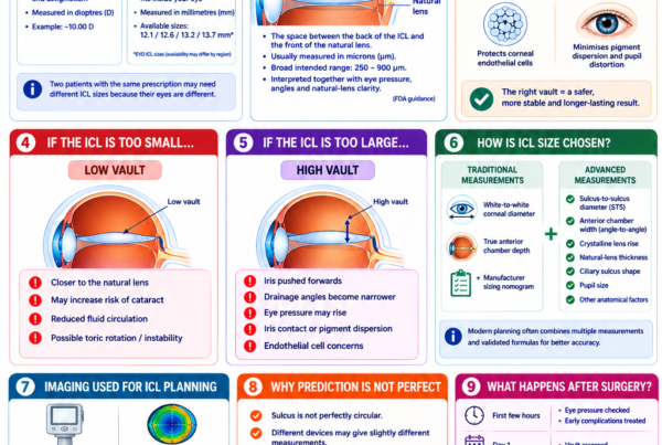
Very Low Vault
Vault is the space between the back of the ICL and the front of the natural lens.
When vault is very low, the ICL may sit too close to the natural lens.
This may increase concern about:
- Reduced aqueous-fluid circulation
- Contact with the natural lens
- Anterior subcapsular lens opacity
- Cataract formation
FDA professional guidance recommends ICL removal when vault is insufficient and an early anterior subcapsular cataract has developed. [2]
Before cataract develops, exchange for a different-sized ICL may sometimes be considered when the vault is extremely low and the expected benefit outweighs the risks of another operation.
Very High Vault
An ICL that is too large may produce excessive vault.
This can:
- Narrow the drainage angles
- Interfere with aqueous-fluid outflow
- Raise eye pressure
- Increase contact with the back of the iris
- Cause pigment dispersion
- Bring the ICL closer to the corneal endothelium
- Contribute to inflammation or pupil distortion
Removal or exchange may be required when excessive vault produces angle narrowing, raised pressure or another clinically important complication. [2]
High vault alone does not automatically require removal. The decision also depends on:
- Eye pressure
- Drainage-angle anatomy
- Endothelial cell density
- Pupil appearance
- Iris contact
- Symptoms
- Whether the measurements are changing over time
FDA guidance notes that vault outside the intended range may not require exchange or removal when the patient has no associated symptoms or complications. [2]
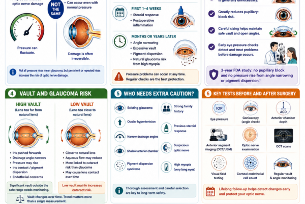
Raised Eye Pressure
An ICL may need to be removed or exchanged when raised eye pressure is caused by:
- Excessive vault
- Persistent drainage-angle narrowing
- Pigment dispersion
- Pupillary block
- Abnormal lens orientation
- Chronic iris contact
Not every pressure increase requires removal.
Early postoperative pressure elevation may result from:
- Retained surgical viscoelastic
- Postoperative inflammation
- A steroid response
These causes can often be managed with medication, modification of steroid treatment or removal of retained viscoelastic.
Removal becomes more relevant when the pressure problem is directly caused by the ICL’s physical size, position or interaction with the drainage structures.
Corneal Endothelial Cell Loss
The corneal endothelium is the layer of cells lining the inner surface of the cornea. These cells help keep the cornea clear.
ICL removal may be considered when:
- Endothelial cell density is falling excessively
- High vault brings the ICL too close to the cornea
- The drainage angle is becoming increasingly narrow
- Corneal swelling begins to develop
- Continued implantation is judged to pose an unacceptable long-term risk
A study of eyes undergoing ICL explantation found that higher vault, narrower anterior chamber angles and increased iris pigmentation were associated with greater endothelial cell loss. [4]
Removing the ICL may reduce an ongoing source of endothelial stress. However, cells that have already been permanently lost do not reliably regenerate.
Incorrect ICL Power
A significant residual prescription may remain because of:
- Measurement variation
- An incorrect lens power
- Limited available ICL powers
- Unexpected effective lens position
- Refractive change after surgery
- Toric lens rotation
- Astigmatism that was not fully corrected
Management may include:
- Spectacles
- Contact lenses
- Observation
- Corneal laser enhancement
- ICL exchange
Exchange is generally considered when the residual prescription significantly affects daily vision and a replacement ICL is expected to provide a meaningful benefit.
Toric ICL Rotation
A toric ICL corrects astigmatism and must remain aligned with its intended axis.
Significant rotation can cause:
- Residual astigmatism
- Blurred vision
- Ghosting
- Shadowing around letters
- Reduced night-time visual quality
The first treatment is often repositioning rather than complete removal.
Exchange may be considered when:
- The lens repeatedly rotates
- The physical size appears unsuitable
- Repositioning does not remain stable
- A different cylinder power or lens configuration is required
Persistent Inflammation or Iris Irritation
The ICL may occasionally produce ongoing interaction with the iris or other internal structures.
Possible effects include:
- Persistent inflammation
- Recurrent uveitis
- Iris pigment release
- Pupil distortion
- Posterior synechiae
- Raised eye pressure
- Deposits on the lens
Treatment may initially include medication and assessment of the lens position and vault.
Removal may be considered when inflammation persists despite appropriate treatment or when the ICL is clearly contributing to the problem.
Infection
Severe intraocular infection, known as endophthalmitis, is rare but potentially sight-threatening.
Treatment may require:
- Antibiotic injections into the eye
- Laboratory sampling
- Intensive topical treatment
- Vitrectomy
- ICL removal
Removal may help eliminate an implant surface on which microorganisms or inflammatory material could remain.
Trauma or ICL Displacement
Significant trauma may cause:
- ICL displacement
- Toric rotation
- Iris injury
- Natural-lens injury
- Abnormal vault
- Pupil distortion
- Raised eye pressure
- Retinal injury
A displaced ICL may be repositioned, exchanged or removed depending on:
- Lens integrity
- Eye pressure
- Natural-lens condition
- Iris damage
- Retinal findings
- Visual symptoms
Patient Preference
A patient may request removal because of:
- Persistent glare or halos
- Dissatisfaction with vision
- Anxiety about having an implant
- A desire to return to spectacles
- Intolerance of residual optical symptoms
Elective removal may be considered, but the patient should understand that:
- The original or current myopia will largely return
- Strong spectacles or contact lenses may again be required
- Visual symptoms may not necessarily disappear
- Removal is another intraocular operation
- The eye may have changed since the original surgery
The cause of glare, halos or visual dissatisfaction should be investigated before assuming that ICL removal will solve the symptoms.
How Common Is ICL Removal or Exchange?
Reported rates vary according to:
- The ICL model
- Length of follow-up
- Patient age
- Lens-sizing methods
- Surgeon experience
- The definitions used in each study
- Whether repositioning is counted separately
In a seven-year study of 2,283 central-port ICL procedures, 46 lenses—approximately 2%—required exchange or explantation. Incorrect vault was the most frequent indication. [3]
In the three-year United States FDA EVO ICL study, lens exchange was uncommon and was mainly performed for excessive vault. [6]
These figures come from specific clinical populations and should not be interpreted as a guaranteed rate for every patient or centre.
The overall message is that ICL removal or exchange is uncommon but remains a recognised component of long-term ICL care.
Does an Abnormal Vault Always Require Removal?
No.
Vault should not be interpreted as a number in isolation.
The surgeon also considers:
- Eye pressure
- Drainage-angle width
- Corneal endothelial cell density
- Natural-lens clarity
- Pupil shape
- Iris contact
- Symptoms
- Changes over time
An asymptomatic patient with stable pressure, open drainage angles, healthy endothelial cells and a clear natural lens may only require observation.
Removal or exchange becomes more likely when abnormal vault is accompanied by:
- Cataract
- Progressive endothelial cell loss
- Angle narrowing
- Raised eye pressure
- Pigment dispersion
- Pupil distortion
- Significant visual symptoms
How Is an ICL Removed?
ICL removal is normally performed in an operating theatre using sterile technique.
The precise method depends on:
- The ICL model
- The reason for removal
- Whether another ICL will be inserted
- Whether cataract surgery is also being performed
- The condition of the cornea, iris and natural lens
- The surgeon’s technique
A typical procedure includes the following steps.
Anaesthesia and Preparation
The eye is prepared with:
- Anaesthetic eyedrops
- Pupil-dilating medication when required
- Antiseptic cleaning
- A sterile eyelid speculum
Sedation or additional anaesthesia may be offered when appropriate.
Opening the Corneal Incision
The surgeon may reopen the original corneal incision or make a new small incision.
A side-port incision may also be created for surgical instruments.
Protecting Internal Eye Structures
A surgical viscoelastic may be placed inside the eye to:
- Protect the corneal endothelium
- Maintain space
- Separate the ICL from the natural lens
- Reduce unintended contact with the iris
Freeing the ICL
The surgeon carefully releases the ICL’s supporting footplates from behind the iris and brings the lens forwards into the anterior chamber.
Care is taken to avoid damage to:
- The natural lens
- The iris
- The corneal endothelium
- The drainage angle
Removing the Lens
The soft ICL may be:
- Folded
- Rolled
- Grasped and withdrawn
- Cut into smaller sections
- Removed through a specialised small-incision technique
Published surgical techniques have demonstrated that posterior chamber phakic lenses can be removed through small corneal incisions, reducing the need for a large wound. [10]
Completing the Procedure
The surgeon then:
- Removes excess viscoelastic
- Checks the natural lens
- Assesses the iris and pupil
- Confirms that the wound is secure
- Checks or arranges monitoring of eye pressure
When an exchange is planned, the replacement ICL may be implanted during the same operation.
Does the Corneal Incision Leave a Scar?
A small peripheral corneal scar or incision mark may remain after ICL implantation or removal.
The incision is located near the edge of the cornea rather than across the central visual axis. It is therefore unlikely to interfere significantly with central vision in an uncomplicated case.
Small clear-corneal incisions generally:
- Seal without stitches
- Heal over the following weeks
- Become minimally visible
- Produce little long-term visual disturbance
A small amount of surgically induced astigmatism may occur, particularly if the incision is enlarged or does not heal symmetrically. However, modern small-incision construction is intended to minimise this effect. [1,11]
The cornea is therefore not literally untouched, but it remains substantially closer to its original structure than after LASIK, PRK or SMILE, where central corneal tissue is intentionally reshaped or removed.
Is ICL Removal Painful?
ICL removal is normally performed with anaesthetic medication.
Patients may notice:
- Bright lights
- Movement
- Mild pressure
- Water around the eye
- Temporary blurring
Sharp pain is not expected during uncomplicated surgery.
Mild grittiness, watering or light sensitivity may occur afterwards.
Severe or increasing pain requires urgent assessment.
Can the Same ICL Be Put Back Into the Eye?
When an exchange is required, a new sterile ICL is generally implanted.
An explanted ICL is a single-use medical device and should not be resterilised and reimplanted. FDA professional instructions state that the lens should not be reused or resterilised. [2]
If an ICL is only repositioned and remains within the sterile surgical field, the existing lens may remain in the eye.
What Happens to Vision After Simple ICL Removal?
If the ICL is removed without another corrective lens being implanted, the myopia and astigmatism it was correcting will largely return.
A highly myopic patient may once again experience:
- Markedly blurred unaided distance vision
- Dependence on strong spectacles
- Dependence on contact lenses
- Image minification with high-minus spectacles
- Difficulty functioning without optical correction
In an uncomplicated eye, the prescription after removal is expected to be close to the underlying prescription that the eye would have had without the ICL.
This may be similar to the original preoperative prescription, but it may not be identical because the eye can change over time.
Possible reasons for a difference include:
- Natural progression or regression of myopia
- A change in corneal astigmatism
- Cataract development
- Changes in the natural crystalline lens
- Previous corneal laser enhancement
- Surgical effects
- Measurement variation
Appropriate spectacles or contact lenses should generally restore vision close to the preoperative corrected level, provided the cornea, natural lens, retina and optic nerve remain healthy.
Does ICL Removal Restore the Eye to Its Preoperative State?
In most uncomplicated cases, ICL removal restores the eye largely to its preoperative optical and anatomical state.
Once the ICL is removed:
- Its refractive correction is eliminated
- The original or current underlying myopia largely returns
- The natural crystalline lens remains
- The central cornea remains substantially unchanged
- Glasses or contact lenses can usually be used again
A minor peripheral incision scar may remain, but this usually heals well and is not visually significant.
The eye may not return completely to its original state when there has been:
- Natural prescription change
- Cataract
- Significant endothelial cell loss
- Iris damage
- Pupil distortion
- Glaucoma or optic nerve injury
- Infection or severe inflammation
- Previous corneal laser treatment
- Retinal or macular disease
- Another surgical complication
ICL removal can remove the optical effect and physical presence of the lens. It cannot reverse unrelated eye disease or damage that has already occurred.
The FDA therefore appropriately cautions that the residual effects after removal are not fully known, even though the additive nature of ICL means that uncomplicated removal generally brings the eye close to its previous state. [1]
What Happens to Vision After an ICL Exchange?
When the original ICL is replaced with a more appropriate lens, the new ICL provides the refractive correction.
The intended benefit may be:
- Improved vault
- Wider drainage angles
- Reduced pressure risk
- Better toric stability
- Improved astigmatism correction
- Reduced residual myopia or hyperopia
- Better visual quality
Vision may initially be blurred because of:
- Pupil dilation
- Postoperative inflammation
- Mild corneal swelling
- Tear-film disturbance
- Temporary pressure changes
The final result depends on the replacement lens, the eye’s anatomy, healing and the health of the rest of the eye.
What Happens During Cataract Surgery?
When an ICL patient develops a visually significant cataract:
- The ICL is removed.
- The cloudy natural lens is removed.
- A cataract intraocular lens is implanted.
The new cataract lens can usually correct much of the patient’s myopia and astigmatism.
Potential lens strategies include:
- Monofocal correction
- Toric correction
- Monovision
- Selected presbyopia-correcting lenses
Suitability depends on:
- Corneal health
- Retinal and macular health
- Optic nerve health
- Lifestyle
- Visual priorities
- The degree and source of astigmatism
Studies have reported favourable visual outcomes after combined ICL removal and cataract surgery. [5,12]
Will Halos or Glare Disappear After Removal?
Halos or glare may improve when they are directly caused by:
- The ICL optic
- The central port
- Lens decentration
- Toric misalignment
- Inappropriate vault
- Light reflecting from the implant
However, symptoms may continue when they arise from:
- Large pupils
- Dry eye
- Residual refractive error
- Corneal irregularity
- Cataract
- Retinal or macular disease
- Previous laser surgery
- Other optical aberrations
The cause of the symptoms should be investigated before deciding that removal will provide relief.
Will Eye Pressure Return to Normal After Removal?
Eye pressure may improve when the ICL is causing:
- Drainage-angle narrowing
- Pupillary block
- Pigment dispersion
- Iris crowding
- Excessive vault
Pressure may remain elevated when the patient also has:
- Primary glaucoma
- A steroid response
- Permanent drainage-angle damage
- Peripheral anterior synechiae
- Optic nerve susceptibility unrelated to the ICL
Removing the lens cannot reverse optic nerve damage that has already occurred.
Will Endothelial Cell Loss Stop After Removal?
Removal may reduce continued endothelial stress when the ICL’s position, vault or effect on the drainage angle is contributing to cell loss.
Endothelial measurements may stabilise after removal in many patients, but continued monitoring remains necessary. [4]
Corneal endothelial cells have very limited regenerative capacity. An apparent increase in measured cell density after removal may reflect:
- Measurement variation
- Changes in cell shape
- Redistribution of the remaining cells
It should not necessarily be interpreted as true regrowth of lost endothelial cells.
What Are the Risks of ICL Removal?
ICL removal is generally feasible, but it is another operation inside the eye.
Potential risks include:
- Corneal endothelial cell loss
- Corneal swelling
- A small corneal scar
- Surgically induced astigmatism
- Iris injury
- Bleeding
- Pupil distortion
- Natural-lens injury
- Cataract
- Raised eye pressure
- Low eye pressure
- Inflammation
- Infection
- Wound leakage
- Residual prescription
- Need for further surgery
- Loss of best-corrected vision
Risk may be greater when:
- The ICL has been present for many years
- There are iris adhesions
- The anterior chamber is shallow
- Endothelial cell density is already low
- Cataract is present
- The ICL is abnormally positioned
- Infection or severe inflammation is present
- Previous surgery has altered the anatomy
Can the Natural Lens Be Damaged During Removal?
Yes, although careful technique reduces this risk.
The ICL lies immediately in front of the natural crystalline lens. Surgical instruments and the explanted lens must therefore be moved without touching it.
Accidental contact may cause:
- Anterior subcapsular lens opacity
- Cataract
- The need for cataract surgery
Surgical experience is especially important when freeing the ICL from behind the iris and bringing it into the anterior chamber.
What Is Recovery Like After ICL Removal?
Recovery depends on whether the procedure involves:
- Simple removal
- ICL exchange
- Toric repositioning
- Combined cataract surgery
- Treatment of infection or inflammation
The First Day
Patients may experience:
- Blurred vision
- Mild grittiness
- Watering
- Redness
- Light sensitivity
- Fluctuating vision
The surgeon usually checks:
- Eye pressure
- Corneal clarity
- Inflammation
- Pupil appearance
- Natural-lens clarity
- Wound integrity
- Replacement ICL position when applicable
The First Week
Antibiotic and anti-inflammatory eyedrops are commonly prescribed.
Patients are generally advised to:
- Avoid rubbing the eye
- Keep contaminated water out of the eye
- Avoid swimming
- Avoid strenuous exercise
- Use an eye shield when directed
- Attend all follow-up visits
The First Month
Vision and refraction usually become more stable.
The surgeon may assess:
- Corneal healing
- Endothelial health
- Eye pressure
- Natural-lens clarity
- Residual prescription
- Replacement ICL position
- Vault
- Toric alignment
Recovery after combined cataract surgery generally follows a cataract-surgery recovery timeline rather than a simple ICL-removal timeline.
Can Another ICL Be Implanted Later?
Possibly.
A new ICL may be considered when:
- The original lens was the wrong size
- The optical power was unsuitable
- A toric lens repeatedly rotated
- The eye remains anatomically suitable
- Endothelial cell density is adequate
- The natural lens remains clear
- The original reason for removal has resolved
Replacement may occur:
- During the same procedure
- After the eye has healed
- After infection or inflammation has completely resolved
A replacement ICL is less likely to be appropriate when removal was required because of:
- Significant endothelial cell loss
- Persistently narrow drainage angles
- Cataract
- Glaucoma
- Inadequate internal eye space
- Recurrent inflammation
Can LASIK, SMILE or PRK Be Performed After ICL Removal?
Potentially, but only when the cornea is suitable.
Assessment should include:
- Corneal thickness
- Corneal topography and tomography
- Dry-eye status
- Residual refractive error
- Previous laser surgery
- Corneal biomechanical risk
- Visual potential
Laser surgery may be considered when:
- The remaining prescription is within a suitable range
- Adequate corneal tissue is available
- Corneal tomography is normal
- The eye surface is healthy
A highly myopic patient who originally received ICL because laser treatment was unsuitable may remain unsuitable for corneal laser surgery after removal.
Can the ICL Be Removed Because the Prescription Has Changed?
Yes, but removal is not always the first option.
A changed prescription may be managed with:
- Spectacles
- Contact lenses
- Corneal laser enhancement
- Toric repositioning
- ICL exchange
- Observation
The cause of the refractive change should first be identified.
Possible causes include:
- Myopia progression
- Toric rotation
- Cataract
- Corneal change
- Macular disease
- Measurement variability
Should an ICL Be Removed Before It Causes Problems?
Routine preventive removal is generally not recommended when:
- Vision is good
- Vault is clinically acceptable or stable
- Eye pressure is normal
- The drainage angles remain open
- Endothelial cell density is satisfactory
- The natural lens is clear
- The patient has no significant symptoms
Removing a well-functioning lens would expose the patient to another intraocular procedure without a clear medical benefit.
Regular monitoring is intended to identify changes early enough for treatment or elective removal before permanent damage develops.
How Long Can an ICL Stay Inside the Eye?
The ICL is designed to remain inside the eye long term.
There is no routine expiry date after implantation.
Long-term studies of central-port ICLs have reported favourable visual and refractive stability at ten years in appropriately selected patients. [13]
Removal may eventually become necessary because of:
- Cataract
- Vault changes
- Endothelial concerns
- Eye-pressure problems
- Refractive change
- Another eye operation
The lens should not be removed solely because a particular number of years has passed.
Why Is Long-Term Follow-Up Important?
Regular follow-up helps identify whether repositioning, exchange or removal is becoming necessary before permanent damage occurs.
Reviews may assess:
- Visual acuity
- Refraction
- ICL position
- Vault
- Toric alignment
- Eye pressure
- Drainage-angle anatomy
- Corneal endothelial cell density
- Natural-lens clarity
- Retinal and macular health
- Optic nerve health
FDA patient guidance recommends continued monitoring of cataract, eye pressure and the corneal endothelium for as long as the ICL remains inside the eye. [1]
When Should You Seek Urgent Eye Care?
Seek urgent assessment if you have an ICL and develop:
- Severe or increasing eye pain
- Rapidly worsening vision
- Sudden loss of vision
- Marked redness
- Significant light sensitivity
- Severe headache
- Nausea or vomiting
- A cloudy cornea
- A white or grey spot on the eye
- A distorted pupil
- New flashes or floaters
- A curtain-like shadow
- Significant trauma
These symptoms may indicate:
- Raised eye pressure
- Infection
- Severe inflammation
- Cataract-related complications
- ICL displacement
- Retinal tear or detachment
- Another sight-threatening problem
Frequently Asked Questions About ICL Removal
Can an ICL Be Removed at Any Time?
An ICL can generally be removed when medically necessary.
Removal may become more complex when there are:
- Iris adhesions
- Cataract
- Inflammation
- Endothelial damage
- Altered internal anatomy
The decision should be based on a clear balance of the expected benefits and surgical risks.
Is ICL Removal Easier Than the Original Surgery?
Not necessarily.
The ICL is soft and removable, but the surgeon must avoid damaging the cornea, iris, drainage angle and natural lens.
Removal may be more complex when the ICL has been present for many years or when a complication is already present.
Is ICL Removal Reversible?
ICL surgery is largely reversible.
Removing the lens usually:
- Eliminates its refractive effect
- Allows the original or current myopia to return
- Preserves the natural crystalline lens
- Leaves the central cornea substantially unchanged
A small peripheral corneal incision or scar remains, but this generally heals well and does not significantly affect central vision.
The eye may not return exactly to its preoperative condition if natural changes or complications have occurred.
Will My Original Prescription Return?
The original myopia and astigmatism will largely return when the ICL is removed without replacement.
The measured prescription may differ slightly because the eye may have changed naturally since the original procedure.
Will the Cornea Be Scarred?
A small incision is made near the edge of the cornea to insert and remove the ICL.
This may leave a minor peripheral scar or incision mark. It generally heals well and is unlikely to affect central vision.
A small astigmatic change may occur, particularly if the incision needs to be enlarged.
Will My Vision Be the Same as Before ICL Surgery?
In an uncomplicated eye, vision with the appropriate spectacles or contact lenses should generally be similar to the preoperative corrected level.
Vision may differ if there has been:
- A prescription change
- Cataract
- Retinal disease
- Corneal disease
- Optic nerve damage
- A surgical complication
Will My Vision Be Blurry Immediately After Removal?
Yes, particularly when no replacement correction is provided.
Temporary blur may also result from:
- Pupil dilation
- Inflammation
- Corneal swelling
- Tear-film disturbance
Can the ICL Be Exchanged for a Stronger or Weaker Lens?
Yes, provided the eye remains suitable and an appropriate replacement lens is available.
Can the ICL Be Exchanged for a Different Size?
Yes.
A smaller lens may be used for excessive vault.
A larger lens may be considered for inadequate vault or rotational instability in selected cases.
Can a Toric ICL Be Repositioned Without Removal?
Yes.
When the power and size are otherwise appropriate, a toric ICL can often be rotated back to its intended astigmatic axis.
Does Cataract Always Require ICL Removal?
When cataract surgery is required, the ICL is normally removed so that the natural lens can be safely extracted and replaced with a cataract intraocular lens.
Can I Keep the ICL if I Have Mild Cataract?
Possibly.
An early cataract that does not affect vision may be monitored.
The decision depends on:
- Cataract type
- Vault
- Visual symptoms
- Rate of progression
- Age
- Other eye findings
Does Removing the ICL Stop Cataract Progression?
Removal may eliminate a contributing factor when low vault is causing natural-lens contact.
However, an existing cataract may remain or continue to progress. Age-related cataract risk also remains.
Will Halos Disappear After Removal?
They may improve when the ICL is the main cause.
They may persist when caused by:
- Pupil size
- Dry eye
- Cataract
- Corneal irregularity
- Retinal disease
- Another optical issue
Can the ICL Be Removed if Eye Pressure Is High?
Yes, when its size, position or vault is contributing to the pressure problem.
Medication or another procedure may be sufficient when the pressure increase has a different cause.
Can ICL Removal Damage the Cornea?
It can cause:
- Additional endothelial cell loss
- Temporary corneal swelling
- A peripheral incision scar
- A small astigmatic change
Significant corneal damage is uncommon with careful surgery, but the risk cannot be eliminated.
Can I Have Another ICL in the Future?
Possibly, depending on why the original lens was removed and whether the internal eye anatomy remains suitable.
Will Insurance Cover ICL Removal?
Coverage depends on:
- The insurer
- The policy
- The medical reason for removal
- Whether the procedure is considered medically necessary
Removal for cataract, glaucoma, endothelial damage or another complication may be treated differently from elective removal requested because of preference or dissatisfaction.
Key Takeaway
An ICL can usually be surgically removed, repositioned or exchanged.
Because it is an additional lens placed behind the iris rather than a procedure that permanently removes central corneal tissue, uncomplicated removal generally restores the eye largely to its preoperative optical state.
After simple removal:
- The refractive correction provided by the ICL is lost
- The original or current myopia and astigmatism largely return
- Spectacles or contact lenses can usually be used again
- The natural crystalline lens remains in place
- The central corneal structure remains substantially unchanged
A small peripheral corneal incision or scar remains. This generally heals well and is unlikely to affect central vision significantly.
The eye may not return exactly to its original condition if there has been:
- Natural refractive progression
- Cataract
- Endothelial cell loss
- Glaucoma
- Iris or pupil injury
- Infection
- Retinal disease
- Previous additional surgery
Removal may be required because of:
- Cataract
- Excessively low or high vault
- Raised eye pressure
- Endothelial cell loss
- Incorrect refractive power
- Recurrent toric rotation
- Persistent inflammation
- Infection
- Trauma or displacement
ICL surgery can therefore reasonably be described as largely reversible, while recognising that removal is still another intraocular operation and carries its own risks.
Regular long-term examinations help detect vault, pressure, endothelial and cataract changes early, when treatment or elective exchange is generally safer and more effective.
References
- US Food and Drug Administration. EVO and EVO+ Visian Implantable Collamer Lens: Patient Information Booklet. 2022.
- US Food and Drug Administration. EVO and EVO+ Visian Implantable Collamer Lens: Instructions for Use. 2022.
- Alhamzah A, et al. Indications for Exchange or Explantation of Phakic Implantable Collamer Lens With Central Port in Patients With and Without Keratoconus. 2021.
- Yoon HY, et al. Causes and Outcomes of Implantable Collamer Lens Explantation in Patients With Corneal Endothelial Cell Loss. 2024.
- Kamiya K, et al. A Multicentre Study on Clinical Outcomes of Simultaneous Implantable Collamer Lens Removal and Phacoemulsification With Intraocular Lens Implantation in Eyes Developing Cataract. 2025.
- Parkhurst G, et al. Three-Year Results From the United States FDA Prospective Multicentre Clinical Study of the EVO Implantable Collamer Lens. 2025.
- Choi H, et al. Size Exchange and Vault Response in Posterior Chamber Phakic Intraocular Lens Implantation. 2026.
- AlSabaani NA, et al. Causes of Phakic Implantable Collamer Lens Explantation or Exchange. 2016.
- Kaur M, et al. Indications for Explantation of Implantable Collamer Lens. 2018.
- Coskunseven E, et al. Tuck-and-Pull Technique for Posterior Chamber Phakic Intraocular Lens Explantation. 2021.
- Borkenstein AF, Borkenstein EM, Malyugin B. Clear Corneal Incision, an Important Step in Modern Cataract Surgery: A Review. 2023.
- Del Risco NE, et al. Visual Outcomes of Cataract Surgery in Patients With Previous Implantable Collamer Lens Surgery. 2024.
- Alfonso-Bartolozzi B, et al. Ten-Year Follow-up of Posterior Chamber Phakic Intraocular Lenses With Central-Port Design. 2024.
- Vargas V, et al. Safety and Visual Outcomes Following Posterior Chamber Phakic Intraocular Lens Removal and Cataract Surgery. 2020.