Author: Dr Val Phua
Estimated reading time: 16 minutes
Implantable Collamer Lens surgery—commonly known as ICL surgery—can provide long-lasting correction of myopia and astigmatism without permanently removing central corneal tissue.
The lens is implanted behind the iris and in front of the natural crystalline lens. It is designed to remain inside the eye for many years and does not have a routine expiry date after implantation.
Modern central-port ICLs have shown favourable safety, visual outcomes and refractive stability in studies extending to eight and ten years. Recent systematic reviews have also found that serious or vision-threatening complications appear uncommon in appropriately selected patients. However, ICL surgery remains an operation inside the eye, and no implant should be considered completely free of long-term risk.
The main issues that require long-term monitoring are:
- Cataract development
- Corneal endothelial cell loss
- Eye-pressure and drainage-angle changes
- ICL vault and position
- Toric ICL rotation
- Glare or halos
- The need for repositioning, exchange or removal
- Retinal and macular disease associated with high myopia
An excellent visual result during the first year is reassuring, but it does not remove the need for regular eye examinations throughout life.
What Does “Long-Term Safety” Mean?
Long-term safety refers to whether an ICL continues to function without causing clinically significant problems over many years.
Doctors assess:
- Whether vision remains clear
- Whether the refractive correction remains stable
- Whether the natural lens stays clear
- Whether eye pressure remains healthy
- Whether the drainage angles remain open
- Whether corneal endothelial cells remain adequate
- Whether vault remains acceptable
- Whether the ICL requires repositioning, exchange or removal
- Whether any complication causes permanent loss of vision
A procedure can have excellent average long-term results while still producing complications in a small number of patients.
Long-term safety therefore does not mean that every patient will have an identical result. It means that serious adverse events are uncommon when appropriate patients are selected, surgery is performed carefully and follow-up is maintained.
Is There a Difference Between Older ICLs and Modern EVO ICLs?
Yes.
Not all ICL studies evaluate the same lens design.
Earlier ICL Models
Earlier posterior-chamber ICLs did not have a central opening.
Patients commonly required a laser peripheral iridotomy before surgery to create an alternative pathway for fluid to move from behind the iris into the front of the eye.
Earlier lens models were associated with concerns including:
- Pupillary-block glaucoma
- Anterior subcapsular cataract
- Low-vault contact with the natural lens
- Greater uncertainty regarding long-term endothelial cell loss
These older studies remain important, but their complication rates should not automatically be applied to current EVO ICL surgery.
Modern Central-Port EVO ICLs
Modern EVO ICLs contain a small central opening that allows aqueous fluid to circulate through the lens.
This design generally:
- Eliminates the need for routine preoperative peripheral iridotomy
- Improves aqueous circulation around the natural lens
- Reduces pupillary-block risk
- Appears to reduce anterior subcapsular cataract risk compared with earlier non-port models
The United States FDA three-year study found that the central port permitted physiological aqueous flow and was associated with a low incidence of anterior subcapsular cataract and no pupillary block requiring peripheral iridotomy in the studied group.
Modern central-port ICLs have improved the safety profile, but they have not eliminated every possible complication.
What Do Long-Term Studies Show?
Available evidence includes studies with follow-up periods of:
- Five years
- Seven years
- Eight years
- Ten years
- More than ten years for some earlier lens models
Collectively, these studies generally report:
- Good preservation of corrected vision
- Stable refractive results
- Stable average eye pressure
- Gradual reduction in vault over time
- Modest average endothelial cell change
- A low incidence of serious adverse events
- Occasional cataract, pressure, sizing or exchange-related complications
A 2025 meta-analysis of central-port ICL studies concluded that the lenses demonstrated high efficacy, predictability and safety for moderate-to-high myopia, with a low incidence of serious adverse events. The authors nevertheless called for continued long-term follow-up and broader geographic data.
A 2026 systematic review examining possible harms similarly concluded that complications were uncommon and vision-threatening events appeared very rare. However, it cautioned that many available studies had relatively short follow-up or potential sources of bias, which may underestimate complications that develop many years later.
What Have Eight-Year Studies Found?
An eight-year study of central-port ICL implantation for moderate-to-high myopia reported favourable long-term safety, effectiveness and refractive stability.
The study evaluated:
- Unaided and corrected vision
- Refractive error
- Eye pressure
- Endothelial cell density
- Vault
- Postoperative complications
The overall results supported the long-term use of the central-port lens in appropriately selected eyes.
As with all retrospective long-term studies, the findings should be interpreted in the context of:
- Patient selection
- Loss of some patients to follow-up
- Surgical techniques used at the time
- Lens-sizing methods
- The relatively limited number of eyes still examined at the final visit
What Have Ten-Year Central-Port Studies Found?
A 2024 study evaluating central-port ICLs at ten years reported good long-term clinical outcomes, including preservation of corrected visual acuity and a favourable complication profile in the studied population.
Importantly, this provided direct ten-year evidence for the central-port design rather than relying only on earlier ICL models.
The study supports the view that a properly positioned central-port ICL can remain safe and effective over a decade.
However, ten-year evidence still does not represent lifetime evidence.
Many patients receive an ICL in their twenties or thirties. The lens may therefore remain inside the eye for several decades—far longer than the follow-up duration of most published studies.
Does the ICL Power Wear Out?
No.
The optical power of the ICL does not normally fade or become weaker with time.
The lens does not:
- Dissolve
- Lose its refractive power
- Require routine replacement because it has expired
- Need cleaning like a surface contact lens
However, vision can change even when the ICL remains optically stable.
Possible causes include:
- Continued myopia progression
- Natural changes in the crystalline lens
- Presbyopia
- Cataract
- Toric ICL rotation
- Corneal changes
- Dry eye
- Retinal or macular disease
- Glaucoma
A future prescription does not necessarily mean that the ICL itself has deteriorated.
How Stable Is Vision Over Time?
Long-term studies generally show stable refractive correction after central-port ICL implantation.
Some patients may develop a small myopic shift over time because of:
- Continuing axial elongation
- Natural-lens changes
- Cataract development
- Measurement variation
- Age-related ocular changes
A mild return of myopia does not automatically require ICL exchange.
Depending on its severity, management may include:
- Observation
- Spectacles for selected tasks
- Contact lenses
- Corneal laser enhancement in suitable eyes
- ICL exchange
- Cataract surgery when lens changes are present
ICL surgery corrects the prescription measured at the time of surgery. It does not guarantee that the eye itself will never change again.
Does ICL Surgery Cause Cataract?
Cataract is one of the most important long-term concerns following ICL implantation.
The ICL sits in front of the natural crystalline lens. Cataract may develop because of:
- Normal ageing
- Low vault
- Contact or close proximity between the ICL and natural lens
- Reduced fluid circulation
- Surgical contact with the natural lens
- Very high myopia
- Older age at implantation
- Individual anatomical factors
The cataract most specifically associated with ICL surgery is an anterior subcapsular cataract, which forms at the front surface of the natural lens.
Is Cataract Less Common With Modern EVO ICLs?
Current evidence suggests that central-port ICLs have reduced anterior subcapsular cataract risk compared with earlier non-port designs.
The central opening allows aqueous fluid to circulate more naturally around the anterior surface of the crystalline lens.
The three-year United States FDA study reported a very low incidence of anterior subcapsular cataract and no pupillary-block events requiring peripheral iridotomy.
Ten-year central-port studies have also reported favourable lens-safety outcomes.
Nevertheless, modern EVO ICL does not prevent:
- Age-related nuclear cataract
- Cortical cataract
- Cataract caused by low vault
- Cataract caused by surgical contact with the natural lens
- Cataract associated with high myopia
Who Has a Higher Long-Term Cataract Risk?
Risk may be higher in patients who:
- Are older at implantation
- Have very high myopia
- Have a shallow anterior chamber
- Develop persistently low vault
- Already have early natural-lens changes
- Keep the ICL for many years
A long-term study found that older age and lower postoperative vault were associated with cataract development. The reported cataract rate from that cohort should not be applied directly to every modern EVO ICL patient because the study population, lens generations and follow-up characteristics differ.
This is one reason ICL suitability should be considered particularly carefully in patients over 40.
What Happens if Cataract Develops?
If cataract becomes visually significant:
- The ICL is removed.
- The cloudy natural lens is removed.
- A cataract intraocular lens is implanted.
The new cataract lens can usually correct much of the myopia and astigmatism.
Previous ICL surgery generally does not prevent cataract surgery, but the surgeon must consider:
- The degree of high myopia
- Axial length
- Retinal health
- Corneal measurements
- Previous ICL power
- Astigmatism
- The desired refractive target
What Is Corneal Endothelial Cell Loss?
The corneal endothelium is a single layer of cells lining the inner surface of the cornea.
These cells pump fluid out of the cornea and help keep it transparent.
Endothelial cells naturally decrease with age and have very limited regenerative ability.
Additional cell loss may occur after any operation inside the eye.
If too many cells are lost, the cornea may develop:
- Swelling
- Cloudiness
- Glare
- Reduced vision
- Painful epithelial blisters
- Corneal decompensation
Severe corneal decompensation may require corneal transplantation.
Does ICL Cause Continuous Endothelial Cell Loss?
Some endothelial cell reduction occurs after ICL surgery.
Part of this may represent:
- An initial surgical effect
- Normal age-related cell loss
- Measurement variation
- Ongoing anatomical interaction between the ICL and the front of the eye
Long-term central-port studies have generally reported modest average endothelial cell changes, although individual patients may lose more cells than the study average.
The FDA notes that endothelial cell loss can continue over time and recommends periodic endothelial monitoring for as long as the ICL remains implanted.
Who Is at Greater Risk of Endothelial Cell Loss?
Possible risk factors include:
- A low preoperative endothelial cell count
- A shallow anterior chamber
- Excessively high vault
- Narrow drainage angles
- An ICL positioned too close to the cornea
- Increasing age
- Surgical trauma
- Persistent inflammation
- Multiple intraocular procedures
High vault may bring the ICL closer to the iris and corneal endothelium and may contribute to both angle narrowing and endothelial stress.
How Is Endothelial Safety Monitored?
Specular microscopy is used to measure:
- Endothelial cell density
- Average cell size
- Variation in cell size
- Variation in cell shape
The FDA provides age- and anterior-chamber-depth-related minimum endothelial cell criteria before implantation. It recommends postoperative monitoring at intervals determined by the treating ophthalmologist.
A single measurement should not be interpreted in isolation.
The surgeon considers:
- The trend over time
- Measurement repeatability
- The patient’s age
- Corneal thickness and clarity
- Vault
- Anterior chamber depth
- Whether the lens remains anatomically suitable
Does ICL Surgery Cause Raised Eye Pressure?
Eye pressure can rise after ICL surgery.
Early causes include:
- Retained surgical viscoelastic
- Postoperative inflammation
- Steroid response
- Pupillary block
- Excessive vault
- Angle narrowing
Longer-term causes may include:
- Persistent angle crowding
- Pigment dispersion
- Peripheral anterior synechiae
- Steroid response
- Glaucoma developing independently of the ICL
The FDA recommends checking eye pressure within one to six hours after implantation because early pressure elevation may need prompt treatment.
Is Long-Term Glaucoma Common After ICL?
Published central-port studies generally report stable average eye pressure over medium- and long-term follow-up.
However, average study results cannot exclude glaucoma in an individual patient.
Highly myopic patients already have an increased glaucoma risk, even without ICL surgery. Their optic nerves and OCT scans can also be more difficult to interpret.
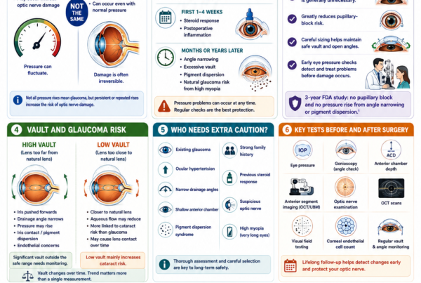
Long-term monitoring should therefore include:
- Eye pressure
- Drainage-angle examination
- Optic nerve assessment
- OCT when appropriate
- Visual-field testing when indicated
The FDA states that the longer-term risks of glaucoma, pigment dispersion and peripheral anterior synechiae are not fully established.
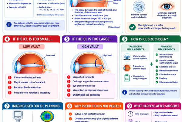
What Is Vault and Why Does It Matter Long Term?
Vault is the distance between the back of the ICL and the front of the natural crystalline lens.
An appropriate vault maintains separation from the natural lens without excessively crowding the front of the eye.
Low Vault May Increase the Risk of:
- Natural-lens contact
- Anterior subcapsular cataract
- Reduced aqueous circulation
High Vault May Increase the Risk of:
- Narrow drainage angles
- Raised eye pressure
- Iris contact
- Pigment dispersion
- Pupil distortion
- Endothelial concerns
FDA professional guidance describes a broad intended vault range but also states that a measurement outside this range does not automatically require exchange or removal when no associated complication is present.
Does Vault Change With Time?
Yes.
Vault commonly decreases gradually over the years.
Possible reasons include:
- Thickening of the natural crystalline lens
- Age-related anatomical changes
- Changes in pupil size
- Changes in the ciliary body
- Natural variation in measurement
A gradual reduction does not necessarily indicate a problem.
The significance depends on whether low vault is accompanied by:
- Natural-lens opacity
- Contact between the ICL and natural lens
- Visual symptoms
- Other anatomical concerns
This is why vault should be monitored as a trend rather than judged from one measurement alone.
Can the ICL Move or Rotate Years Later?
A spherical ICL usually remains stable behind the iris.
A toric ICL must remain aligned with its intended astigmatic axis. Rotation can reduce the cylinder correction and cause:
- Blurred vision
- Ghosting
- Residual astigmatism
- Reduced night-time clarity
Most clinically significant toric rotation occurs relatively early, but later movement can occur after:
- Trauma
- Anatomical change
- Lens-sizing problems
- Additional eye surgery
Treatment may include:
- Observation
- Spectacle correction
- Surgical repositioning
- ICL exchange
- Corneal laser enhancement in suitable eyes
Can an ICL Cause Pigment Dispersion?
The back surface of the iris contains pigment.
If the ICL repeatedly rubs against the iris, pigment may be released and accumulate within the drainage angle.
This may contribute to:
- Increased angle pigmentation
- Raised eye pressure
- Pigmentary glaucoma
- Inflammation
Risk may be influenced by:
- Excessive vault
- Lens position
- Iris anatomy
- Pupil movement
- Lens sizing
Clinically significant pigment-related pressure problems appear uncommon in modern central-port studies, but long-term monitoring remains important because the FDA states that this risk is not yet completely established.
Does ICL Surgery Increase Retinal-Detachment Risk?
Most ICL patients have moderate, high or very high myopia.
High myopia itself increases the lifetime risk of:
- Lattice degeneration
- Retinal holes
- Retinal tears
- Posterior vitreous detachment
- Rhegmatogenous retinal detachment
Correcting the prescription with an ICL does not shorten the elongated eye or remove these underlying retinal risks.
Ten-year comparative research found no significant increase in retinal-detachment prevalence in ICL-operated eyes compared with similar highly myopic eyes that had not undergone ICL surgery.
Another long-term comparison between central-port ICL patients and highly myopic rigid gas-permeable contact lens wearers similarly did not find evidence that ICL added significant long-term retinal-detachment risk.
These findings are reassuring, but they do not mean that retinal detachment cannot occur after ICL surgery.
Why Is a Dilated Retinal Examination Still Important?
ICL surgery corrects focus. It does not treat:
- Lattice degeneration
- Retinal holes
- Retinal tears
- Posterior staphyloma
- Myopic macular degeneration
- Myopic traction maculopathy
- Myopic choroidal neovascularisation
A highly myopic patient with excellent unaided vision after ICL surgery still requires regular retinal and macular examinations.
Seek urgent assessment for:
- Sudden new floaters
- Flashes of light
- A curtain or shadow
- Sudden peripheral visual-field loss
- Sudden central distortion
- Sudden reduction in vision
Can ICL Surgery Cause Chronic Inflammation?
Mild inflammation is expected immediately after surgery and usually resolves with prescribed anti-inflammatory eyedrops.
Chronic or recurrent inflammation is uncommon but may occur because of:
- Iris contact
- Pigment dispersion
- Abnormal lens position
- Infection
- Pre-existing inflammatory eye disease
- An unusual immune response
Possible symptoms include:
- Redness
- Eye pain
- Light sensitivity
- Blurred vision
- Floaters
- Raised eye pressure
Persistent inflammation may require:
- Additional medication
- Investigation for infection
- ICL repositioning
- ICL exchange or removal
Patients with a history of uveitis require careful individual assessment because safety has not been established for all such patients.
Can Infection Occur Years Later?
Postoperative endophthalmitis usually develops during the early period after intraocular surgery.
Late infection is much less common but may theoretically occur after:
- Trauma
- Another intraocular procedure
- Wound-related problems
- Severe inflammation
Any patient with an ICL who develops increasing pain, redness and rapidly worsening vision requires urgent assessment.
Can the ICL Cause Glare or Halos Long Term?
Yes.
Patients may experience:
- Halos around headlights
- Glare
- Starbursts
- Rings or arcs of light
- Difficulty with night driving
Possible contributing factors include:
- A large pupil
- The optical edge of the lens
- The central port
- Residual myopia or astigmatism
- Toric misalignment
- Dry eye
- Vault
- Other optical aberrations
Many patients adapt, and symptoms often become less noticeable over time.
However, a small proportion may continue to notice night-vision symptoms despite excellent eye-chart vision.
The FDA advises that patients whose pupils are larger than the ICL optic in dim lighting may be more likely to notice glare or halos.
Does the Central Port Affect Vision?
The central port is small and designed to maintain aqueous circulation.
Most patients do not consciously see it.
Some patients may notice transient rings, arcs or halos, particularly in dim lighting. These symptoms often decrease with neural adaptation.
The central port’s safety advantage is that it facilitates physiological aqueous flow and reduces the need for a peripheral iridotomy.
Can the ICL Material Deteriorate Inside the Eye?
The Collamer lens is designed for long-term biocompatibility.
Published long-term studies have not shown routine degradation, clouding or loss of optical power of the lens material.
The lens generally does not need replacement because the material has worn out.
Removal or exchange is more likely to be required because of:
- Cataract
- Vault
- Incorrect power
- Toric rotation
- Eye-pressure problems
- Endothelial concerns
- Another eye operation
Can an ICL Stay Inside the Eye for Life?
Potentially, yes.
There is no routine requirement to remove a well-functioning ICL after a fixed number of years.
An ICL may remain in place when:
- Vision is satisfactory
- Eye pressure is healthy
- Drainage angles remain acceptable
- Vault remains clinically safe
- Endothelial cell density remains adequate
- The natural lens remains clear
- There is no significant inflammation
- The patient continues regular follow-up
However, many patients will eventually develop cataract as part of normal ageing.
When cataract surgery becomes necessary, the ICL is generally removed and the natural lens is replaced with a cataract intraocular lens.
How Often Does an ICL Need to Be Removed or Exchanged?
ICL exchange or removal is uncommon.
A seven-year retrospective series of 2,283 central-port ICL implantations reported exchange or explantation in approximately 2% of lenses. Incorrect vault was the most common indication.
The three-year United States FDA EVO study reported a low exchange rate, primarily related to excessive vault.
Possible reasons for later intervention include:
- Excessively high or low vault
- Cataract
- Significant residual prescription
- Toric rotation
- Raised eye pressure
- Endothelial cell loss
- Inflammation
- Trauma
- Patient intolerance
Study-specific exchange rates should not be interpreted as a guaranteed rate for every surgeon or patient.
Is ICL Removal Safe After Many Years?
The ICL can usually be removed or exchanged.
Removal may be combined with cataract surgery when the natural lens has become cloudy.
However, removal is another intraocular procedure and carries risks including:
- Endothelial cell loss
- Corneal swelling
- Iris injury
- Natural-lens injury
- Cataract
- Raised eye pressure
- Inflammation
- Infection
- Residual prescription
ICL surgery is therefore best described as largely reversible, rather than risklessly or perfectly reversible.
In uncomplicated cases, removal usually restores the eye close to its underlying preoperative optical state. The original or current myopia largely returns, and a small peripheral corneal incision generally heals well. Existing cataract, endothelial loss, glaucoma or retinal disease will not necessarily reverse after removal.
Does ICL Prevent Presbyopia?
No.
Presbyopia is the age-related loss of the natural lens’s ability to focus at near.
The ICL corrects myopia and astigmatism but does not prevent ageing of the natural lens.
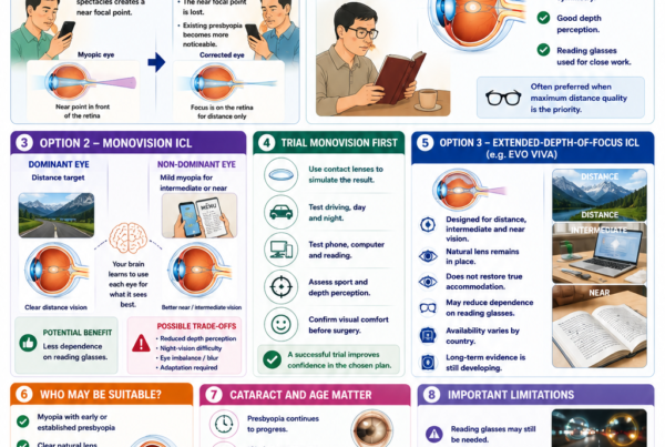
Patients approaching or over 40 may eventually need:
- Reading glasses
- Progressive spectacles
- A monovision strategy
- Cataract or lens surgery later in life
How Does Age Affect Long-Term Safety?
Age influences:
- Cataract risk
- Endothelial cell reserve
- Anterior chamber depth
- Natural-lens thickness
- Presbyopia
- The likely number of years the ICL will remain inside the eye
Younger Patients
A younger patient may have:
- A clearer natural lens
- Greater endothelial reserve
- More years of useful spectacle independence
- A longer lifetime during which monitoring is needed
- A greater possibility of future myopia progression
Patients Over 40
An older patient may have:
- Early presbyopia
- Early cataract
- A thicker natural lens
- Reduced anterior chamber space
- A shorter expected interval before cataract surgery
- A higher cataract risk
ICL may still be appropriate after 40 in selected patients, but counselling should include the likelihood of reading-glasses use and future cataract surgery.
Does Very High Myopia Increase Long-Term Risk?
Very high myopia may increase the likelihood of:
- Refractive progression
- Cataract
- Glaucoma
- Retinal tears
- Retinal detachment
- Myopic macular degeneration
- Myopic traction maculopathy
Some of these risks arise from the elongated eye rather than the ICL.
FDA guidance notes that patients with higher myopia may experience lower efficacy or higher rates of certain adverse events.
A good postoperative visual result should not create the impression that the underlying high-myopia-related risks have disappeared.
What Factors Improve Long-Term Safety?
Careful Patient Selection
A suitable patient should generally have:
- Stable myopia
- Adequate anterior chamber depth
- Open drainage angles
- Adequate endothelial cell density
- A clear natural lens
- Acceptable eye pressure
- No active eye inflammation or infection
- Realistic expectations
- Willingness to attend long-term follow-up
Accurate Lens Sizing
Correct sizing reduces the risk of:
- Excessive vault
- Inadequate vault
- Angle narrowing
- Natural-lens contact
- Toric instability
- Lens exchange
Modern planning may incorporate:
- White-to-white diameter
- Anterior chamber depth
- Angle-to-angle measurements
- Sulcus-to-sulcus measurements
- Crystalline lens rise
- Anterior-segment OCT
- Ultrasound biomicroscopy
No sizing formula predicts postoperative vault perfectly.
Careful Surgical Technique
Important factors include:
- Strict sterile preparation
- Avoiding natural-lens contact
- Minimising iris manipulation
- Correct ICL orientation
- Accurate toric alignment
- Complete removal of excess viscoelastic
- Early pressure monitoring
Appropriate Early Follow-Up
Early reviews assess:
- Vision
- Eye pressure
- Inflammation
- Corneal clarity
- Pupil shape
- ICL position
- Vault
- Toric alignment
Lifelong Monitoring
Regular examinations should continue even after the first postoperative year.
What Follow-Up Is Needed Long Term?
The exact schedule should be individualised.
A stable patient may require a review approximately yearly, while patients with borderline anatomy or developing changes may require more frequent assessments.
Long-term reviews may include:
- Unaided and corrected vision
- Refraction
- Eye pressure
- Slit-lamp examination
- ICL position
- Vault
- Toric alignment
- Drainage-angle assessment
- Endothelial cell count
- Natural-lens examination
- Optic nerve examination
- OCT or visual-field testing when indicated
- Dilated retinal and macular examination
FDA guidance recommends continued monitoring of cataract, eye pressure, drainage-angle anatomy and endothelial health for as long as the ICL remains implanted.
Who May Need More Frequent Monitoring?
Closer review may be appropriate when there is:
- Low vault
- High vault
- Narrow drainage angles
- Low or declining endothelial cell density
- Raised eye pressure
- Glaucoma risk
- Early cataract
- Toric instability
- Previous inflammation
- Very high myopia
- Peripheral retinal degeneration
- New visual symptoms
What Symptoms Require Urgent Assessment?
Seek urgent eye care for:
- Severe or increasing eye pain
- Rapidly worsening vision
- Sudden vision loss
- Marked redness
- Significant light sensitivity
- Severe headache
- Nausea or vomiting
- A cloudy cornea
- A white or grey spot on the eye
- A distorted pupil
- Sudden new floaters
- Flashes of light
- A curtain or shadow
- Significant trauma
These symptoms may indicate:
- Raised eye pressure
- Infection
- Severe inflammation
- Corneal disease
- Cataract-related complications
- ICL displacement
- Retinal tear or detachment
Frequently Asked Questions About Long-Term ICL Safety
Is ICL Safe After Ten Years?
Published ten-year studies of central-port ICLs have reported favourable visual, refractive and safety outcomes in appropriately selected patients.
These results are reassuring but do not guarantee that every patient will remain complication-free.
Is There Evidence Beyond Ten Years?
There is longer-term evidence for earlier ICL models, including studies extending beyond ten years.
Direct evidence for modern central-port EVO lenses is strongest up to approximately eight to ten years.
Because many patients may keep an ICL for several decades, ongoing research remains important.
Does the Lens Need to Be Changed Every Ten Years?
No.
There is no routine replacement interval.
A stable ICL can remain inside the eye beyond ten years when the eye remains healthy.
Can the ICL Expire Inside the Eye?
The lens does not have a routine expiry date once implanted.
Its material and optical power are designed for long-term implantation.
Does Everyone Eventually Develop Cataract?
Most people develop some natural-lens change with age, whether or not they have an ICL.
Not every ICL patient develops an ICL-related cataract.
Modern central-port lenses appear to have reduced anterior subcapsular cataract risk, but age-related cataract can still occur.
Does Low Vault Always Cause Cataract?
No.
Some patients with low vault remain free of cataract for many years.
Risk depends on:
- The degree of low vault
- Whether the ICL touches the natural lens
- Age
- Aqueous circulation
- Lens design
- Duration of follow-up
Does High Vault Always Require ICL Exchange?
No.
High vault may be observed when:
- Eye pressure is normal
- Drainage angles remain acceptable
- Endothelial cells are stable
- There are no significant symptoms
Exchange becomes more likely when high vault causes angle narrowing, pressure problems, pigment dispersion, pupil distortion or endothelial concerns.
Does Everyone Lose Corneal Endothelial Cells?
Some cell reduction occurs naturally with age and after intraocular surgery.
Most patients remain well above the level required to maintain corneal clarity, but endothelial density should still be monitored.
Can ICL Cause Glaucoma Years Later?
It can contribute to glaucoma when it causes persistent angle narrowing, pigment dispersion or repeated pressure elevation.
Glaucoma can also develop independently because high myopia itself increases risk.
Does ICL Increase Retinal-Detachment Risk?
Current comparative studies have not shown a clear increase beyond the underlying risk associated with high myopia.
The retinal risk nevertheless remains because ICL surgery does not shorten the elongated eye.
Can the ICL Move Years Later?
Significant movement is uncommon.
Toric rotation or displacement may occur after trauma or because of anatomical or sizing factors.
Is ICL Safer Long Term Than LASIK or SMILE?
The procedures have different risk profiles.
ICL avoids:
- Permanent central corneal tissue removal
- LASIK flap complications
- Corneal ectasia caused by laser tissue removal
- Much of the central corneal nerve disruption associated with laser surgery
ICL introduces:
- Cataract risk
- Vault-related risks
- Eye-pressure risks
- Endothelial cell concerns
- Intraocular inflammation and infection risk
- The need for implant-specific monitoring
The safest option depends on the patient’s prescription, cornea, internal eye anatomy, age and lifestyle.
Can the ICL Be Removed if a Long-Term Problem Develops?
Usually, yes.
It may be repositioned, exchanged or removed.
Removal may stop an ongoing implant-related problem but cannot necessarily reverse damage that has already occurred.
Will My Vision Return to the Preoperative State After Removal?
In an uncomplicated eye, removal generally returns the eye close to its underlying preoperative optical state.
The original or current myopia largely returns, and spectacles or contact lenses can usually be used again.
A small peripheral corneal incision or scar remains but generally heals well.
Do I Need Eye Checks Even if My Vision Is Perfect?
Yes.
Some complications, including endothelial cell loss, angle narrowing or raised eye pressure, may initially cause few symptoms.
Key Takeaway
Modern central-port ICL surgery has a favourable long-term safety profile in carefully selected patients.
Five-, seven-, eight- and ten-year studies generally show:
- Stable vision
- Predictable refractive correction
- Stable average eye pressure
- Modest average endothelial cell changes
- Low rates of serious complications
- A low need for exchange or removal
The central-port EVO design has reduced concerns about pupillary block and anterior subcapsular cataract compared with earlier non-port ICLs.
However, ICL surgery is not free from long-term risk.
Important issues include:
- Cataract
- Corneal endothelial cell loss
- Eye-pressure and drainage-angle changes
- High or low vault
- Toric rotation
- Glare and halos
- The need for repositioning, exchange or removal
ICL surgery corrects myopia but does not cure the elongated highly myopic eye. The lifetime risks of retinal detachment, myopic macular disease, glaucoma and cataract remain.
An ICL does not need routine replacement because a fixed number of years has passed. It can potentially remain inside the eye for life when:
- Vision remains satisfactory
- The natural lens remains clear
- Eye pressure and drainage angles remain healthy
- Endothelial cell density remains adequate
- Vault remains clinically safe
- Regular follow-up continues
The strongest long-term safety strategy is not simply choosing a modern lens. It is combining careful patient selection, accurate sizing, appropriate surgery, early postoperative checks and lifelong eye monitoring.
References
- US Food and Drug Administration. EVO and EVO+ Visian Implantable Collamer Lens: Patient Information Booklet. 2022.
- US Food and Drug Administration. EVO and EVO+ Visian Implantable Collamer Lens: Instructions for Use. 2022.
- Packer M, et al. Evaluation of the EVO/EVO+ Sphere and Toric Visian Implantable Collamer Lens. 2022. PMID: 35645557.
- Parkhurst G, et al. Three-Year Results From the United States FDA Prospective Multicentre Clinical Study of the EVO Implantable Collamer Lens. 2025. PMID: 40937096.
- Kamiya K, et al. Eight-Year Outcomes of Implantation of Posterior Chamber Phakic Intraocular Lens With a Central Port for Moderate-to-High Ametropia. 2021. PMID: 34977099.
- Fernández-Vega-Cueto L, et al. Seven-Year Follow-up of Posterior Chamber Phakic Intraocular Lens With Central-Port Design. 2021. PMID: 34112241.
- Alfonso-Bartolozzi B, et al. Ten-Year Follow-up of Posterior Chamber Phakic Intraocular Lenses With Central-Port Design. 2024. PMID: 38085219.
- Larivoir NB, et al. Efficacy, Predictability and Safety of Phakic Implantable Collamer Lenses With a Central Port: A Meta-analysis. 2025. PMID: 41369665.
- Passaro ML, et al. Potential Harms of Posterior Chamber Phakic Intraocular Lenses: A Systematic Review and Meta-analysis of Complication Incidence. 2026. PMID: 41611001.
- Kim YH, et al. Long-Term Outcome and Related Risk Factors in Implantable Collamer Lens Surgery. 2025. PMID: 40007092.
- Papa-Vettorazzi MR, et al. Long-Term Efficacy and Safety Profiles After Posterior Chamber Phakic Intraocular Lens Implantation. 2022. PMID: 34670948.
- Shimizu K, et al. Long-Term Comparison of Posterior Chamber Phakic Intraocular Lens With and Without a Central Hole. 2016. PMID: 27057883.
- Arrevola-Velasco L, et al. Ten-Year Prevalence of Rhegmatogenous Retinal Detachment Following ICL Implantation for High Myopia. 2023. PMID: 36730449.
- Xu W, et al. Long-Term Outcomes of Retinal Detachment in Phakic Eyes After ICL Implantation. 2020. PMID: 33425935.
- Alhamzah A, et al. Indications for Exchange or Explantation of Phakic Implantable Collamer Lenses With a Central Port. 2021. PMID: 34804861.
- Wan Q, et al. Long-Term Observation of V4c Implantable Collamer Lenses in Moderate-to-Extreme High Myopia. 2024. PMID: 38519715.
- Thompson V, et al. Implantable Collamer Lens Procedure Planning: A Review of Sizing and Vault. 2024. PMID: 38601168.
- Packer M. The Implantable Collamer Lens With a Central Port: Review of the Literature. Clinical Ophthalmology. 2018.