Author: Dr Val Phua
Estimated reading time: 16 minutes
Implantable Collamer Lens surgery—commonly known as ICL surgery—requires more than selecting the correct lens power.
The surgeon must also choose the correct physical size of the ICL.
These are two separate decisions:
- ICL power determines how much myopia and astigmatism the lens corrects.
- ICL size determines how the lens fits inside the eye.
An ICL with the correct optical power but an unsuitable physical size may still produce problems.
A lens that is too small may sit too close to the natural crystalline lens. A lens that is too large may sit too far forwards and crowd the iris or drainage angles.
The aim of ICL sizing is to achieve an appropriate vault—the space between the back of the ICL and the front of the natural lens.
Accurate sizing helps reduce the risks of:
- Cataract
- Raised eye pressure
- Narrowing of the drainage angles
- Pigment dispersion
- Corneal endothelial cell loss
- Toric ICL rotation
- Lens exchange or removal
Modern imaging, refined sizing formulas and artificial-intelligence models have improved ICL planning. However, no measurement or formula can predict postoperative vault perfectly because the ICL rests within internal structures that vary in size, shape and behaviour between individuals.
What Does ICL Size Mean?
ICL size refers to the lens’s overall diameter from one end to the other, including its supporting footplates.
The lens does not attach to the cornea or natural lens. Its footplates rest within the ciliary sulcus behind the iris.
Under current United States FDA labelling, available EVO ICL overall diameters include:
- 12.1 mm
- 12.6 mm
- 13.2 mm
- 13.7 mm
Availability may differ according to the lens model and country.
A difference of 0.5 or 0.6 mm may appear small, but it can significantly affect how the lens curves and sits inside the eye.
If the ICL Is Relatively Small
The lens may sit flatter, producing a lower vault.
If the ICL Is Relatively Large
The lens may be compressed more strongly within the eye, causing it to bow forwards and produce a higher vault.
However, the relationship is not completely predictable. Internal anatomy, lens power, ciliary sulcus shape and the orientation of implantation also influence vault.
Is ICL Size the Same as ICL Power?
No.
A patient may hear two different sets of numbers during ICL planning.
ICL Optical Power
The optical power is measured in dioptres and corrects:
- Myopia
- Astigmatism
- The spherical equivalent of the prescription
For example, one lens may have a spherical power of −10.00 D.
ICL Physical Size
The physical size is measured in millimetres.
For example, the surgeon may select a 12.6 mm or 13.2 mm ICL.
Two patients with the same spectacle prescription may require different physical ICL sizes because their internal eye dimensions are different.
Similarly, two patients with similar-sized eyes may require different lens powers because their prescriptions are different.
Where Does the ICL Sit?
The ICL is positioned:
- Behind the iris
- In front of the natural crystalline lens
- Within the posterior chamber
- With its supporting footplates resting in the ciliary sulcus
The ciliary sulcus is an internal groove-like region behind the iris.
Unlike the white-to-white corneal diameter, the ciliary sulcus cannot be seen directly during a routine external examination. It must be estimated or imaged using specialised measurements.
The shape of the sulcus also varies between individuals. It may be horizontally oval, vertically oval or asymmetrical rather than perfectly circular. This is one reason why a simple external measurement cannot predict the fit of every ICL perfectly.
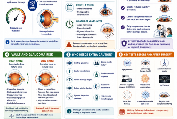
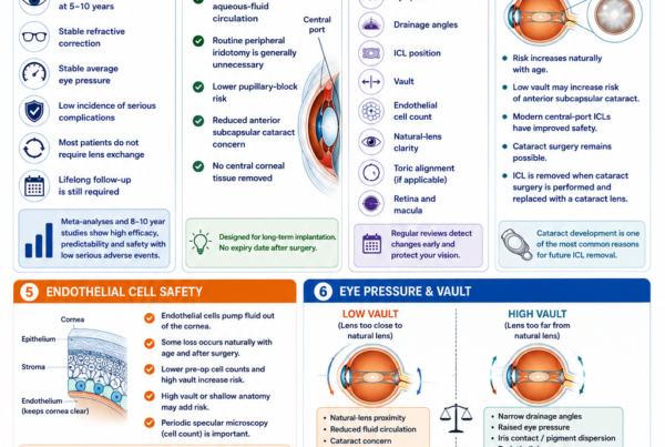
What Is ICL Vault?
Vault is the central distance between:
- The back surface of the ICL
- The front surface of the natural crystalline lens
Vault is usually measured in micrometres, or microns.
One millimetre equals 1,000 microns.
FDA professional guidance describes an intended postoperative vault of approximately one central corneal thickness. It considers approximately 250 to 900 microns—about 50% to 150% of central corneal thickness—a broadly desirable range. However, a vault outside this range does not automatically require lens exchange or removal when the eye remains otherwise healthy.
The ideal vault is not a single exact number that applies to every eye.
The surgeon also considers:
- The patient’s age
- Natural-lens thickness
- Eye pressure
- Drainage-angle width
- Endothelial cell count
- ICL position
- Pupil shape
- Evidence of iris contact
- Whether the vault is stable or changing
- Whether cataract or other complications are developing
Why Is Vault Important?
The ICL must remain sufficiently separated from the natural lens without excessively crowding the front of the eye.
If Vault Is Too Low
A very low vault may increase concern about:
- Proximity or contact with the natural lens
- Reduced aqueous-fluid circulation
- Anterior subcapsular cataract
- Progressive reduction in vault as the natural lens thickens with age
If Vault Is Too High
A very high vault may:
- Push the iris forwards
- Narrow the drainage angles
- Raise eye pressure
- Cause iris contact
- Release iris pigment
- Contribute to pupil distortion
- Bring the ICL closer to the corneal endothelium
Correct sizing aims to balance these competing concerns.
Does Low Vault Always Cause Cataract?
No.
Some patients with relatively low vault remain stable for many years without developing cataract.
The significance depends on:
- The actual distance between the ICL and natural lens
- Whether physical contact is present
- The patient’s age
- Natural-lens clarity
- Changes in vault over time
- The central-port design
- Aqueous-fluid circulation
A five-year study comparing eyes with low vault and eyes with conventionally normal vault reported comparable safety, refractive efficacy and visual outcomes in its selected population. This suggests that low vault should be interpreted together with clinical findings rather than treated as an automatic indication for exchange.
However, a very low vault accompanied by anterior subcapsular lens opacity or direct contact remains clinically important.
Does High Vault Always Require Lens Exchange?
No.
Some eyes with high vault maintain:
- Normal eye pressure
- Adequately open drainage angles
- Stable endothelial cell counts
- A clear natural lens
- Good vision
- No significant symptoms
Observation may be appropriate when the anatomy remains safe.
Exchange becomes more likely when high vault is associated with:
- Progressive angle narrowing
- Raised eye pressure
- Pigment dispersion
- Pupil distortion
- Endothelial concerns
- Significant glare or discomfort
FDA guidance specifically notes that a vault outside the intended range may not necessarily require exchange or removal in the absence of symptoms or associated complications.
How Was ICL Size Traditionally Chosen?
Traditional ICL sizing has primarily used:
- Horizontal white-to-white corneal diameter
- Anterior chamber depth
The manufacturer’s sizing nomogram then recommends one of the available ICL diameters.
During the original United States clinical study, the calculated lens length was based on adding approximately 1.1 to 1.6 mm to the horizontal white-to-white measurement, with the amount influenced by anterior chamber depth. The current professional instructions contain a table matching white-to-white and true anterior chamber depth to a recommended lens diameter.
This method is simple, widely available and clinically established.
However, it has an important limitation: the ICL rests in the ciliary sulcus, while white-to-white measures the visible cornea.
What Is White-to-White?
White-to-white, commonly abbreviated as WTW, is the horizontal distance between the visible borders of the cornea.
It is measured from one corneal limbus to the other.
The limbus is the transition between:
- The clear cornea
- The white sclera
WTW can be measured using:
- Corneal topography devices
- Optical biometers
- Anterior-segment OCT
- Digital photography
- Manual callipers
It is non-contact and easy to obtain.
Why Is White-to-White Used?
White-to-white is used because it provides a convenient external estimate of the eye’s overall anterior dimensions.
Advantages include:
- Rapid measurement
- No contact with the eye
- Good repeatability on many modern devices
- Wide availability
- Integration with established manufacturer nomograms
However, WTW is only a surrogate for the internal ciliary sulcus diameter.
A large cornea does not always correspond to a proportionately large sulcus, and a smaller cornea does not always indicate a small internal sulcus.
What Are the Limitations of White-to-White?
It Measures the Cornea, Not the Ciliary Sulcus
The ICL footplates sit within the sulcus behind the iris.
WTW measures a visible external structure that is anatomically separate from the lens’s final resting position.
Devices Can Produce Different Results
Different instruments may define the corneal border differently.
A patient may therefore receive slightly different WTW values from:
- An optical biometer
- A topographer
- An anterior-segment camera
- Manual measurement
Interdevice measurement bias can influence the recommended ICL size when the eye lies close to the boundary between two lens sizes.
WTW and Sulcus Size Do Not Always Match
Research has shown that the difference between WTW and sulcus-to-sulcus diameter can be greater in eyes with unusual WTW measurements or anterior chamber depths. These patients may have a higher risk of an unexpected vault when sizing relies mainly on WTW.
It Does Not Describe Sulcus Shape
The ciliary sulcus may be:
- Oval
- Vertically elongated
- Horizontally elongated
- Asymmetrical
- Different between the two eyes
One horizontal external measurement cannot fully represent this three-dimensional anatomy.
What Is Anterior Chamber Depth?
Anterior chamber depth, or ACD, describes the space between the cornea and the natural lens.
For ICL suitability and FDA labelling, the relevant measurement is the true ACD:
- From the back surface of the cornea
- To the front surface of the crystalline lens
Some devices instead measure from the front surface of the cornea. In that situation, corneal thickness must be subtracted to obtain the true ACD.
Under current United States labelling, EVO ICL requires a true anterior chamber depth of at least 3.0 mm. Local indications may differ.
Why Does Anterior Chamber Depth Affect Sizing?
Anterior chamber depth provides information about how much internal space is available.
A deeper eye may accommodate a larger or more anteriorly vaulted lens differently from a shallower eye.
A shallow anterior chamber can increase concern about:
- Narrow drainage angles
- Endothelial cell proximity
- Iris crowding
- Excessive postoperative vault
ACD is therefore important for both:
- Determining whether ICL surgery is suitable
- Selecting the lens size
However, two eyes with the same ACD may still have different sulcus widths and postoperative vaults.
What Is Sulcus-to-Sulcus Measurement?
Sulcus-to-sulcus, or STS, is the internal distance between opposite sides of the ciliary sulcus.
Because the ICL footplates rest in the sulcus, STS appears intuitively more directly related to lens fit than WTW.
STS can be measured in:
- The horizontal meridian
- The vertical meridian
- Oblique meridians
Research has shown that including STS in sizing formulas can improve vault predictability compared with relying on traditional WTW-based sizing alone.
How Is Sulcus-to-Sulcus Measured?
STS is most commonly assessed using ultrasound biomicroscopy, or UBM.
UBM uses very-high-frequency ultrasound to image structures behind the iris, including:
- The ciliary sulcus
- Ciliary body
- Zonules
- Posterior iris surface
- Natural lens
- Internal chamber dimensions
Advantages of UBM
- Direct visualisation behind the iris
- Measurement of horizontal and vertical STS
- Assessment of ciliary sulcus shape
- Identification of ciliary body abnormalities
- Evaluation of lens position and orientation
Limitations of UBM
- Requires contact or an immersion interface
- More operator-dependent than non-contact optical imaging
- Patient positioning can influence measurements
- Image quality and landmark identification may vary
- Different UBM devices or operators may produce different values
- It requires additional time and expertise
STS is useful, but it is not a perfect solution on its own.
Is STS Always Better Than WTW?
Not necessarily in every clinical setting.
Some studies have shown better vault prediction when STS is included. Others have found that modern optical measurements and multivariable formulas can perform similarly or better without direct STS measurement.
The practical value of STS depends on:
- UBM image quality
- Operator experience
- Repeatability
- Whether horizontal and vertical measurements are obtained
- The formula into which the measurement is entered
- The patient’s anatomy
The best planning approach generally combines several measurements rather than treating one dimension as infallible.
Why Are Horizontal and Vertical STS Different?
The ciliary sulcus is often not perfectly circular.
In many eyes:
- Vertical STS is greater than horizontal STS
- The sulcus is elliptical
- The degree of asymmetry differs between patients
This has implications for:
- Lens size
- Lens orientation
- Toric stability
- Postoperative vault
A lens implanted horizontally may behave differently from one placed vertically or obliquely within the same eye.
Recent research continues to explore how horizontal-to-vertical sulcus differences can guide individualised sizing and orientation.
What Is Angle-to-Angle or Anterior Chamber Width?
Angle-to-angle, also called anterior chamber width, is the internal distance between opposite drainage-angle landmarks.
It is commonly measured with anterior-segment OCT.
Unlike WTW, angle-to-angle is an internal measurement.
Although the ICL footplates do not rest directly at the drainage angles, anterior chamber width provides information about the size of the front of the eye and can contribute to vault prediction.
What Is Crystalline Lens Rise?
Crystalline lens rise, sometimes abbreviated as CLR, describes how far the natural lens rises above a reference line across the anterior chamber.
A more anteriorly positioned or more prominent natural lens may reduce the available space between the ICL and crystalline lens.
CLR can therefore influence postoperative vault.
Anterior-segment OCT-based sizing research has found anterior chamber width and crystalline lens rise to be significant predictors of postoperative vault.
What Is Lens Thickness?
The natural crystalline lens gradually thickens with age.
A thicker lens may:
- Occupy more internal space
- Sit closer to the ICL
- Contribute to lower vault
- Reduce anterior chamber depth
Lens thickness is therefore relevant both before surgery and during long-term follow-up.
Studies have identified crystalline lens thickness, ICL size and horizontal and vertical STS as factors that influence postoperative vault.
What Other Measurements May Affect ICL Sizing?
Modern formulas may incorporate:
- White-to-white diameter
- True anterior chamber depth
- Angle-to-angle distance
- Sulcus-to-sulcus diameter
- Crystalline lens rise
- Natural-lens thickness
- Ciliary body inner diameter
- Zonule-to-zonule distance
- Anterior chamber angle
- Pupil diameter
- ICL optical power
- Patient age
- Sex
- Preoperative refraction
A study developing new sizing parameters found that vault prediction was influenced by several internal structures, including STS, ciliary body dimensions, zonular measurements and lens rise.
Why Does ICL Optical Power Affect Vault?
Higher-powered myopic ICLs do not have exactly the same shape or thickness profile as lower-powered lenses.
The physical design of the optic can affect how the lens bends and vaults inside the eye.
This means postoperative vault depends not only on overall diameter but also on:
- ICL power
- Lens model
- Toric design
- Internal eye anatomy
A sizing formula that ignores lens power may be less accurate for certain prescriptions.
Does Age Affect Vault?
Yes.
Age may influence vault through changes in:
- Natural-lens thickness
- Anterior chamber depth
- Ciliary body anatomy
- Pupil behaviour
- Tissue flexibility
Vault commonly decreases gradually as the natural lens thickens with age.
An older patient may therefore require different interpretation from a younger patient with the same immediate postoperative vault.
Age should also be considered when deciding whether a borderline low vault is likely to remain safe over the expected duration of implantation.
Does Pupil Size Affect Vault?
Vault can change slightly as the pupil constricts and dilates.
Pupil movement alters:
- Iris configuration
- Ciliary muscle activity
- Contact between the iris and ICL
- Position of the ICL
Vault measured under bright-light conditions may therefore differ from vault measured in dim lighting.
This is one reason that a single measurement should not be viewed as an absolute, unchanging property.
Can Implantation Orientation Change Vault?
Yes.
Because the ciliary sulcus is frequently elliptical, changing the ICL’s orientation may alter the amount of compression applied to the lens.
For example, implanting a non-toric ICL vertically rather than horizontally may reduce vault in an eye whose vertical sulcus diameter is larger than its horizontal diameter.
A customised approach using STS dimensions, ciliary sulcus morphology and intended lens orientation has been reported to improve the proportion of eyes achieving a desirable vault.
What About Toric ICLs?
A toric ICL must be aligned with the intended astigmatic axis.
The surgeon cannot freely choose any orientation solely to optimise vault because rotating the lens changes the astigmatism correction.
The lens design allows limited planned rotation for axis alignment, but excessive rotation from the intended orientation is not desirable. FDA instructions state that the EVO toric ICL is designed to require no more than 22.5 degrees of rotation from its manufactured orientation.
Sizing and toric alignment must therefore be considered together.
What Imaging Devices Are Used for ICL Planning?
Optical Biometry
Optical biometers may measure:
- White-to-white
- Anterior chamber depth
- Corneal thickness
- Lens thickness
- Axial length
Advantages include speed, repeatability and non-contact measurement.
Corneal Topography or Tomography
These devices assess:
- Corneal shape
- Corneal diameter
- Astigmatism
- Corneal thickness
- Keratoconus risk
They may also provide WTW and pupil measurements.
Anterior-Segment OCT
Anterior-segment OCT provides non-contact cross-sectional imaging of:
- Anterior chamber depth
- Angle-to-angle distance
- Crystalline lens rise
- Drainage-angle anatomy
- Postoperative vault
Advantages include:
- High resolution
- Rapid imaging
- Good patient comfort
- Repeatable postoperative vault measurement
Limitations include reduced visibility of some structures located behind the pigmented iris.
Ultrasound Biomicroscopy
UBM can image structures behind the iris, including the ciliary sulcus.
It may be particularly helpful when:
- WTW and internal dimensions do not appear to match
- The eye is between two lens sizes
- Previous surgery produced an unexpected vault
- Sulcus shape or ciliary body anatomy needs assessment
- Customised orientation is being considered
How Does the Surgeon Choose Between Two Sizes?
Some patients fall clearly within the range for one ICL size.
Others lie near the boundary between two sizes.
For example, measurements may support either:
- A 12.6 mm lens
- A 13.2 mm lens
The surgeon then considers the likely consequences of each choice.
Choosing the Smaller Size May:
- Reduce the risk of excessive vault
- Preserve wider drainage angles
- Increase the risk of lower vault
- Potentially affect toric stability in some anatomies
Choosing the Larger Size May:
- Increase separation from the natural lens
- Improve stability in some eyes
- Increase the risk of high vault
- Narrow the drainage angles
The final decision may incorporate:
- WTW
- ACD
- STS
- Anterior chamber width
- Lens rise
- Lens thickness
- Age
- ICL power
- Sulcus shape
- Surgeon experience
- Expected implantation orientation
There is no universal rule that the larger or smaller option is always safer.
What Is a Sizing Nomogram?
A nomogram is a structured calculation or table used to recommend an ICL size based on preoperative measurements.
Examples include:
- Manufacturer WTW–ACD nomograms
- UBM-based formulas
- Anterior-segment OCT formulas
- Multivariable regression models
- Machine-learning models
- Surgeon-specific nomograms
A comparative study of multiple available nomograms found that predictive performance varied and that no formula eliminated unexpected postoperative vault completely.
Why Do Different Formulas Recommend Different Sizes?
Different formulas may use different combinations of:
- External measurements
- Internal measurements
- Lens power
- Patient demographics
- Imaging platforms
- Historical surgical outcomes
They may also have been developed using different:
- Ethnic populations
- Age groups
- ICL models
- Surgical techniques
- Measurement devices
A formula trained in one population may not perform identically in another.
This is why external validation is important before a new formula is adopted widely.
Can Artificial Intelligence Improve ICL Sizing?
Artificial-intelligence and machine-learning models are increasingly being studied for ICL sizing and vault prediction.
These systems may analyse:
- Multiple numerical measurements
- Anterior-segment OCT data
- Ultrasound images
- Patient age and prescription
- ICL power and size
- Interactions between anatomical variables
Studies have reported that machine-learning approaches can outperform traditional linear models or manufacturer nomograms within their study datasets.
Deep-learning systems have also been developed to interpret raw OCT or ultrasound images directly and estimate postoperative vault.
Does AI Guarantee the Correct ICL Size?
No.
AI models have several limitations:
- They may be trained mainly on one ethnic or geographic population
- They may use measurements from a particular device
- They may not perform identically with another ICL model
- Retrospective datasets may contain selection bias
- A model can reproduce errors in its training data
- External validation may be limited
- Clinical judgement remains necessary
AI is best viewed as a decision-support tool rather than a replacement for the surgeon.
A 2025 AI model showed promising robustness using a relatively small number of parameters, while a separate externally validated image-based model also reported improved vault prediction. These developments are encouraging, but they still require continued validation before being treated as universal standards.
Why Can Vault Still Be Unexpected?
Even with careful measurement, postoperative vault may differ from prediction because of:
- Measurement error
- Differences between devices
- Sulcus asymmetry
- Ciliary body morphology
- Lens orientation
- ICL power
- Pupil size
- Natural-lens position
- Tissue flexibility
- Postoperative settling
- Changes in lighting conditions
- Individual biological variation
The ICL is a flexible lens resting within dynamic living tissue, not a rigid implant fixed to a precisely measurable frame.
Can the Two Eyes Have Different ICL Sizes?
Yes.
The two eyes may differ in:
- White-to-white diameter
- Sulcus-to-sulcus diameter
- Anterior chamber depth
- Lens rise
- Ciliary sulcus shape
- Astigmatic axis
- Lens power
Many patients receive the same ICL size in both eyes, but this should not be assumed automatically.
Each eye should be measured and planned individually.
Can the First Eye Help Determine the Size for the Second Eye?
Potentially.
The two eyes of one person often have similar anatomy.
In staged surgery, the early postoperative vault in the first eye may provide useful information when selecting the ICL size for the second eye.
A 2025 study found that first-eye vault could assist second-eye size selection and prediction. However, this strategy is only relevant when surgery is performed sequentially and the eyes are sufficiently similar.
It does not replace independent measurements of both eyes.
When Is Vault Checked After Surgery?
FDA professional guidance recommends assessing vault approximately 24 hours after implantation.
Vault may also be checked at:
- One week
- One month
- Three to six months
- Yearly follow-up
- Any visit when symptoms or pressure changes occur
The exact schedule depends on:
- Initial vault
- Eye pressure
- Drainage-angle appearance
- Natural-lens clarity
- Endothelial cell density
- Patient age
- Other risk factors
Does Vault Change During the First Day?
Yes.
Vault can change during the first hours and days as:
- The pupil changes size
- Surgical medication wears off
- Viscoelastic is removed
- Inflammation settles
- The ICL adjusts within the sulcus
Research has shown that vault may decrease and subsequently rise during the early postoperative period rather than remaining fixed immediately after implantation.
A very early measurement should therefore be interpreted together with subsequent follow-up.
Does Vault Decrease Over the Years?
Vault commonly decreases gradually over time.
Possible reasons include:
- Natural-lens thickening
- Changes in anterior chamber depth
- Age-related anatomical changes
- Ciliary body changes
A gradual reduction does not necessarily indicate incorrect initial sizing.
The clinical importance depends on whether it is associated with:
- Lens opacity
- Natural-lens contact
- Symptoms
- Other anatomical changes
Long-term monitoring is particularly important in younger patients because the ICL may remain inside the eye for several decades.
How Is Vault Measured After Surgery?
Slit-Lamp Estimation
The surgeon can estimate vault by comparing it with central corneal thickness.
This provides a rapid clinical assessment.
Anterior-Segment OCT
OCT provides a numerical vault measurement in microns and can also assess:
- Drainage angles
- Iris configuration
- ICL position
- Natural-lens relationship
Ultrasound Biomicroscopy
UBM may be used when more detailed assessment of:
- Footplate position
- Ciliary sulcus
- ICL orientation
- Iris contact
is required.
What Happens if the ICL Is Too Small?
A relatively undersized ICL may produce:
- Low vault
- Increased proximity to the natural lens
- Anterior subcapsular cataract concern
- Reduced lens stability
- Toric rotation in some cases
Management depends on the findings.
Observation May Be Appropriate When:
- The natural lens is clear
- There is no ICL–lens contact
- Vision is good
- Vault is stable
- Eye pressure is normal
Exchange May Be Considered When:
- Vault is extremely low
- The ICL contacts the natural lens
- An anterior subcapsular opacity develops
- A toric lens repeatedly rotates
- The anticipated long-term risk is unacceptable
What Happens if the ICL Is Too Large?
An oversized ICL may produce:
- Excessive vault
- Drainage-angle narrowing
- Raised eye pressure
- Iris contact
- Pigment dispersion
- Pupil distortion
- Endothelial concerns
Management may include:
- Observation
- More frequent pressure and angle monitoring
- Reorientation of a non-toric ICL
- Exchange for a smaller lens
- Removal
A 2026 size-exchange study found that downsizing reliably reduced excessive vault, while exchanging for the same size produced little vault change. This supports selecting a genuinely smaller lens when excessive size is the underlying problem.
Can an ICL Be Rotated to Change Vault?
A non-toric ICL may sometimes be rotated from a horizontal to a more vertical orientation.
If the vertical sulcus is larger than the horizontal sulcus, this can reduce compression and lower the vault.
This strategy is anatomy-dependent and is not suitable in every case.
A toric ICL cannot usually be rotated freely because its optical axis must remain aligned with the patient’s astigmatism.
How Common Is Exchange for Sizing Problems?
ICL exchange is uncommon but recognised.
A seven-year series found that approximately 2% of implanted central-port ICLs required exchange or removal, with inappropriate vault being the most common reason.
More recent high-volume data have reported lower exchange rates, although rates vary according to:
- Lens model
- Patient selection
- Follow-up duration
- Sizing method
- Definition of exchange
- Surgical centre
Inappropriate sizing and off-label use remain important contributors to reported adverse events, reinforcing the need for careful planning and postoperative vault assessment.
Is Lens Exchange Dangerous?
ICL exchange is generally feasible, but it is another operation inside the eye.
Potential risks include:
- Corneal endothelial cell loss
- Iris injury
- Natural-lens injury
- Cataract
- Raised eye pressure
- Inflammation
- Infection
- Residual refractive error
The aim of accurate primary sizing is to minimise the need for secondary surgery.
However, when an unsuitable vault poses a greater long-term risk, exchange may be safer than leaving the original lens in place.
Can Perfect Sizing Eliminate All ICL Risks?
No.
Accurate sizing reduces several important risks but cannot prevent complications unrelated to size.
These include:
- Infection
- Steroid response
- Retinal disease associated with high myopia
- Natural cataract formation
- Myopia progression
- Inflammation
- Surgical trauma
Sizing is one essential component of safe ICL surgery, not the only one.
What Tests Should Patients Expect Before ICL Surgery?
A comprehensive assessment may include:
Refraction
To determine the optical ICL power.
Corneal Topography and Tomography
To assess corneal shape and exclude progressive corneal disease.
White-to-White Measurement
To estimate the overall horizontal anterior eye dimension.
True Anterior Chamber Depth
To confirm sufficient space for implantation.
Anterior Chamber Width or Angle-to-Angle Distance
To assess internal anterior-segment size.
Crystalline Lens Rise and Thickness
To estimate how much the natural lens projects forwards.
Sulcus-to-Sulcus Measurement
UBM may be used to image the intended resting location of the ICL.
Drainage-Angle Assessment
Gonioscopy or imaging confirms that the angles are sufficiently open.
Endothelial Cell Count
To confirm adequate corneal endothelial reserve.
Pupil Size
To help assess possible glare and halo risk.
Dilated Retinal Examination
Particularly important in moderate and high myopia.
What Questions Should You Ask About ICL Sizing?
Useful questions include:
- What ICL size do you recommend?
- Which measurements were used?
- What are my white-to-white and anterior chamber depth?
- Did you measure my anterior chamber width?
- Was sulcus-to-sulcus imaging necessary?
- Is my eye close to the boundary between two sizes?
- What postoperative vault do you expect?
- How will vault be measured after surgery?
- Are my drainage angles sufficiently open?
- How does my age affect the expected long-term vault?
- Would a toric lens limit the possible implantation orientation?
- What would happen if my vault were higher or lower than expected?
- How often will the vault be monitored?
Frequently Asked Questions About ICL Sizing
How Does the Surgeon Know Which ICL Size to Use?
The surgeon combines measurements of the cornea and internal eye, usually including WTW and ACD, with an established sizing nomogram.
Additional imaging such as anterior-segment OCT or UBM may refine the decision.
Are There Different ICL Sizes?
Yes.
Under United States FDA labelling, the available overall diameters are 12.1, 12.6, 13.2 and 13.7 mm. Available models may differ between markets.
Is the Biggest ICL Better?
No.
A lens that is too large may cause excessive vault, angle narrowing and raised eye pressure.
Is a Smaller ICL Safer?
Not automatically.
A lens that is too small may sit too close to the natural lens and may be less rotationally stable.
What Is a Good ICL Vault?
A broadly desirable range is often described as approximately 250 to 900 microns, but the ideal vault is individual.
A measurement outside that range does not automatically mean that the lens is unsafe.
Is 500 Microns the Perfect Vault?
Approximately 500 microns is often used as a convenient reference because it is close to average central corneal thickness.
It is not a universal perfect number.
Is Low Vault Dangerous?
It may increase cataract concern, particularly when the ICL is extremely close to or touching the natural lens.
Some relatively low-vault eyes remain safe for years with careful monitoring.
Is High Vault Dangerous?
It may narrow the drainage angles and increase pressure, pigment or endothelial concerns.
Some high-vault eyes remain stable without requiring exchange.
Can Vault Change After Surgery?
Yes.
It can vary with pupil size during the day and may gradually decrease as the natural lens thickens with age.
Can WTW Alone Choose the Correct Size?
WTW and ACD form the basis of traditional sizing, but they do not fully describe the ciliary sulcus.
Additional internal measurements may be helpful, especially in borderline or unusual eyes.
Is UBM Required for Every Patient?
Not necessarily.
Some surgeons achieve reliable results using modern optical imaging and validated nomograms.
UBM may be particularly valuable when the anatomy is unusual, measurements disagree or previous sizing was unexpected.
Is Sulcus-to-Sulcus More Accurate?
It measures anatomy closer to the ICL’s resting location and may improve vault prediction when measured reliably.
However, it remains operator-dependent and does not eliminate prediction error.
Can AI Choose My ICL Size?
AI may support the surgeon by combining multiple measurements and predicting vault.
It should not replace clinical judgement or postoperative monitoring.
Can the Two Eyes Need Different Sizes?
Yes.
Each eye should be measured and planned independently.
Can the Lens Be Exchanged if the Size Is Wrong?
Yes.
An oversized lens may be exchanged for a smaller one, while an undersized or rotationally unstable lens may be exchanged for a larger one when appropriate.
Does Lens Exchange Restore a Normal Vault?
It often moves the vault in the intended direction, but the exact change cannot be guaranteed.
Does Toric ICL Sizing Differ?
The same anatomical principles apply, but toric lenses must also remain aligned with the intended astigmatic axis.
The surgeon must balance vault, physical stability and rotational alignment.
Is ICL Sizing More Important for High Myopia?
Sizing is important at every prescription.
Very-high-powered lenses may have different optical geometry, and highly myopic patients may also have unusual anterior-segment dimensions.
Does Correct Sizing Guarantee No Cataract?
No.
It reduces vault-related cataract risk but cannot prevent natural age-related cataract or accidental surgical contact with the natural lens.
Do I Need Long-Term Vault Checks?
Yes.
Vault can change over time, and some vault-related complications initially cause few symptoms.
Key Takeaway
ICL sizing determines how the implant fits inside the eye.
It is separate from ICL power:
- Power corrects myopia and astigmatism.
- Size influences lens position, stability and vault.
Traditional sizing uses:
- Horizontal white-to-white diameter
- True anterior chamber depth
Modern planning may also incorporate:
- Sulcus-to-sulcus diameter
- Anterior chamber width
- Crystalline lens rise
- Natural-lens thickness
- Ciliary body anatomy
- ICL power
- Pupil size
- Patient age
- Implantation orientation
The aim is to achieve an appropriate vault between the ICL and natural lens.
A lens that is too small may produce low vault and cataract concern. A lens that is too large may produce high vault, angle narrowing and raised eye pressure.
No sizing formula is perfect because the ICL rests within complex, dynamic and partly hidden anatomy. Anterior-segment OCT, UBM, multivariable formulas and artificial intelligence can improve prediction, but they cannot eliminate individual variation.
The safest approach combines:
- Careful patient selection
- Accurate and repeatable measurements
- Use of a validated sizing method
- Experienced clinical judgement
- Postoperative vault and pressure checks
- Long-term monitoring
An unexpected vault does not automatically mean that the operation has failed or that the ICL must be exchanged. The decision depends on the complete clinical picture, including eye pressure, drainage angles, endothelial cells, natural-lens clarity, symptoms and changes over time.
References
- US Food and Drug Administration. EVO and EVO+ Visian Implantable Collamer Lens: Instructions for Use. 2022.
- Thompson V, et al. Implantable Collamer Lens Procedure Planning: A Review of Sizing and Vault. 2024. PMID: 38601168.
- Moshirfar M, et al. Comparative Evaluation of Multiple Nomograms for Predicting Postoperative Vault After Implantable Collamer Lens Surgery. 2024. PMID: 37702514.
- Reinstein DZ, et al. Comparison of Postoperative Vault Height Predictability Using White-to-White and Sulcus Diameter-Based ICL Sizing. 2013. PMID: 23311739.
- Kojima T, et al. Optimisation of an Implantable Collamer Lens Sizing Method Using High-Frequency Ultrasound Biomicroscopy. 2012. PMID: 22245462.
- Guber I, et al. Correcting Interdevice Bias of Horizontal White-to-White and Sulcus-to-Sulcus Measurements for ICL Size Selection. 2016. PMID: 26454242.
- Nakamura T, et al. Implantable Collamer Lens Sizing Method Based on Swept-Source Anterior-Segment OCT. 2018. PMID: 29294311.
- Chen X, et al. Effect of the Difference Between White-to-White and Sulcus-to-Sulcus Diameters on Vault After ICL Implantation. 2021. PMID: 34460084.
- Zhu QJ, et al. Validation of a Vault Prediction Model Based on Sulcus-to-Sulcus Measurements. 2022.
- Reinstein DZ, et al. New Sizing Parameters and Model for Predicting Postoperative Vault for the Implantable Collamer Lens. 2022. PMID: 35536711.
- Wei R, et al. Outcomes of EVO ICL Using a Customised Non-Horizontal Implantation Orientation. 2022. PMID: 35415777.
- Russo A, et al. Predictability of ICL Vault Using Machine Learning of Preoperative Anterior-Segment OCT Measurements. 2023. PMID: 36913536.
- Di Y, et al. Predicting Implantable Collamer Lens Vault Using Machine-Learning Models. 2024. PMID: 38224328.
- Zhu J, et al. Enhancing Vault Prediction and ICL Sizing Through Machine Learning. 2024. PMID: 38466764.
- Hirabayashi MT, et al. Vault Accuracy Using Deep-Learning Technology and Preoperative Ultrasound Imaging. 2024. PMID: 38651696.
- Yamashita K, et al. Enhanced ICL Sizing Accuracy Using Advanced Optical Measurements. 2024. PMID: 39094990.
- Wu W, et al. Development and Validation of a Novel Vault Prediction Formula. 2024. PMID: 39152392.
- Sheng P, et al. Using Artificial Intelligence to Predict Implantable Collamer Lens Vault. 2025. PMID: 40985791.
- Li H, et al. Using First-Eye Early Vault to Determine ICL Size for the Second Eye. 2025. PMID: 39325572.
- Zéboulon P, et al. Validation of a New Image-Based Implantable Collamer Lens Sizing Algorithm. 2025–2026. PMID: 40827890.
- Silva R, et al. Long-Term Efficacy and Safety of Low Versus Normal Vault Following ICL Implantation. 2026. PMID: 40790614.
- Choi H, et al. Size Exchange and Vault Response in Posterior-Chamber Phakic Intraocular Lens Implantation. 2026. PMID: 42017508.