Author: Dr Val Phua
Estimated reading time: 19 minutes
A cataract is clouding of the eye’s natural crystalline lens.
The natural lens normally sits behind the coloured iris and focuses light onto the retina. In a healthy young eye, the lens is transparent. As a cataract develops, the lens becomes progressively cloudy, yellowed or optically irregular, preventing light from passing through and focusing clearly.
A cataract may cause:
- Blurred or hazy vision
- Increasing glare
- Halos around lights
- Difficulty driving at night
- Colours appearing faded
- Reduced contrast
- Frequent changes in spectacle prescription
- Double or ghosted vision from one eye
- Increasing short-sightedness
Cataracts are very common with increasing age. They may develop in one or both eyes, although they do not spread from one eye to the other. Cataracts are also a major global cause of avoidable visual impairment because many patients still lack access to surgery.
Early cataracts may be managed with brighter lighting, anti-glare measures and updated spectacles. However, surgery is currently the only established treatment that removes a cataract. During cataract surgery, the cloudy natural lens is removed and replaced with a clear artificial intraocular lens.
Where Is the Natural Lens?
The natural crystalline lens is located:
- Behind the cornea
- Behind the iris
- In front of the vitreous gel and retina
The cornea provides most of the eye’s focusing power. The natural lens provides additional focusing power and, when younger, can change shape to focus on objects at different distances.
Light normally travels through:
- The tear film
- The cornea
- The pupil
- The natural lens
- The vitreous
- The retina
A cataract interferes with this optical pathway by absorbing and scattering light.
Is a Cataract a Film Growing Over the Eye?
No.
A cataract is not a membrane, skin or film growing across the outside of the eye.
It is clouding inside the natural lens.
This distinction is important because:
- Cataracts cannot be washed away
- Ordinary eyedrops cannot remove the cloudy lens
- Cataracts cannot be scraped off the corneal surface
- Laser vision correction such as LASIK does not remove cataracts
Cataract surgery treats the condition by removing the cloudy natural lens itself and replacing it with an artificial lens.
How Does a Cataract Form?
The natural lens is made largely of water and highly organised proteins.
The lens must remain transparent for light to pass through without excessive scattering. With ageing, the lens proteins and fibres undergo structural and chemical changes. Proteins may break down, aggregate or lose their orderly arrangement, gradually creating cloudy areas within the lens.
Age-related lens changes commonly begin long before a person notices symptoms. Over time:
- The lens becomes less transparent
- It may become harder and thicker
- Its colour may change from clear to yellow or brown
- Light scatter increases
- Contrast and colour perception decline
The National Eye Institute describes age-related cataract as developing when proteins within the lens break down and clump together, progressively clouding a larger area of the lens.
Why Does a Cataract Cause Blurred Vision?
A clear natural lens focuses light accurately onto the retina.
A cataract may:
- Scatter light in multiple directions
- Prevent some light from reaching the retina
- Create several different focal points
- Alter the focusing power of the lens
- Reduce the contrast between objects and their background
The effect is not always simple blur.
A person may still read relatively small letters on a bright eye chart but struggle with:
- Headlights
- Dim restaurants
- Wet roads
- Low-contrast print
- Faces in poor lighting
- Bright sunlight
- Driving at dusk
The main optical effect of cataract is increased light scatter, which contributes to glare, reduced contrast sensitivity and reduced visual acuity.
What Are the Symptoms of a Cataract?
Cataracts usually develop gradually and painlessly.
Common symptoms include:
- Cloudy, blurry or hazy vision
- Colours appearing faded or yellowed
- Difficulty seeing in dim light
- Headlights appearing excessively bright
- Halos around lights
- Glare in bright sunlight
- Difficulty reading small print
- Frequent changes in spectacle prescription
- Double vision from one eye
- Reduced contrast
- Increasing short-sightedness
- Poor night-driving vision
Symptoms may affect one eye more than the other.
Is Cataract Vision Always Blurry?
No.
Early cataracts may affect the quality of vision before reducing high-contrast visual acuity significantly.
A patient may describe:
- Good vision in the clinic but poor vision while driving at night
- Difficulty with headlights despite reading 6/6
- Hazy vision in sunlight
- Reduced colour brightness
- A constant need for more light
- Trouble seeing golf balls, shuttlecocks or tennis balls against certain backgrounds
- Greater difficulty when it rains
This occurs because visual acuity is only one component of vision. Cataracts may also impair contrast sensitivity, colour perception and glare tolerance.
Can Cataracts Cause Glare and Halos?
Yes.
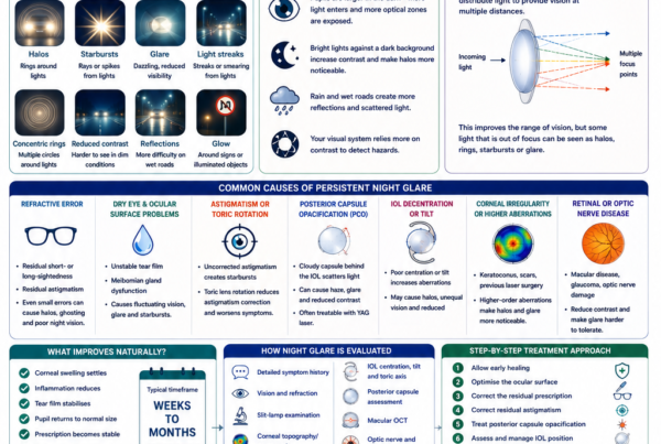
Light entering a cataractous lens is scattered instead of being focused cleanly.
This can make:
- Car headlights appear larger
- Streetlights develop halos
- Bright signs appear hazy
- Sunlight feel uncomfortable
- Reflections from wet roads more distracting
Glare may be particularly troublesome with cortical and posterior subcapsular cataracts because the opacity may interfere with light passing through the pupil under particular lighting conditions.
Can a Cataract Cause Double Vision?
Yes.
A cataract can sometimes produce monocular diplopia, meaning double or multiple images remain even when the other eye is covered.
The irregular lens may split incoming light into several focal points, causing:
- A shadow beside letters
- Several images of the moon
- Ghosted television subtitles
- Multiple points around a light
Binocular double vision that disappears when either eye is covered has different possible causes, including eye-muscle or neurological disorders, and requires a separate assessment.
Can a Cataract Make Someone More Short-Sighted?
Yes.
Certain cataracts, particularly nuclear sclerosis, may increase the focusing power of the natural lens and cause a myopic shift.
A patient may notice:
- Distance vision becoming blurrier
- The spectacle prescription becoming increasingly short-sighted
- Temporary improvement in unaided reading
- A need to remove distance glasses to read
This temporary improvement in near vision is sometimes called second sight.
It does not mean the cataract is improving. The lens is changing its optical power as it becomes more sclerotic.
Do Cataracts Cause Pain or Redness?
Ordinary age-related cataracts do not usually cause:
- Eye pain
- Marked redness
- Discharge
- Sudden severe light sensitivity
A painful red eye or sudden vision loss should not be attributed automatically to cataract.
Possible urgent causes include:
- Acute glaucoma
- Corneal infection
- Uveitis
- Retinal disease
- Eye injury
A very advanced swollen lens can occasionally contribute to inflammation or glaucoma, but this is different from the usual gradual presentation of an uncomplicated age-related cataract.
What Are the Main Types of Age-Related Cataract?
Age-related cataracts are often classified according to the part of the lens affected.
The three main morphological patterns are:
- Nuclear cataract
- Cortical cataract
- Posterior subcapsular cataract
Mixed cataracts containing more than one pattern are also common.
Nuclear Sclerotic Cataract
A nuclear cataract affects the central nucleus of the natural lens.
Over time, the central lens becomes:
- Harder
- More yellow
- More brown
- More optically dense
Possible symptoms include:
- Gradually blurred distance vision
- Reduced colour brightness
- Reduced contrast
- Increasing myopia
- Frequent spectacle changes
- Difficulty in dim lighting
Nuclear cataracts commonly progress slowly.
A person may initially notice that reading becomes easier without spectacles because the lens-induced myopic shift provides a nearer focal point.
Cortical Cataract
A cortical cataract develops in the outer lens cortex.
The opacities often appear as:
- White spokes
- Wedges
- Radial streaks
- Peripheral areas extending towards the centre
Possible symptoms include:
- Glare
- Halos
- Difficulty with headlights
- Reduced contrast
- Variable vision depending on pupil size and lighting
A cortical cataract can be visually significant even when the central lens initially appears relatively clear.
Posterior Subcapsular Cataract
A posterior subcapsular cataract forms near the back surface of the natural lens, close to the visual axis.
Because of its central position, even a relatively small opacity may produce disproportionate symptoms.
Possible symptoms include:
- Glare
- Halos
- Reduced near vision
- Difficulty reading
- Poor vision in bright conditions
- Rapid decline in visual quality
Posterior subcapsular cataracts are associated with several conditions, including diabetes, intraocular inflammation and corticosteroid exposure. Long-term combined inhaled and oral corticosteroid exposure has been associated with an increased risk of posterior subcapsular and nuclear cataracts.
Can a Patient Have More Than One Type?
Yes.
Many patients have mixed cataracts, such as:
- Nuclear and cortical cataract
- Nuclear and posterior subcapsular cataract
- All three components
The symptoms depend on:
- The location of the opacity
- Its density
- Pupil size
- Lighting
- The health of the other eye
- Other ocular conditions
The cataract grade alone does not determine how much difficulty a patient experiences.
What Is an Age-Related Cataract?
Age-related cataract is the commonest form of cataract.
Ageing increases the likelihood of:
- Protein aggregation
- Oxidative damage
- Lens hardening
- Yellowing or browning
- Reduced transparency
Cataracts become increasingly common with age, although the exact age at which symptoms develop varies greatly.
Some people develop visually significant cataracts in their fifties, while others maintain relatively clear lenses well into their seventies or eighties.
What Is a Traumatic Cataract?
A traumatic cataract develops after injury to the eye.
Possible causes include:
- A blunt impact
- Penetrating injury
- A sharp object
- Fireworks
- Sports injuries
- Electrical injury
- Chemical injury
A cataract may develop:
- Immediately
- Over several weeks
- Months later
- Many years after the original injury
Trauma can also damage:
- The lens capsule
- Zonular fibres holding the lens
- Iris
- Cornea
- Retina
- Optic nerve
Surgery may therefore be more complex than ordinary age-related cataract surgery.
What Is a Congenital or Pediatric Cataract?
A child may be born with a cataract or develop one during childhood.
Causes may include:
- Genetic conditions
- Developmental abnormalities
- Infection during pregnancy
- Metabolic disease
- Trauma
- Inflammation
- Steroid treatment
A central visually significant cataract in a young child can prevent normal visual development and cause amblyopia.
Unlike many adult cataracts, which can be monitored until they interfere with function, a visually significant pediatric cataract may require early treatment to protect visual development.
Small peripheral cataracts that do not interfere with vision may sometimes be monitored.
What Is a Radiation Cataract?
Exposure to certain forms of radiation can damage the natural lens and increase cataract risk.
Relevant exposures may include:
- Ultraviolet radiation
- Ionising radiation
- Radiation treatment near the head or upper body
- Occupational radiation exposure
Appropriate ultraviolet protection and occupational safety measures may reduce avoidable exposure.
What Is a Secondary Cataract?
The term secondary cataract commonly refers to posterior capsule opacification after cataract surgery.
However, it is not a true new cataract.
During cataract surgery:
- The cloudy natural lens material is removed
- The outer capsular bag is largely retained
- The artificial intraocular lens is placed inside the capsule
Months or years later, lens epithelial cells may cause the back capsule to become cloudy.
This is called:
- Posterior capsule opacification
- PCO
- After-cataract
- Secondary cataract
Symptoms may resemble the original cataract:
- Hazy vision
- Glare
- Halos
- Reduced contrast
PCO is treated with a YAG laser capsulotomy, which creates an opening in the cloudy posterior capsule.
What Increases the Risk of Cataracts?
Age is the most important risk factor.
Other recognised risk factors include:
- Diabetes
- Smoking
- Excessive alcohol exposure
- Long-term corticosteroid use
- Eye trauma
- Previous eye surgery
- Intraocular inflammation
- Radiation exposure
- Prolonged ultraviolet exposure
- Family history
Cataracts and Diabetes
People with diabetes may develop cataracts:
- At a younger age
- More rapidly
- In both eyes
- Alongside diabetic retinal disease
The Beaver Dam Eye Study found that diabetes was associated with the development and progression of cortical and posterior subcapsular lens opacities, with glycaemic level appearing relevant to risk.
Blur in a patient with diabetes should not automatically be attributed to cataract.
Other possible causes include:
- Fluctuating blood glucose
- Diabetic retinopathy
- Diabetic macular oedema
- Corneal surface disease
- Glaucoma
A dilated retinal examination and OCT may be required.
Cataracts and Steroids
Corticosteroids can increase cataract risk, particularly posterior subcapsular cataract.
Relevant steroid exposure may include:
- Oral steroids
- Inhaled steroids
- Injected steroids
- Long-term topical medication
- Steroid eyedrops
Risk depends on factors such as:
- Dose
- Duration
- Route of administration
- Individual susceptibility
Steroid medication should not be stopped suddenly or without consulting the prescribing doctor.
Cataracts and Smoking
Smoking is associated with a higher risk of age-related cataract, particularly nuclear cataract.
Stopping smoking has benefits for:
- Eye health
- Cardiovascular health
- Lung health
- Cancer risk
- Surgical recovery
Smoking cessation cannot reverse an existing cataract, but it may reduce further avoidable health risk.
Cataracts and Sunlight
Long-term ultraviolet exposure may contribute to cataract formation.
Protective measures include:
- Sunglasses that block ultraviolet radiation
- A broad-brimmed hat
- Avoiding direct prolonged sun exposure where practical
- Appropriate occupational eye protection
These measures may reduce cumulative exposure but cannot guarantee that cataracts will never develop.
Cataracts and Eye Inflammation
Repeated or chronic uveitis can cause cataract.
The cataract may arise from:
- Inflammation itself
- Steroid treatment
- Adhesions around the pupil
- Abnormal aqueous humour
- Damage to the lens capsule
Cataract surgery in an eye with uveitis is generally planned when inflammation is adequately controlled.
These eyes may have a greater risk of:
- Postoperative inflammation
- Macular oedema
- Pressure rise
- Posterior capsule opacification
- Recurrent uveitis
Cataracts After Previous Eye Surgery
Cataract may develop or progress after procedures such as:
- Vitrectomy
- Retinal detachment surgery
- Glaucoma surgery
- Other intraocular operations
After vitrectomy, nuclear cataract progression is particularly common in older phakic patients.
Previous surgery may also affect:
- Intraocular lens calculations
- Zonular stability
- Pupil dilation
- Retinal visual potential
- The complexity of cataract surgery
Can Cataracts Be Prevented?
There is currently no guaranteed method of preventing all age-related cataracts.
However, potentially helpful measures include:
- Avoiding smoking
- Controlling diabetes
- Protecting the eyes from ultraviolet exposure
- Wearing protective eyewear during hazardous activities
- Using steroids only when medically necessary
- Maintaining regular eye examinations
- Managing intraocular inflammation appropriately
The National Eye Institute recommends ultraviolet protection, smoking cessation, eye protection and healthy dietary habits as sensible eye-health measures.
These measures may reduce modifiable risks but cannot stop the natural ageing of the lens.
Can Diet or Supplements Reverse Cataracts?
No vitamin, supplement or food has been proven to dissolve an established age-related cataract.
A balanced diet may support general health, but it should not be presented as a substitute for surgery when a cataract is functionally significant.
Patients should be cautious about claims that:
- Special eyedrops dissolve cataracts
- Supplements restore lens transparency
- Eye exercises remove cataracts
- Herbal products eliminate cataracts
Surgery remains the only established method of removing the cloudy natural lens.
Are There Cataract Eyedrops?
There are currently no routinely approved eyedrops that reliably reverse age-related cataracts in humans.
Research continues into mechanisms that might prevent or reduce lens-protein aggregation, but experimental findings should not be confused with proven clinical treatment.
Eyedrops may be prescribed for other conditions occurring alongside cataract, such as:
- Dry eye
- Glaucoma
- Allergy
- Inflammation
They do not remove the cataract itself.
How Is a Cataract Diagnosed?
Cataracts are diagnosed during an eye examination.
The assessment may include:
- Visual acuity
- Refraction
- Glare symptoms
- Slit-lamp examination
- Eye-pressure measurement
- Pupil dilation
- Retinal examination
- OCT when indicated
A comprehensive examination is important because blurred vision may arise from more than one condition.
Visual Acuity
Vision is measured:
- Without correction
- With the current spectacles
- After refraction
The doctor assesses whether updated glasses restore satisfactory vision.
A patient whose vision remains reduced despite the best refraction may have cataract or another structural problem.
Refraction
Cataract may cause:
- Increasing myopia
- Astigmatic change
- Unequal prescriptions between the eyes
- Frequent changes in spectacle power
An updated prescription may help temporarily during the early stages.
Repeated rapid changes suggest that the lens is becoming optically unstable.
Slit-Lamp Examination
A slit lamp allows the ophthalmologist to examine the lens under magnification.
The doctor assesses:
- Cataract type
- Cataract density
- Lens colour
- Position of the opacity
- Pupil dilation
- Corneal health
- Anterior chamber
- Zonular stability
The appearance of the cataract is considered together with the patient’s symptoms.
A dense-looking cataract in a person who functions well may not require immediate surgery. A smaller posterior subcapsular cataract may cause major glare and justify earlier intervention.
Dilated Fundus Examination
Dilating the pupil allows examination of the:
- Retina
- Macula
- Optic nerve
- Retinal blood vessels
- Peripheral retina
This is important because cataract may coexist with:
- Diabetic retinopathy
- Macular degeneration
- Epiretinal membrane
- Retinal tears
- Glaucoma
- Myopic macular disease
When a cataract is very dense, the retina may be difficult or impossible to view directly.
OCT
OCT provides cross-sectional imaging of the retina and optic nerve.
Macular OCT may identify:
- Epiretinal membrane
- Macular oedema
- Macular degeneration
- Vitreomacular traction
- Myopic macular disease
These conditions may limit the visual result after cataract surgery and should be discussed during counselling.
Corneal Topography or Tomography
Corneal mapping may be recommended when:
- Significant astigmatism is present
- A toric IOL is being considered
- The patient previously underwent LASIK, PRK or SMILE
- Keratoconus is suspected
- A presbyopia-correcting IOL is being considered
- Corneal irregularity may limit visual quality
Biometry
Biometry measures the dimensions and optical characteristics of the eye.
These measurements help calculate the power of the artificial intraocular lens.
Biometry may include:
- Axial length
- Corneal curvature
- Anterior chamber depth
- Lens thickness
- Total corneal astigmatism
Accurate measurements are especially important when the aim is to reduce dependence on spectacles.
When Should Cataract Surgery Be Performed?
There is no single cataract grade or visual-acuity number at which every patient must undergo surgery.
Surgery is generally considered when the cataract interferes with activities that matter to the patient, such as:
- Driving
- Reading
- Using a computer
- Working
- Cooking
- Walking safely
- Recognising faces
- Sports
- Watching television
- Managing medication
- Functioning in dim lighting
The National Eye Institute recommends considering surgery when cataract-related vision loss interferes with normal daily activities.
Must a Cataract Be “Ripe” Before Surgery?
No.
Patients do not need to wait until a cataract is extremely dense or vision is severely impaired.
The concept of waiting for a cataract to become “ripe” arose partly from older surgical techniques, when removing a harder and more mature lens could sometimes be preferable.
Modern cataract surgery is usually based on:
- Symptoms
- Functional impairment
- Surgical risk
- Visual needs
- Other eye conditions
- Patient preference
Waiting too long may occasionally make surgery more difficult because a very dense cataract can require:
- More ultrasound energy
- More manipulation
- A longer procedure
- Greater stress on the cornea and zonules
However, surgery should not be rushed when the cataract causes minimal difficulty.
Can Surgery Be Recommended Even if the Cataract Is Mild?
Yes, in selected circumstances.
Surgery may be considered when:
- Glare prevents safe driving
- The patient’s occupation requires higher visual quality
- The prescription changes repeatedly
- The cataract prevents retinal examination or treatment
- Significant anisometropia affects binocular vision
- The lens contributes to angle closure or raised pressure
- The patient has another lens-related problem
The decision should be individualised rather than based solely on the apparent cataract grade.
Can Cataract Surgery Be Delayed?
Often, yes.
For most ordinary age-related cataracts, surgery is elective rather than an emergency.
Early symptoms may be managed using:
- Updated spectacles
- Brighter lighting
- Magnification
- Anti-glare sunglasses
- Avoiding night driving when vision is unsafe
However, delaying surgery may be inappropriate when:
- Vision is unsafe for driving
- Falls or accidents are occurring
- The cataract blocks retinal treatment
- Lens-induced glaucoma develops
- Significant inflammation occurs
- A child’s visual development is at risk
What Happens During Cataract Surgery?
Modern cataract surgery commonly uses phacoemulsification.
A typical procedure includes:
- Anaesthetic medication is given.
- Small corneal incisions are created.
- An opening is made in the front lens capsule.
- The cataract is divided and removed.
- The remaining soft lens material is aspirated.
- The capsular bag is cleaned.
- A folded artificial intraocular lens is inserted.
- The IOL unfolds inside the capsular bag.
- The incisions are checked and usually seal without stitches.
The patient is generally awake but should not see the surgical details clearly. Most patients notice lights, colours or movement.
Is the Whole Lens Removed?
The cloudy lens contents are removed, but most of the transparent outer capsule is preserved.
The capsular bag supports the artificial intraocular lens.
Preserving the capsule allows the IOL to sit in the normal anatomical lens position.
When the capsule or zonular support is inadequate, another method of lens fixation may be required.
Does Cataract Surgery Use a Laser?
Standard cataract surgery primarily uses:
- Fine surgical instruments
- Fluidics
- Ultrasound phacoemulsification
A femtosecond laser may be used for selected steps, such as:
- Corneal incisions
- Capsulotomy
- Lens fragmentation
- Astigmatic corneal incisions
A femtosecond laser does not eliminate the need to remove the lens material and implant an IOL.
Randomised studies have generally found that conventional phacoemulsification and femtosecond laser-assisted cataract surgery both provide good outcomes, without consistent evidence that femtosecond surgery produces superior vision for every routine patient.
The surgeon’s planning, technique and the patient’s eye often matter more than whether a femtosecond laser is used.
Is Cataract Surgery Painful?
Cataract surgery is usually performed with:
- Topical anaesthetic drops
- Local anaesthetic
- Sedation when appropriate
Patients may notice:
- Pressure
- Water around the eye
- Bright lights
- Movement
- Mild awareness of instruments
Sharp pain should not normally occur.
A patient who is anxious should discuss this before surgery so that the anaesthetic and sedation plan can be individualised.
How Long Does Cataract Surgery Take?
The actual surgical time is often relatively short for an uncomplicated case, but the entire visit takes longer because of:
- Registration
- Pupil dilation
- Preparation
- Anaesthesia
- Surgery
- Recovery
- Postoperative checks
More complex cataracts may take longer.
Complexity may increase with:
- Very dense cataract
- Small pupil
- Weak zonules
- Previous trauma
- Previous vitrectomy
- Corneal disease
- Glaucoma
- Pseudoexfoliation
- Previous eye surgery
What Is an Intraocular Lens?
An intraocular lens, or IOL, is the artificial lens implanted after the cataract is removed.
The IOL:
- Replaces much of the focusing power of the natural lens
- Remains permanently inside the eye
- Does not require cleaning by the patient
- Cannot usually be felt
- Is intended to remain for life
The choice of IOL affects:
- Distance vision
- Near and intermediate vision
- Astigmatism correction
- Spectacle dependence
- Contrast
- Glare and halos
No IOL can perfectly reproduce all the functions of a young natural lens.
What Is a Monofocal IOL?
A monofocal IOL has one main focal range.
It is commonly targeted for:
- Distance
- Intermediate
- Near
Most patients choose distance correction and use reading glasses for near work.
Potential advantages include:
- Good image quality
- Good contrast
- Fewer lens-related halos than multifocal designs
- Predictable optical performance
A monofocal lens does not normally provide full unaided vision at every distance.
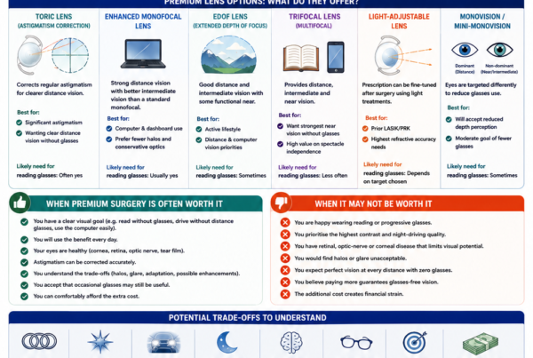
What Is Monovision?
Monovision means targeting:
- One eye mainly for distance
- The other eye for intermediate or near vision
This may reduce dependence on glasses.
Possible disadvantages include:
- Reduced depth perception
- Eye imbalance
- Difficulty driving at night
- Incomplete near vision
- Need for adaptation
Patients who have never experienced monovision may benefit from a contact lens simulation before surgery where practical.
What Is a Toric IOL?
A toric IOL corrects regular corneal astigmatism.
It must be:
- Selected using appropriate calculations
- Positioned at the intended axis
- Rotationally stable after surgery
A randomised trial found that toric IOL implantation in patients with corneal astigmatism produced better unaided distance vision, less residual astigmatism and greater distance spectacle independence than a non-toric control lens.
A toric IOL does not guarantee freedom from glasses.
Residual astigmatism may remain because of:
- Measurement variability
- Lens rotation
- Posterior corneal astigmatism
- Surgically induced astigmatism
- Irregular corneal shape
What Is a Multifocal or Trifocal IOL?
Multifocal and trifocal IOLs divide or distribute light to provide several focal ranges.
They may improve unaided:
- Distance vision
- Intermediate vision
- Near vision
Their main aim is to reduce spectacle dependence.
Potential disadvantages include:
- Halos
- Glare
- Starbursts
- Reduced contrast
- Difficulty in dim lighting
- Neuroadaptation
- Less tolerance of residual prescription or dry eye
Clinical trials show that multifocal IOLs generally provide better unaided near vision and greater spectacle independence than monofocal lenses, but photic phenomena are more common.
These lenses require careful patient selection.
What Is an Extended-Depth-of-Focus IOL?
An extended-depth-of-focus, or EDOF, IOL aims to extend the range of relatively clear vision rather than producing several distinct focal points.
Depending on the design, it may improve:
- Distance vision
- Intermediate vision
- Some functional near vision
Reading glasses may still be required for small print.
EDOF lenses can still cause:
- Halos
- Glare
- Reduced contrast
- Residual spectacle dependence
Different EDOF technologies have different optical profiles and should not be regarded as interchangeable.
Which IOL Is Best?
There is no single best lens for every patient.
The choice depends on:
- Corneal health
- Corneal astigmatism
- Macular health
- Optic nerve health
- Glaucoma
- Previous LASIK or other refractive surgery
- Pupil size
- Night-driving requirements
- Occupation
- Reading habits
- Tolerance of halos
- Willingness to use glasses
- Expectations
A lens providing greater spectacle independence may involve a greater chance of halos or reduced contrast.
A patient who prioritises night driving may make a different choice from someone who prioritises unaided reading.
Will Cataract Surgery Eliminate the Need for Glasses?
Not always.
Glasses may still be required for:
- Reading
- Computer work
- Night driving
- Residual myopia or hyperopia
- Residual astigmatism
- Fine visual tasks
- Imbalance between the eyes
Spectacle independence depends on:
- IOL type
- IOL calculation accuracy
- Corneal astigmatism
- Healing
- Retinal and optic nerve health
- The visual target
The aim should be a realistic refractive plan rather than a guarantee of complete freedom from spectacles.
How Successful Is Cataract Surgery?
Cataract surgery is among the most frequently performed and effective operations.
The National Eye Institute states that approximately nine out of ten patients see better after surgery, although the final result depends on the health of the rest of the eye.
Studies have also shown meaningful improvements in:
- Visual function
- Independence
- Vision-related quality of life
- Ability to perform daily activities
Cataract removal cannot restore vision lost permanently from:
- Advanced glaucoma
- Macular degeneration
- Diabetic retinal damage
- Optic nerve disease
- Amblyopia
- Corneal scarring
What Are the Risks of Cataract Surgery?
Cataract surgery is generally safe, but no operation is risk-free.
Possible risks include:
- Infection
- Inflammation
- Corneal swelling
- Raised or reduced eye pressure
- Cystoid macular oedema
- Bleeding
- Retinal tear or detachment
- Posterior capsule rupture
- Retained lens material
- Intraocular lens displacement
- Residual refractive error
- Glare or halos
- Posterior capsule opacification
- Rare permanent loss of vision
Risk varies according to:
- Cataract density
- Corneal health
- Pupil size
- Zonular strength
- Previous surgery
- Diabetes
- Glaucoma
- Retinal disease
- Medication
- Surgical complexity
What Is Endophthalmitis?
Endophthalmitis is a serious infection inside the eye.
Possible symptoms include:
- Increasing pain
- Rapidly worsening vision
- Marked redness
- Increasing light sensitivity
- Eyelid swelling
- Discharge
It requires urgent treatment.
Antiseptic preparation, sterile surgical technique and appropriate medication are used to reduce risk, but infection cannot be eliminated completely.
What Is Cystoid Macular Oedema?
Cystoid macular oedema is swelling at the central retina after surgery.
It may cause:
- Blurred central vision
- Distortion
- Reduced contrast
- Slower-than-expected recovery
Treatment may include:
- Anti-inflammatory eyedrops
- Steroid medication
- Injections in selected cases
- Treatment of underlying retinal disease
Patients with diabetes, uveitis or retinal vascular disease may have an increased risk.
Can Cataract Surgery Cause Retinal Detachment?
Retinal detachment is an uncommon but recognised risk after cataract surgery.
Risk may be higher in patients who are:
- Highly myopic
- Younger
- Male
- Previously affected by retinal tears
- Previously affected by retinal detachment
- Complicated by posterior capsule rupture
Warning symptoms include:
- New flashes
- A sudden shower of floaters
- A curtain or shadow
- Sudden peripheral visual-field loss
These symptoms require urgent retinal assessment.
Can the IOL Move?
An intraocular lens may rarely become:
- Decentered
- Tilted
- Rotated
- Dislocated
This is more likely when the capsular or zonular support is weak.
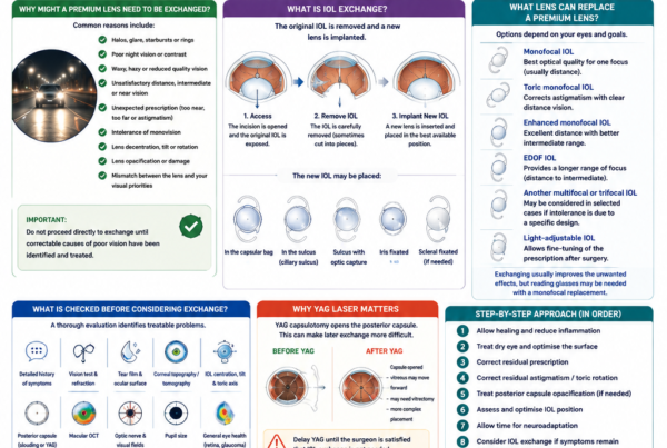
A rotated toric lens may reduce astigmatic correction.
A significantly displaced lens may require:
- Repositioning
- Fixation
- Exchange
- Another corrective procedure
What Is Posterior Capsule Opacification?
Posterior capsule opacification is one of the commonest later causes of cloudy vision after cataract surgery.
It may develop:
- Within months
- Several years later
- Gradually or asymmetrically
Symptoms include:
- Hazy vision
- Glare
- Halos
- Reduced contrast
- A sense that the cataract has returned
The cataract itself cannot return because the natural lens has been removed.
PCO is treated using YAG laser capsulotomy.
What Happens After Cataract Surgery?
After surgery, patients commonly use:
- Antibiotic drops
- Steroid or anti-inflammatory drops
- Lubricating drops when needed
An eye shield may be recommended during sleep.
Patients may be advised temporarily to avoid:
- Eye rubbing
- Contaminated water
- Swimming
- Heavy lifting
- Direct trauma
- Dusty environments
- Eye makeup
Instructions vary according to the surgeon and the individual eye.
How Quickly Does Vision Recover?
Many patients notice improved vision within the first few days.
Vision may initially be affected by:
- Pupil dilation
- Corneal swelling
- Inflammation
- Tear-film instability
- Residual prescription
- Retinal disease
Comfort often improves after the first one or two days, while complete healing may take several weeks. The National Eye Institute notes that many eyes are fully healed by approximately eight weeks.
Why Do Colours Look Brighter After Surgery?
A cataractous lens often develops a yellow or brown tint.
The brain gradually adapts to this altered colour perception.
After the cloudy lens is replaced with a clear IOL:
- White objects may appear brighter
- Blue colours may appear more vivid
- Colours may initially seem unusually cool
- The two eyes may perceive colour differently between surgeries
This commonly becomes less noticeable as the visual system adapts.
What Symptoms Require Urgent Review After Surgery?
Seek urgent assessment for:
- Sudden or rapidly worsening vision
- Severe or increasing pain
- Marked redness
- Significant discharge
- Increasing light sensitivity
- Nausea with eye pain
- New flashes
- A sudden increase in floaters
- A curtain-like shadow
- Eye trauma
These symptoms may indicate:
- Infection
- Acute pressure rise
- Retinal tear or detachment
- Significant inflammation
- Corneal complication
Can Cataract Return After Surgery?
No.
Once the natural cataractous lens has been removed, that cataract cannot grow back.
Posterior capsule opacification may later make vision cloudy, but this is scar-like clouding of the retained capsule rather than regrowth of the natural lens.
Can Cataracts Spread From One Eye to the Other?
No.
Cataracts commonly develop in both eyes because both lenses undergo similar ageing or systemic influences.
However, a cataract does not spread or transfer from one eye to the other.
Can Cataracts Develop at Different Speeds?
Yes.
One eye may develop cataract more quickly because of:
- Previous trauma
- Inflammation
- Steroid exposure
- Previous surgery
- Retinal surgery
- Differences in anatomy
- Natural biological variation
It is common for one eye to require surgery before the other.
Is Cataract Surgery an Emergency?
Routine age-related cataract surgery is usually elective.
It may become more urgent when:
- The cataract causes unsafe vision
- The lens causes glaucoma
- Inflammation develops
- Retinal disease cannot be examined or treated
- A child’s visual development is threatened
- The lens is unstable after trauma
Should Both Eyes Be Operated on at the Same Time?
Cataract surgery is commonly performed one eye at a time.
This allows:
- The first eye to begin healing
- The refractive result to be reviewed
- The second-eye plan to be adjusted when needed
- Avoidance of simultaneous bilateral complications
Same-day bilateral cataract surgery is performed in selected centres and selected patients using strict separation protocols, but it is not appropriate for every case.
Is Cataract Surgery the Same as Refractive Lens Exchange?
The surgical steps are similar because both procedures remove the natural lens and implant an IOL.
The difference is the main indication.
Cataract Surgery
The natural lens has become cloudy and is interfering with vision.
Refractive Lens Exchange
The lens may still be relatively clear, but it is removed mainly to correct:
- High refractive error
- Presbyopia
- Spectacle dependence
Because refractive lens exchange removes a clear natural lens, its risk-benefit calculation differs from surgery for a visually significant cataract.
Can LASIK Treat a Cataract?
No.
LASIK reshapes the cornea.
It does not remove or clear the natural lens.
Performing LASIK when cataract is the true cause of blur may:
- Fail to improve visual quality
- Leave glare unchanged
- Complicate later IOL calculations
- Delay the correct treatment
The natural lens should be assessed before laser vision correction, especially in patients in their forties, fifties or older.
Can Someone Have Cataract Surgery After LASIK?
Yes.
Cataract surgery is routinely performed after LASIK, PRK or SMILE.
However, previous corneal laser treatment changes the relationship between measured corneal curvature and actual corneal focusing power.
This can make IOL calculation more challenging.
Helpful information includes:
- Pre-LASIK spectacle prescription
- Previous corneal measurements
- Type of laser treatment
- Historical records
- Current corneal tomography
Patients should disclose previous refractive surgery even if it occurred decades earlier.
How Is Cataract Different From Presbyopia?
Cataract
- Clouding of the natural lens
- Causes blur, glare and reduced contrast
- May affect distance and near vision
- Treated definitively with cataract surgery
Presbyopia
- Age-related loss of natural-lens flexibility
- Mainly affects near focusing
- The lens may remain clear
- Managed with reading glasses, contact lenses or selected surgical strategies
A patient may have both cataract and presbyopia.
How Is Cataract Different From Glaucoma?
Cataract
- Affects the natural lens
- Commonly causes blurred vision and glare
- Vision can often be restored by removing the cloudy lens
Glaucoma
- Damages the optic nerve
- Often causes peripheral visual-field loss
- Vision lost from glaucoma cannot usually be restored
Cataract surgery may improve lens-related blur but cannot reverse established glaucomatous optic nerve damage.
How Is Cataract Different From Macular Degeneration?
Cataract
- Clouding of the lens
- Often causes general blur, glare and faded colours
- Usually treatable with surgery
Macular Degeneration
- Damages the central retina
- May cause central distortion or missing areas
- Cataract surgery does not remove the retinal disease
Both conditions may occur together.
Cataract surgery can still improve vision-related quality of life in many patients with macular degeneration, although the final visual potential depends on the severity of retinal damage.
Frequently Asked Questions About Cataracts
What Is a Cataract?
A cataract is clouding of the eye’s natural crystalline lens.
Is a Cataract on the Surface of the Eye?
No.
It develops inside the lens behind the iris.
Are Cataracts Part of Normal Ageing?
Most cataracts are age-related, although trauma, diabetes, inflammation, radiation and medication can cause earlier cataract.
Does Everyone Develop Cataracts?
The likelihood increases greatly with age, although not everyone becomes visually impaired at the same age.
Can Young People Develop Cataracts?
Yes.
Possible causes include:
- Genetics
- Diabetes
- Steroids
- Trauma
- Uveitis
- Previous surgery
- Radiation
Can Babies Be Born With Cataracts?
Yes.
Visually significant congenital cataracts require early specialist assessment because they can interfere with normal visual development.
Do Cataracts Hurt?
Ordinary age-related cataracts are usually painless.
Pain or redness suggests another condition or a complicated lens problem.
Can Cataracts Cause Headaches?
Cataracts do not directly cause most headaches.
Visual strain from blur or changing prescription may contribute to discomfort, but persistent headaches require an appropriate assessment.
Can Cataracts Cause Floaters?
No.
Floaters arise mainly from the vitreous.
A cataract may make floaters harder or easier to notice, but it does not create ordinary vitreous floaters.
Can Cataracts Cause Flashes?
No.
New flashes usually arise from the vitreous or retina and require assessment for a retinal tear.
Can Cataracts Cause Blindness?
Advanced cataracts can cause severe visual impairment.
The vision loss is often reversible with surgery if the retina, optic nerve and other structures remain healthy.
Can Cataracts Be Treated With Glasses?
Updated spectacles may temporarily improve early cataract-related refractive change.
They cannot remove the cloudy lens.
Can Cataracts Be Removed With a Laser Alone?
No.
The cataractous lens material must be removed surgically.
A femtosecond laser may assist selected surgical steps, but it does not replace lens removal.
Can YAG Laser Remove a Cataract?
No.
YAG laser treats posterior capsule opacification after cataract surgery.
It does not remove the original natural-lens cataract.
Should I Wait Until the Cataract Is Ripe?
No.
Surgery is usually based on functional difficulty, safety and ocular health rather than waiting for extreme lens maturity.
Can Cataract Surgery Be Done Too Early?
Surgery should not be performed simply because a mild cataract is visible.
The expected visual benefit should justify the surgical risk.
Can Cataract Surgery Be Done Too Late?
Yes.
A very advanced cataract may make surgery and retinal assessment more difficult.
Waiting is also inappropriate when vision is unsafe or the lens causes another medical problem.
Will Cataract Surgery Give Me Perfect Vision?
Not necessarily.
The result depends on:
- Corneal health
- Retinal health
- Optic nerve health
- IOL calculation
- Astigmatism
- Healing
- Lens choice
Will I Need Glasses After Surgery?
Possibly.
The need depends on the refractive target and IOL chosen.
Which Lens Is Best?
The best IOL depends on the individual eye and the patient’s visual priorities.
There is no universally best lens.
Can I Choose Near Vision?
Yes.
A monofocal lens may be targeted for distance, intermediate or near vision.
The consequences for spectacle use and binocular balance should be discussed.
Can Astigmatism Be Corrected During Surgery?
Regular corneal astigmatism may be reduced using:
- A toric IOL
- Corneal relaxing incisions
- Other refractive planning
Irregular astigmatism is more difficult to correct.
Will a Multifocal Lens Eliminate Glasses?
It may reduce spectacle dependence but does not guarantee freedom from glasses.
Halos, glare and reduced contrast may occur.
Does the Artificial Lens Wear Out?
Modern IOLs are designed to remain in the eye permanently.
They do not ordinarily need routine replacement.
Can the Artificial Lens Develop a Cataract?
No.
The artificial lens itself does not develop an ordinary cataract.
Posterior capsule opacification may occur behind it.
Can Cataract Come Back?
The natural cataract cannot return after removal.
Posterior capsule opacification can mimic it and is treated with YAG laser.
Can Cataract Surgery Cause Dry Eye?
Temporary dry-eye symptoms can occur because of:
- Surgical incisions
- Antiseptic preparation
- Eyedrops
- Reduced corneal sensation
- Pre-existing eyelid-gland disease
Dryness should be treated because it can affect comfort and visual quality.
When Can I Drive After Cataract Surgery?
Driving may resume when:
- Vision meets legal requirements
- The surgeon confirms that the eye is safe
- The patient feels comfortable
- Glare and binocular balance are acceptable
When Should I Seek Urgent Care?
Urgent assessment is needed for:
- Sudden loss of vision
- Severe pain
- Marked redness
- Significant discharge
- New flashes or many floaters
- A curtain-like shadow
- Eye trauma
Key Takeaway
A cataract is clouding of the natural crystalline lens inside the eye.
It can cause:
- Blurred or hazy vision
- Glare
- Halos
- Faded colours
- Poor night vision
- Reduced contrast
- Frequent spectacle changes
- Increasing myopia
- Monocular double vision
The main age-related cataract patterns are:
- Nuclear sclerotic cataract
- Cortical cataract
- Posterior subcapsular cataract
Other forms include:
- Traumatic cataract
- Pediatric or congenital cataract
- Radiation-related cataract
- Cataract related to inflammation, medication or surgery
Risk increases with:
- Age
- Diabetes
- Smoking
- Steroid exposure
- Ultraviolet exposure
- Eye trauma
- Inflammation
- Previous surgery
- Family history
Early cataracts may be managed temporarily with:
- Updated spectacles
- Brighter lighting
- Magnification
- Anti-glare measures
Surgery is the only established method of removing a cataract.
Cataract surgery involves:
- Removing the cloudy natural lens
- Preserving the capsular support when possible
- Implanting a clear artificial intraocular lens
The timing of surgery should be based on:
- Functional difficulty
- Driving and occupational needs
- Visual quality
- Other eye disease
- Safety
- Patient preference
A cataract does not need to become “ripe” before surgery.
Intraocular lens options may include:
- Monofocal
- Toric
- Multifocal or trifocal
- Extended-depth-of-focus
- Monovision strategies
No lens is ideal for every patient.
Cataract surgery usually improves vision substantially, but the final outcome also depends on the health of the:
- Cornea
- Retina
- Macula
- Optic nerve
- Rest of the eye
Sudden vision loss, severe pain, increasing redness, discharge, flashes, many floaters or a curtain-like shadow should not be attributed to an ordinary cataract and requires urgent assessment.
References
- National Eye Institute. Cataracts. Updated November 2025.
- National Eye Institute. Types of Cataract. Updated August 2025.
- National Eye Institute. Cataract Surgery. Updated December 2024.
- World Health Organization. Blindness and Vision Impairment. Updated February 2026.
- Brown NA, Hill AR. Cataract: The Relation Between Myopia and Cataract Morphology. The Morphology of Cataract and Visual Performance. 1993. PMID: 8325426.
- Chylack LT Jr, et al. The Lens Opacities Classification System III. Archives of Ophthalmology. 1993. PMID: 8512486.
- Klein BEK, Klein R, Lee KE. Diabetes, Cardiovascular Disease, Selected Cardiovascular Disease Risk Factors, and the Five-Year Incidence of Age-Related Cataract: The Beaver Dam Eye Study. 1998. PMID: 9860001.
- Cumming RG, et al. Use of Inhaled and Oral Corticosteroids and the Long-Term Risk of Cataract. 2009. PMID: 19243828.
- Desai P, et al. Gains From Cataract Surgery: Visual Function and Quality of Life. 1996. PMID: 8976696.
- Chan CWN, et al. Evaluation of Quality of Life in Patients With Cataract Before and After Cataract Surgery. 2003. PMID: 14522296.
- Visser N, et al. Toric Versus Aspherical Control Intraocular Lenses in Patients With Cataract and Corneal Astigmatism: A Randomised Clinical Trial. JAMA Ophthalmology. 2014. PMID: 25256624.
- Shah S, et al. Visual Outcomes After Cataract Surgery: Multifocal Versus Monofocal Intraocular Lenses. 2015. PMID: 26465253.
- Häring G, et al. Subjective Photic Phenomena With Refractive Multifocal and Monofocal Intraocular Lenses. 2001. PMID: 11226790.
- Roberts HW, et al. A Randomised Controlled Trial Comparing Femtosecond Laser-Assisted Cataract Surgery Versus Conventional Phacoemulsification. 2019. PMID: 30413333.
- Forooghian F, et al. Visual Acuity Outcomes After Cataract Surgery in Patients With Age-Related Macular Degeneration: AREDS Report. 2009. PMID: 19700198.