Author: Dr Val Phua
Estimated reading time: 20 minutes
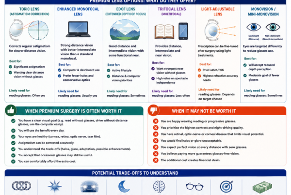
Premium intraocular lenses can provide a wider range of unaided vision after cataract surgery. Depending on the design, they may reduce the need for glasses when driving, using a computer, looking at a phone or reading.
However, some patients notice unwanted visual effects after surgery, particularly at night.
These may include:
- Halos around headlights
- Glare from bright lights
- Starbursts
- Concentric rings
- Light streaks
- A glow around illuminated signs
- Reduced contrast in dim conditions
- Difficulty seeing through reflections on wet roads
- A sensation that vision is clear but not completely crisp
These symptoms are often grouped under the term dysphotopsia.
Night glare can occur after any cataract operation, including surgery with a monofocal lens. However, halos and other photic phenomena are generally reported more often with multifocal and trifocal lenses than with conventional monofocal IOLs because presbyopia-correcting designs manipulate or distribute light to provide more than one focal range.
Night glare does not automatically mean that the operation has failed or that the lens must be removed.
In many patients, symptoms improve as:
- Corneal swelling settles
- Inflammation reduces
- The tear film stabilises
- The pupil returns to its usual size
- The prescription becomes stable
- Both eyes are treated
- The brain adapts to the new optical system
However, glare should not simply be dismissed as something the patient must tolerate. Persistent symptoms may have a correctable cause, including dry eye, residual refractive error, astigmatism, posterior capsule opacification or IOL decentration. Referral studies identify residual prescription and dry eye among the most frequent causes of dissatisfaction after presbyopia-correcting IOL implantation.
The Quick Answer
Is Night Glare Normal After Premium-Lens Surgery?
Some night glare, halos or starbursts are common during the early recovery and adaptation period after multifocal, trifocal and some EDOF lens procedures.
These symptoms may be particularly noticeable:
- During the first few weeks
- Before both eyes have been operated on
- When the pupil is large
- In very dark environments
- Around LED headlights
- In rain
- When the ocular surface is dry
- When residual astigmatism is present
A prospective neuroadaptation study found that glare and halo symptoms were more pronounced after multifocal than monofocal implantation and improved between the early postoperative period and three months, although they remained more noticeable in the multifocal group.
Does Night Glare Always Improve?
No.
Many patients become less aware of the symptoms over weeks or months, but complete disappearance cannot be guaranteed.
Long-term studies generally show high overall satisfaction after multifocal implantation, while also finding that glare and halos remain more noticeable than with accommodating or monofocal-like optics in some patients.
When Should Night Glare Be Investigated?
Arrange an examination when glare:
- Is severe
- Prevents safe night driving
- Is worsening rather than improving
- Occurs mainly in one eye
- Is associated with blurred or double vision
- Develops after an initially clear period
- Is accompanied by eye pain or redness
- Is associated with a significant change in vision
- Persists despite adequate healing time
The aim is to determine whether the glare is an expected optical effect of the lens or a symptom of a separate, potentially treatable problem.
Does the Lens Need to Be Exchanged?
Usually not.
Many patients improve after treatment of:
- Dry eye
- Residual short-sightedness or long-sightedness
- Astigmatism
- Capsule clouding
- Lens-position problems
Lens exchange is reserved for selected patients with persistent, disabling symptoms after correctable causes have been addressed and sufficient time for adaptation has been allowed. Studies of dissatisfied patients show that only a minority require exchange, although exchange can improve symptoms in carefully selected cases.
What Does Night Glare Feel Like?
Halos
A halo is a ring or series of rings around a point source of light.
Patients may notice halos around:
- Car headlights
- Streetlights
- Traffic lights
- Illuminated signs
- Decorative lights
- Small LED lamps
Halos may appear as:
- A single glow
- Several concentric circles
- A bright central light surrounded by rings
- A coloured or white luminous border
Halos are among the most frequently reported visual phenomena after multifocal IOL implantation.
Glare
Glare describes visual discomfort or reduced visual performance when a bright light enters the eye.
Patients may say that:
- Headlights feel excessively bright.
- Light spreads across the visual field.
- The surrounding road becomes difficult to see.
- Vision takes time to recover after an oncoming vehicle passes.
- Bright signs obscure nearby objects.
Glare can be uncomfortable even when standard eye-chart vision is excellent.
Starbursts
Starbursts appear as radiating lines extending from a bright light source.
They may resemble:
- A star
- Spokes
- Fine rays
- Long streaks
- A spider-web pattern
Starbursts can be produced by presbyopia-correcting optics but may also be worsened by astigmatism, dry eye or corneal irregularity.
Light Streaks and Smearing
Some patients describe:
- Vertical or diagonal streaks
- Smearing around headlights
- A tail extending from a light
- A flare across the windscreen
These symptoms may indicate:
- Residual astigmatism
- Tear-film instability
- Corneal irregularity
- Lens tilt
- Optical interaction with the IOL design
Waxy or Hazy Vision
A patient may read the smallest letters on the chart yet describe vision as:
- Waxy
- Misty
- Less crisp
- Low contrast
- As though looking through a thin film
This complaint may be associated with multifocal light distribution, but treatable causes such as dry eye, residual prescription and posterior capsule opacification must first be excluded.
Negative Dysphotopsia Is Different
Negative dysphotopsia usually presents as a dark crescent, arc or shadow in the temporal peripheral visual field.
This is different from night glare, halos and starbursts, which are forms of positive dysphotopsia.
Both may occur after cataract surgery, but their mechanisms and management differ. Reviews of pseudophakic dysphotopsia distinguish positive light phenomena from the dark temporal shadow characteristic of negative dysphotopsia.
Why Are Symptoms Worse at Night?
The Pupil Becomes Larger
In darkness, the pupil enlarges to allow more light into the eye.
A larger pupil may:
- Expose a wider area of the IOL optic
- Increase the contribution of peripheral corneal aberrations
- Make diffractive or refractive zones more noticeable
- Increase the effect of lens decentration
- Worsen the effect of residual astigmatism
The relationship between pupil size and symptoms differs among lens designs. Some multifocal optics are more pupil-dependent than others.
Light Sources Have High Contrast
At night, a headlight is a very bright object against a dark background.
This makes optical scatter, rings and aberrations much more noticeable than they would be in daylight.
A small halo around a lamp may be barely visible during the day but appear large and intrusive on a dark road.
The Visual System Relies More on Contrast
Night driving requires the ability to detect:
- Pedestrians in dark clothing
- Lane markings
- Kerbs
- Cyclists
- Objects in the road
- Vehicles emerging from side roads
These are often low-contrast targets.
Premium IOLs may provide excellent high-contrast acuity while still producing differences in contrast sensitivity or photic phenomena that become relevant during night driving. A 2025 review concluded that night-driving difficulty remains an important concern for some multifocal-IOL patients, particularly before neuroadaptation is complete.
Rain and Wet Roads Increase Reflections
Wet surfaces create multiple reflected light sources.
This can make:
- Halos overlap
- Road markings less visible
- Headlight glare spread across the road
- Contrast appear lower
- Driving more visually tiring
A patient who is comfortable driving on a dry, well-lit road may still struggle in rain.
Why Do Premium Lenses Cause Halos?
Multifocal and Trifocal Lenses Divide Light
A multifocal or trifocal lens is designed to provide useful focus at more than one distance.
Depending on the design, incoming light is directed towards:
- Distance
- Intermediate vision
- Near vision
The advantage is a wider range without spectacles.
The compromise is that light from different focal ranges may create simultaneous retinal images. Around a bright point source, these out-of-focus components may be perceived as halos or rings.
Systematic reviews consistently find better unaided near vision and greater spectacle independence with multifocal lenses than with monofocal lenses, but also more glare and halos.
Diffractive Rings Manipulate Light
Many premium lenses use microscopic diffractive steps or rings.
These structures are precisely designed to direct light to different focal points.
They are not defects in the lens.
However, the same optical principle that creates multiple focal ranges can produce:
- Concentric halos
- Rings
- Spider-web patterns
- Light scatter around point sources
Refractive Multifocal Designs Can Also Cause Symptoms
Not every multifocal lens is diffractive.
Refractive multifocal lenses use different optical zones to create a range of focus. Their visual effects may depend more strongly on:
- Pupil size
- Lens centration
- Lighting
- Which zone lies within the pupil
Older refractive multifocal designs were associated with significant night halos in some studies, although modern lenses differ considerably.
Do All Premium Lenses Cause the Same Amount of Glare?
Trifocal and Multifocal Lenses
Trifocal lenses usually provide the strongest unaided near range among commonly used premium-lens categories.
Because they distribute light across distance, intermediate and near foci, halos and glare are recognised trade-offs.
This does not mean that every trifocal patient will be troubled by them. Many patients notice halos but do not consider them severe enough to affect satisfaction.
Diffractive EDOF Lenses
Some EDOF lenses use diffractive optics.
These lenses can also produce:
- Halos
- Rings
- Glare
- Starbursts
EDOF should therefore not be interpreted as meaning “halo-free.”
A systematic review found that EDOF lenses produced more halos than monofocal lenses.
Non-Diffractive EDOF Lenses
Some non-diffractive EDOF lenses are designed to provide distance and intermediate vision with a more monofocal-like visual-disturbance profile.
They may produce fewer or less intense photic phenomena than some diffractive lenses, but:
- Halos may still occur.
- Near vision is usually weaker than with a trifocal lens.
- Results are lens-specific.
- Individual experience varies.
It is unsafe to assume that every lens marketed as EDOF will behave similarly.
EDOF Versus Trifocal Lenses
Comparative evidence is mixed.
A 2023 meta-analysis found better near vision with trifocal lenses but no statistically significant overall difference in reported halos or glare compared with EDOF lenses. This reflects considerable variation among EDOF technologies, trifocal designs, questionnaires and study methods.
A broader network meta-analysis also found that visual-disturbance differences did not consistently reach statistical significance across every lens category, despite the known trade-off between range of vision and optical phenomena.
Enhanced Monofocal Lenses
Enhanced monofocal lenses aim to extend intermediate vision while preserving an optical profile closer to a monofocal lens.
They generally produce fewer multifocal-style halos than trifocal lenses, although glare can still result from:
- Residual prescription
- Dry eye
- Capsule clouding
- Ordinary pseudophakic dysphotopsia
- Other eye disease
Standard Monofocal Lenses
Monofocal lenses generally produce the fewest multifocal-style rings because they direct most light towards one main focus.
However, positive dysphotopsia can occur after uncomplicated monofocal cataract surgery as well. IOL material, optic edge, refractive index, internal reflections and individual anatomy can contribute.
Why Can Two Patients With the Same Lens Have Different Symptoms?
Pupil Size
A patient with larger pupils in darkness may experience a different optical profile from a patient with smaller pupils.
Corneal Aberrations
The IOL does not work in isolation.
The final retinal image depends on the combined optics of:
- Tear film
- Cornea
- Intraocular lens
- Pupil
- Retina
- Visual-processing system
A lens that performs well in a regular cornea may cause more symptoms in an eye with:
- Previous LASIK
- Irregular astigmatism
- Keratoconus
- Corneal scarring
- High higher-order aberrations
Sensory Awareness
Some patients naturally notice visual artefacts more readily.
This does not mean that the symptoms are imagined.
Two patients may have similar optical halos but differ in:
- Awareness
- Tolerance
- Occupation
- Night-driving frequency
- Visual expectations
- Ability to suppress the phenomenon
Lifestyle
A patient who rarely drives after dark may consider mild halos insignificant.
A professional driver may find the same halos unacceptable.
Brain Adaptation
The ability to ignore competing visual information differs among patients.
Neuroadaptation is a genuine neurological process, but it is not complete or equally rapid in everyone.
Early Causes of Night Glare
Dilated Pupil
Dilating drops used during surgery and postoperative examinations can temporarily increase:
- Light sensitivity
- Glare
- Halos
- Blur
Do not judge the permanent night-vision result while the pupil remains dilated.
Corneal Swelling
Mild corneal oedema may cause:
- Misty vision
- Halos
- Glare
- Reduced contrast
- Vision that is worse in the morning
This usually improves as the cornea clears after uncomplicated surgery.
Inflammation
Postoperative inflammation can reduce optical clarity and cause:
- Haze
- Light sensitivity
- Glare
- Fluctuating vision
Continue prescribed anti-inflammatory drops and attend postoperative reviews.
Tear-Film Instability
The ocular surface may be temporarily disturbed by:
- The surgical incision
- Antiseptic exposure
- Postoperative drops
- Reduced blinking
- Meibomian-gland dysfunction
Dryness is particularly important because it can both produce glare and make premium-lens optical effects more noticeable.
Visual Imbalance Between the Eyes
After first-eye surgery, one eye may have:
- A premium IOL
- Clearer colour vision
- A different prescription
- Different halos
while the other eye still has a cataract.
The final binocular result may feel more balanced after the second eye has healed.
Treatable Causes of Persistent Glare
Residual Short-Sightedness or Long-Sightedness
Even a small spherical refractive error can reduce:
- Distance clarity
- Contrast
- Multifocal performance
- Night-driving comfort
A patient may still read the eye chart well but benefit significantly from a precise night-driving prescription.
Residual Astigmatism
Astigmatism may produce:
- Starbursts
- Light streaks
- Ghosting
- Shadowed letters
- Smearing around headlights
Premium lenses, especially multifocal and trifocal IOLs, are sensitive to residual cylinder.
A proper refraction is therefore essential when evaluating night glare.
Toric-Lens Rotation
If a toric premium lens rotates away from its intended axis, astigmatic correction decreases.
Symptoms may include:
- Vision that was initially clear but became blurred
- Increased starbursts
- Ghosting
- Poor night vision
- Unequal vision between the eyes
The toric marks should be examined after dilation when clinically indicated.
Dry Eye
Dry eye is one of the most frequently identified causes of dissatisfaction after presbyopia-correcting IOL implantation.
In one referral series, residual refractive error and dry eye accounted for a large proportion of complaints, and many patients were managed with spectacles, contact lenses or ocular-surface treatment rather than IOL exchange.
Meibomian-Gland Dysfunction
Poor meibomian-gland function destabilises the oily layer of the tear film.
This may cause vision to:
- Clear after blinking
- Blur during driving
- Worsen with air conditioning
- Fluctuate during screen use
- Become more glaring at night
Corneal Irregularity
Topography or tomography may be required when glare is associated with:
- Previous LASIK or PRK
- Keratoconus
- Corneal scars
- Irregular astigmatism
- Decentered laser treatment
IOL Decentration or Tilt
Premium optics generally perform best when the lens is well centred.
Tilt or decentration may worsen:
- Halos
- Asymmetrical glare
- Ghosting
- Reduced contrast
- Unequal visual quality
Long-term trifocal data have also associated more significant halos with greater IOL tilt in some cohorts.
Posterior Capsule Opacification
The capsule behind the IOL may become cloudy months or years after surgery.
Posterior capsule opacification may cause:
- Hazy vision
- Increasing glare
- Reduced contrast
- Worsening halos
- Difficulty reading
- A sensation that the cataract has returned
This can often be treated with YAG laser capsulotomy.
However, YAG should not be performed reflexively in a patient who may require IOL exchange, because opening the posterior capsule can make later lens-exchange surgery more complex.
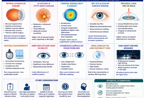
Retinal or Optic-Nerve Disease
Glare may feel more disabling when visual contrast is already reduced by:
- Macular degeneration
- Epiretinal membrane
- Diabetic macular disease
- Glaucoma
- Optic neuropathy
Macular OCT and optic-nerve assessment may be required when visual quality is poorer than expected.
Neuroadaptation
What Is Neuroadaptation?
Neuroadaptation is the brain’s adjustment to a new visual input.
With premium IOLs, the brain may need to learn to:
- Select the clearest focal image
- Suppress unwanted defocused information
- Reduce awareness of halos
- Combine images from the two eyes
- Adapt to altered contrast patterns
Functional imaging research supports measurable changes in visual processing after multifocal implantation.
How Long Does It Take?
There is no single timetable.
Patients may improve over:
- Several weeks
- A few months
- Occasionally longer
The early postoperative result should not always be considered final.
Adaptation may be delayed by:
- Dry eye
- Residual prescription
- Significant astigmatism
- Unequal lenses between the eyes
- Delayed second-eye surgery
- Lens decentration
- Retinal disease
Should Every Patient Simply Wait?
No.
Waiting is reasonable only when:
- The eye is healing normally.
- The prescription is acceptable.
- The ocular surface is being treated.
- No structural problem has been identified.
- Symptoms are stable or improving.
- The patient can function safely.
Severe night-driving difficulty should be investigated rather than being attributed automatically to neuroadaptation.
When Is Night Glare More Concerning?
Sudden Worsening
A sudden increase in glare after a period of stable clear vision may indicate:
- New dry-eye deterioration
- Corneal swelling
- Capsule clouding
- Lens movement
- Retinal or macular disease
Pain or Redness
Night glare alone is usually an optical symptom.
Glare associated with:
- Significant pain
- Increasing redness
- Marked light sensitivity
- Discharge
- Sudden vision loss
requires prompt ophthalmic assessment.
New Flashes, Floaters or a Curtain
These symptoms are not typical premium-lens glare.
Urgent examination is needed for:
- New flashes
- A sudden shower of floaters
- A dark curtain
- Missing peripheral vision
- A fixed shadow
Double Vision
True double vision should be distinguished from monocular ghosting.
- Ghosting that remains with one eye open may be optical.
- Two separate images that disappear when either eye is closed may indicate an eye-alignment problem.
Do not drive until significant double vision has been assessed.
How Is Night Glare Evaluated?
A Detailed Symptom History
The ophthalmologist may ask:
- When did the glare begin?
- Is it improving?
- Is one eye worse?
- Does blinking help?
- Is it present only at night?
- Are there rings, streaks or general haze?
- Can you drive safely?
- Did the symptoms begin after YAG laser?
- Were halos present before surgery?
- Have both eyes been operated on?
Unaided and Corrected Vision
Vision should be tested:
- Without glasses
- With a careful refraction
- At distance
- At intermediate range
- At near
A small prescription may have a large effect on premium-IOL performance.
Slit-Lamp Examination
The examination assesses:
- Tear film
- Corneal surface
- Corneal swelling
- Inflammation
- IOL position
- Capsule clarity
- Pupil
- Wound
Refraction
Refraction identifies:
- Residual sphere
- Residual astigmatism
- Whether glasses improve clarity
- Whether night-driving spectacles may help
Corneal Topography or Tomography
These tests can detect:
- Irregular astigmatism
- Keratoconus
- Previous laser-treatment irregularity
- Corneal asymmetry
- Higher-order optical problems
Macular OCT
OCT may identify:
- Epiretinal membrane
- Macular oedema
- Macular degeneration
- Vitreomacular traction
- Other subtle retinal pathology
IOL Centration and Toric Axis
A dilated examination may be needed to assess:
- IOL centration
- Tilt
- Toric alignment
- Capsular contraction
- Zonular stability
Contrast and Glare Testing
Selected clinics may use:
- Contrast-sensitivity charts
- Glare testing
- Halo simulators
- Patient-reported quality-of-vision questionnaires
- Night-driving questionnaires
The 2025 night-driving review recommends structured assessment because standard visual acuity alone may not capture real-world night performance.
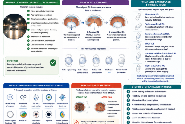
A Step-by-Step Treatment Approach
Step 1: Allow Early Healing
When the operation is recent and the examination is normal, the first step may be:
- Continue postoperative medication.
- Treat inflammation.
- Allow corneal swelling to resolve.
- Monitor the pupil.
- Reassess after the prescription stabilises.
Step 2: Optimise the Ocular Surface
Treatment may include:
- Preservative-free lubricating drops
- Warm compresses
- Eyelid hygiene
- Treatment of meibomian-gland dysfunction
- Anti-inflammatory dry-eye treatment
- Environmental modification
The treatment should match the cause of the ocular-surface disease.
Step 3: Correct the Residual Prescription
A temporary or permanent spectacle prescription may significantly improve:
- Headlight clarity
- Contrast
- Starbursts
- Ghosting
- Driving confidence
Glasses may be used specifically for night driving even when the patient is spectacle-free for most daytime activities.
Step 4: Correct Residual Astigmatism
Options may include:
- Spectacles
- Contact lenses
- Corneal laser enhancement
- Arcuate corneal incisions
- Toric-IOL rotation
- Other refractive treatment
The refraction should be stable before permanent enhancement.
Step 5: Treat Posterior Capsule Opacification
YAG laser capsulotomy may improve glare when genuine capsule clouding is present.
Before proceeding, the surgeon should be satisfied that:
- The capsule is causing the symptoms.
- Lens exchange is unlikely.
- The IOL is acceptably positioned.
- Dry eye and refractive error have been addressed.
Step 6: Assess IOL Position
A decentered, tilted or rotated lens may require:
- Observation
- Repositioning
- Toric-axis rotation
- Capsular management
- Lens exchange
The decision depends on the severity of the optical effect and the stability of the capsule.
Step 7: Allow Neuroadaptation
When all correctable issues have been addressed, a period of observation may be appropriate.
During this period:
- Avoid unsafe night driving.
- Use spectacles when helpful.
- Monitor whether symptoms are improving.
- Complete treatment in the second eye when clinically appropriate.
- Maintain the ocular surface.
Step 8: Consider IOL Exchange
IOL exchange may be considered when:
- Glare remains disabling.
- Night driving is not possible.
- The patient cannot adapt despite adequate time.
- Correctable causes have been treated.
- The lens’s optical design is clearly not tolerated.
- The patient understands the risks and loss of near vision.
Lens Exchange for Persistent Glare
What Lens Is Usually Implanted Instead?
A diffractive multifocal or EDOF lens may be exchanged for:
- A standard monofocal IOL
- A toric monofocal IOL
- An enhanced monofocal IOL
- Occasionally another presbyopia-correcting lens with a different optical profile
Exchanging for a monofocal lens usually reduces multifocal-style photic phenomena but also reduces unaided near vision.
Does Exchange Work?
Case series show that exchange can improve intolerable dysphotopsia, blurred vision and night-driving symptoms in selected patients. In one series of 64 eyes exchanged from diffractive multifocal or EDOF lenses to monofocal IOLs, the main indications included photic phenomena, reduced visual quality and night-vision difficulty.
A separate series of 50 explanted multifocal lenses identified waxy vision, glare, halos and neuroadaptation failure among the common reasons for removal.
What Are the Risks?
Lens exchange is another intraocular operation.
Potential risks include:
- Capsular rupture
- Zonular damage
- Vitreous loss
- Corneal endothelial damage
- Retinal tear or detachment
- Infection
- Macular swelling
- Residual prescription
- Need for an alternative lens position
Why Is Earlier Exchange Sometimes Easier?
Over time, the capsule contracts and adheres to the IOL.
Exchange becomes more difficult after:
- Capsular fibrosis
- Capsular contraction
- YAG laser capsulotomy
- Long periods of implantation
This does not mean that every symptomatic lens should be removed early. It means the decision should not be delayed indefinitely when symptoms are severe, stable and clearly attributable to lens intolerance.
Practical Advice for Night Driving
Do Not Drive Until You Feel Safe
You should not drive at night if:
- Headlights obscure pedestrians.
- Halos overlap nearby vehicles.
- Lane markings disappear in rain.
- You experience double vision.
- Your vision is fluctuating significantly.
- You cannot judge distance confidently.
- Your ophthalmologist has advised against driving.
Begin With Easier Conditions
When resuming night driving:
- Choose a familiar route.
- Begin with a short journey.
- Drive in dry weather.
- Avoid peak traffic.
- Avoid poorly lit unfamiliar roads.
- Consider travelling with another licensed driver.
Use a Precise Night-Driving Prescription
A small pair of spectacles may correct residual:
- Astigmatism
- Short-sightedness
- Long-sightedness
and improve the quality of headlights and road markings.
Keep the Windscreen Clean
Dirty or scratched surfaces can increase apparent glare.
Clean:
- The inside of the windscreen
- The outside of the windscreen
- Mirrors
- Spectacle lenses
Reduce Ocular-Surface Dryness
Before a drive:
- Use prescribed lubricants when appropriate.
- Allow temporary drop-related blur to clear.
- Avoid air-conditioning vents blowing directly towards the eyes.
- Blink consciously.
- Take breaks on long journeys.
Do Not Use Very Dark Lenses at Night
Dark sunglasses reduce the amount of light reaching the eye and may worsen hazard detection.
Tinted night-driving products are not a substitute for proper examination and correction.
Can Night Glare Be Prevented Before Surgery?
Discuss Night Driving Honestly
Tell your ophthalmologist whether you:
- Drive every night
- Drive professionally
- Frequently drive in rain
- Use poorly lit roads
- Operate machinery in dim conditions
- Have low tolerance for halos
The lens with the widest near range may not be the best lens for a patient whose overriding priority is night-distance quality.
Assess the Cornea
Preoperative assessment may include:
- Tear-film evaluation
- Keratometry
- Topography
- Tomography
- Aberrometry
This is particularly important after previous LASIK or when astigmatism is irregular.
Treat Dry Eye Before Measurements
Stable measurements improve:
- IOL-power calculation
- Astigmatism planning
- Toric-lens selection
- Postoperative image quality
Correct Astigmatism
Residual cylinder may magnify glare and starbursts.
A toric version of the selected lens may be needed when regular corneal astigmatism is clinically significant.
Examine the Macula and Optic Nerve
A healthy retina and optic nerve are important for contrast and visual processing.
Choose the Appropriate Optical Design
A patient prioritising reading without glasses may accept a trifocal lens and its potential halo trade-off.
A patient prioritising night driving may prefer:
- A toric monofocal lens
- An enhanced monofocal lens
- A selected EDOF lens
- A conservative mini-monovision strategy
Frequently Asked Questions
Are Halos Normal After Multifocal-Lens Surgery?
They are common, particularly during early recovery and in dark environments.
They often become less noticeable but may not disappear completely.
How Long Should I Wait for Halos to Improve?
Many patients improve over weeks to several months.
The appropriate waiting period depends on:
- Symptom severity
- Whether improvement is occurring
- The health of the eye
- Whether both eyes have been treated
- Whether correctable causes remain
Are Halos Worse With Trifocal Lenses Than EDOF Lenses?
Not always.
Trifocal lenses generally provide better close-reading vision. Some EDOF designs may produce fewer photic phenomena, but pooled studies have not consistently demonstrated a significant difference in halos and glare between every EDOF and trifocal group.
Can a Non-Diffractive EDOF Lens Cause Glare?
Yes.
Its glare profile may be closer to that of a monofocal lens than some diffractive designs, but no premium IOL guarantees zero glare.
Why Is One Eye Worse Than the Other?
Possible reasons include:
- Different residual prescription
- More astigmatism
- Dry eye
- Corneal irregularity
- Lens decentration
- Toric rotation
- Capsule clouding
- Retinal differences
- A different IOL model or target
Can Glasses Help Multifocal-Lens Halos?
Glasses can help when symptoms are worsened by residual prescription or astigmatism.
They cannot completely remove halos caused purely by the multifocal optical design.
Can Dry-Eye Treatment Reduce Glare?
Yes, when tear-film instability is contributing to the symptoms.
Dry eye is a frequent treatable cause of poor visual quality after presbyopia-correcting IOL implantation.
Can YAG Laser Remove Halos?
YAG can improve halos caused by posterior capsule opacification.
It will not correct:
- Dry eye
- Residual astigmatism
- Lens decentration
- Intrinsic multifocal rings
- Neuroadaptation failure
YAG should be delayed when lens exchange remains a realistic possibility.
Will Treating the Second Eye Improve Glare?
It may improve binocular balance and adaptation.
However, severe symptoms in the first eye should still be evaluated before automatically implanting the same lens in the second eye.
Can a Multifocal Lens Be Changed to an EDOF Lens?
Yes, but exchange is another intraocular operation.
Changing from one premium design to another may not guarantee that glare will disappear.
Can a Multifocal Lens Be Changed to a Monofocal Lens?
Yes.
This usually reduces the optical range and increases dependence on reading glasses, but may improve intolerable multifocal-style dysphotopsia.
Can Halos Return Years Later?
New or worsening halos years later may result from:
- Dry eye
- Posterior capsule opacification
- Corneal change
- Refractive change
- IOL movement
- Retinal disease
They should be assessed rather than assumed to be ordinary neuroadaptation.
Does Night Glare Mean the Lens Is Damaged?
Usually not.
The glare often reflects the optical design or another treatable condition rather than physical damage to the IOL.
A Night-Glare Assessment Checklist
Questions About the Symptoms
- Are the symptoms improving or worsening?
- Are they present in one eye or both?
- Do they improve after blinking?
- Are they mainly rings, streaks or general haze?
- Do glasses improve them?
- Can you drive safely?
- Did they start immediately or develop later?
- Were they present before surgery?
Clinical Factors to Assess
- Unaided vision
- Refraction
- Residual astigmatism
- Tear-film stability
- Corneal topography
- IOL centration and tilt
- Toric alignment
- Posterior capsule clarity
- Macular OCT
- Optic-nerve health
- Pupil size
Before Considering Lens Exchange
Confirm that:
- The ocular surface has been treated.
- The prescription is stable.
- Residual refractive error has been assessed.
- Astigmatism has been addressed.
- The posterior capsule has been evaluated.
- Lens position is satisfactory.
- Retinal and optic-nerve disease has been excluded.
- Adequate time for adaptation has been allowed.
- The symptoms remain functionally unacceptable.
- The risks and visual compromises of exchange are understood.
The Bottom Line
Night glare after premium-lens cataract surgery is common enough that it should be discussed before surgery, particularly with patients considering multifocal, trifocal or diffractive EDOF lenses.
Symptoms may include:
- Halos
- Glare
- Starbursts
- Light streaks
- Concentric rings
- Reduced low-light contrast
- Difficulty driving at night
In many patients, these symptoms improve as:
- The eye heals.
- Dryness settles.
- The prescription stabilises.
- Both eyes are treated.
- Neuroadaptation occurs.
However, persistent glare should be investigated for:
- Dry eye
- Residual prescription
- Astigmatism
- Toric-lens rotation
- Corneal irregularity
- IOL decentration or tilt
- Posterior capsule opacification
- Retinal or optic-nerve disease
The first treatment is not automatically lens exchange.
A structured approach usually begins with:
- Completing postoperative healing
- Treating the ocular surface
- Performing an accurate refraction
- Correcting residual astigmatism
- Assessing IOL position
- Treating genuine capsule clouding
- Allowing reasonable neuroadaptation
- Considering IOL exchange only when symptoms remain disabling
Premium lenses provide a wider range of unaided vision by accepting certain optical compromises.
The relevant question is not simply:
“Does this lens cause halos?”
It is:
“Are the halos mild and acceptable, or are they preventing safe and comfortable vision despite proper healing and treatment?”
References
- Pusnik A, Petrovski G, Lumi X. Dysphotopsias or unwanted visual phenomena after cataract surgery. Life (Basel). 2023;13(1):53. doi:10.3390/life13010053. PMID: 36676002.
- Masket S, Fram NR. Pseudophakic dysphotopsia: review of incidence, cause, and treatment of positive and negative dysphotopsia. Ophthalmology. 2021;128(11)–e205. doi:10.1016/j.ophtha.2020.08.009. PMID: 32800744.
- de Silva SR, Evans JR, Kirthi V, Ziaei M, Leyland M. Multifocal versus monofocal intraocular lenses after cataract extraction. Cochrane Database Syst Rev. 2016;12(12). doi:10.1002/14651858.CD003169.pub4. PMID: 27943250.
- Khandelwal SS, Jun JJ, Mak S, Booth MS, Shekelle PG. Effectiveness of multifocal and monofocal intraocular lenses for cataract surgery and lens replacement: a systematic review and meta-analysis. Graefes Arch Clin Exp Ophthalmol. 2019;257(5):863–875. doi:10.1007/s00417-018-04218-6. PMID: 30627791.
- Cho JY, Won YK, Park J, Nam JH, Hong JY, Min S, et al. Visual outcomes and optical quality of accommodative, multifocal, extended depth-of-focus, and monofocal intraocular lenses in presbyopia-correcting cataract surgery: a systematic review and Bayesian network meta-analysis. JAMA Ophthalmol. 2022;140(11):1045–1053. doi:10.1001/jamaophthalmol.2022.3667. PMID: 36136323.
- Karam M, Alkhowaiter N, Alkhabbaz A, et al. Extended depth of focus versus trifocal for intraocular lens implantation: an updated systematic review and meta-analysis. Am J Ophthalmol. 2023;251:52–70. doi:10.1016/j.ajo.2023.01.024. PMID: 36736751.
- Rampat R, Gatinel D. Multifocal and extended depth-of-focus intraocular lenses in 2020. Ophthalmology. 2021;128(11)–e185. doi:10.1016/j.ophtha.2020.09.026. PMID: 32980397.
- Gibbons A, Ali TK, Waren DP, Donaldson KE. Causes and correction of dissatisfaction after implantation of presbyopia-correcting intraocular lenses. Clin Ophthalmol. 2016;10:1965–1970. doi:10.2147/OPTH.S114890. PMID: 27784985.
- Woodward MA, Randleman JB, Stulting RD. Dissatisfaction after multifocal intraocular lens implantation. J Cataract Refract Surg. 2009;35(6):992–997. doi:10.1016/j.jcrs.2009.01.031. PMID: 19465282.
- Zhang L, Lin D, Wang Y, Chen W, Xiao W, Xiang Y, et al. Comparison of visual neuroadaptations after multifocal and monofocal intraocular lens implantation. Front Neurosci. 2021;15:648863. doi:10.3389/fnins.2021.648863. PMID: 34194292.
- Hovanesian JA. Patient-reported outcomes of multifocal and accommodating intraocular lenses: analysis of 117 patients 2–10 years after surgery. Clin Ophthalmol. 2018;12:2297–2304. doi:10.2147/OPTH.S182943.
- Puente-Bustillo S, Holgueras A, López-Miguel A, Maldonado MJ. Visual performance during night driving after intraocular refractive surgery procedures. Expert Rev Med Devices. 2025;22(7):675–684. doi:10.1080/17434440.2025.2512038. PMID: 40418587.
- Alsetri H, Pham D, Masket S, Fram NR, Naids S, Lee A. Diffractive optic intraocular lens exchange: indications and outcomes. J Cataract Refract Surg. 2022;48(6):673–678. doi:10.1097/j.jcrs.0000000000000815. PMID: 34508030.
- Kamiya K, Hayashi K, Shimizu K, Negishi K, Sato M, Bissen-Miyajima H; Survey Working Group of the Japanese Society of Cataract and Refractive Surgery. Multifocal intraocular lens explantation: a case series of 50 eyes. Am J Ophthalmol. 2014;158(2):215–220.e1. doi:10.1016/j.ajo.2014.04.010. PMID: 24792105.
- Hovanesian JA, Jones M, Allen Q. The PanOptix trifocal IOL versus the ReSTOR 2.5 Active Focus and ReSTOR 3.0-add multifocal lenses: a study of patient satisfaction, visual disturbances, and uncorrected visual performance. Clin Ophthalmol. 2021;15:983–995.