Author: Dr Val Phua
Estimated reading time: 19 minutes
Corneal ectasia is a group of conditions in which the cornea becomes progressively weaker, thinner or more irregularly shaped.
Instead of maintaining a smooth, stable dome, an ectatic cornea gradually steepens and bulges forwards. This changes the way light is focused and may cause:
- Increasing myopia
- Increasing or irregular astigmatism
- Blurred or distorted vision
- Ghost images
- Glare, halos and starbursts
- Frequent spectacle-prescription changes
- Reduced vision that ordinary glasses no longer correct fully
The term corneal ectasia includes naturally occurring conditions such as:
- Keratoconus
- Pellucid marginal degeneration
- Keratoglobus
It may also describe weakening that develops after corneal surgery, including:
- Post-LASIK ectasia
- Post-PRK or post-TransPRK ectasia
- Post-SMILE ectasia
- Ectasia after radial keratotomy or other corneal procedures
Post-refractive ectasia is rare but potentially vision-threatening. It has been reported after LASIK, PRK and SMILE, although the published frequency differs among procedures and is difficult to compare because of differences in screening, follow-up, surgical volume and reporting.
Corneal ectasia does not usually cause sudden total blindness. However, progressive corneal distortion can significantly reduce visual quality and make spectacles ineffective.
Early recognition is important because modern treatment can often:
- Slow or stop progression
- Preserve useful vision
- Improve the corneal shape in selected cases
- Restore functional vision with specialised contact lenses
- Reduce the likelihood of needing corneal transplantation
What Is the Cornea?
The cornea is the transparent front window of the eye.
It provides a large proportion of the eye’s focusing power and must maintain a:
- Smooth surface
- Regular curvature
- Stable thickness profile
- Transparent structure
- Predictable biomechanical shape
The cornea is made mainly of organised collagen layers known as lamellae.
These collagen layers resist the outward force generated by the eye’s internal pressure. A healthy cornea maintains a stable dome because its collagen structure and tissue distribution are strong enough to resist deformation.
In corneal ectasia, the balance between:
- Corneal strength
- Corneal thickness
- Tissue distribution
- Internal eye pressure
- External mechanical stress
becomes abnormal.
The weakened region gradually steepens or protrudes, producing irregular astigmatism and worsening optical quality. Reviews of post-refractive ectasia describe progressive corneal steepening, increasing myopia or astigmatism and eventual reduction of both unaided and corrected vision.
What Does “Ectasia” Mean?
“Ectasia” means abnormal expansion or bulging of a tissue.
In the cornea, it refers to progressive deformation rather than simple temporary swelling.
An ectatic cornea may become:
- Steeper
- More asymmetric
- Thinner in a localised region
- More irregular
- Less biomechanically stable
The resulting distortion means that light entering the eye no longer focuses at a single sharp point on the retina.
Is Corneal Ectasia the Same as Keratoconus?
Not exactly.
Keratoconus is one type of corneal ectasia.
Keratoconus generally develops naturally, often beginning during adolescence or early adulthood. It produces progressive corneal thinning and cone-like protrusion, although the shape and position of the cone vary considerably.
The broader term corneal ectasia also includes:
- Pellucid marginal degeneration
- Keratoglobus
- Post-refractive-surgery ectasia
- Other uncommon thinning and protrusion disorders
A patient may therefore have corneal ectasia without having classical keratoconus.
What Is Post-LASIK Ectasia?
Post-LASIK ectasia is progressive corneal weakening that develops after LASIK.
During LASIK:
- A corneal flap is created.
- The flap is lifted.
- An excimer laser removes stromal tissue.
- The flap is repositioned.
The flap and removed tissue reduce the amount of load-bearing anterior corneal stroma.
A structurally healthy cornea with an appropriate treatment plan generally stabilises safely. However, ectasia may develop when:
- An underlying predisposition was already present
- Too much biomechanically important tissue was altered
- The cornea was unusually susceptible despite apparently acceptable screening
- Additional environmental or biological factors contribute after surgery
Post-LASIK ectasia is characterised by progressive corneal thinning or steepening with increasing myopia and astigmatism. Both inherited susceptibility and external factors such as eye rubbing may contribute.
Does LASIK Always Weaken the Cornea?
LASIK changes corneal biomechanics in every treated eye because a flap is created and stromal tissue is removed.
This does not mean that every treated cornea becomes clinically unstable.
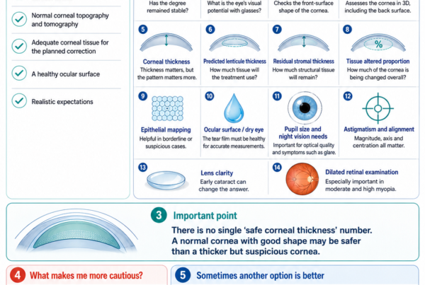
The aim of preoperative screening and surgical planning is to ensure that:
- The cornea is structurally normal
- The amount of tissue altered is appropriate
- Adequate load-bearing tissue remains
- There is no evidence of keratoconus susceptibility
- The treatment profile provides a reasonable long-term safety margin
Ectasia represents abnormal progressive biomechanical failure, not the expected stable structural change after uncomplicated LASIK.
How Common Is Post-LASIK Ectasia?
The true frequency is uncertain.
Published estimates vary according to:
- Patient-selection criteria
- Laser technology
- Flap-creation technique
- Duration of follow-up
- Definition of ectasia
- Availability of postoperative tomography
- Loss of patients to long-term follow-up
A study of 30,167 LASIK-treated eyes identified ten ectasia cases during two to eight years of follow-up, corresponding to an incidence of approximately 0.033% in that cohort. The authors noted that the true rate might be higher because some cases may present later and some patients were lost to follow-up.
A systematic review confirmed that ectasia has been reported after LASIK, PRK and SMILE. Published reporting was more frequent after LASIK, but direct comparisons are limited by unequal procedure numbers and follow-up.
The practical message is:
Post-refractive ectasia is uncommon, but its potential visual consequences make careful screening and early diagnosis essential.
When Can Ectasia Develop?
Post-refractive ectasia can become apparent:
- Within weeks or months
- Several years after surgery
- Occasionally much later
Earlier presentation may suggest:
- Significant pre-existing susceptibility
- Marked biomechanical alteration
- A highly abnormal preoperative cornea that was not recognised
Later presentation may reflect a combination of:
- Subtle predisposition
- Continued biomechanical remodelling
- Eye rubbing
- Hormonal or age-related influences
- Long-term progression of an ectatic disorder
The absence of early problems does not guarantee that ectasia can never develop, although the risk becomes progressively lower when the cornea remains stable over long follow-up.
Can Ectasia Occur After PRK or TransPRK?
Yes.
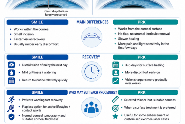
PRK and TransPRK do not create a LASIK flap, so they preserve more anterior load-bearing stromal tissue.
This generally provides a wider structural margin than LASIK for an equivalent refractive correction.
However, PRK still removes stromal tissue.
Post-PRK ectasia has been reported, particularly in eyes with:
- Pre-existing keratoconus susceptibility
- Abnormal or borderline tomography
- High corrections
- Inadequate remaining tissue
- Progressive corneal disease
Avoiding a flap does not make an abnormal cornea safe for laser tissue removal.
Can Ectasia Occur After SMILE?
Yes.
SMILE avoids a hinged LASIK flap but removes a stromal lenticule from within the cornea.
The remaining corneal cap and stromal bed must still provide adequate biomechanical stability.
Cases of ectasia have been reported after SMILE, including some eyes with identifiable preoperative risk factors and some with apparently normal initial screening.
SMILE may preserve anterior corneal fibres differently from LASIK, but it should not be performed in a cornea with:
- Keratoconus
- Suspicious tomography
- Progressive astigmatism
- Inadequate tissue
- Another ectatic disorder
Published reviews confirm that ectasia is possible after all major corneal refractive procedures.
What Causes Corneal Ectasia?
Corneal ectasia usually results from a combination of:
- Intrinsic corneal susceptibility
- Tissue removal or surgical alteration
- Mechanical stress
- Biological and environmental influences
There is rarely one single cause that explains every case.
Pre-existing Keratoconus Susceptibility
Some patients have early or subclinical corneal ectatic disease before refractive surgery.
The cornea may appear relatively normal on a basic front-surface curvature map but show abnormalities in:
- Posterior corneal elevation
- Thickness distribution
- Pachymetric progression
- Epithelial thickness
- Corneal biomechanics
- Asymmetry between the two eyes
A case report of ectasia developing after apparently low-risk LASIK demonstrated that tomography and biomechanical assessment may identify susceptibility that is not captured by older screening scores alone.
Excessive Tissue Alteration
LASIK affects corneal strength through both:
- Flap creation
- Excimer-laser ablation
The amount of tissue affected must be considered relative to the original corneal thickness.
One proposed measure is the percentage of tissue altered, calculated from flap thickness and ablation depth relative to the preoperative corneal thickness.
In a case-control study of eyes with apparently normal preoperative topography, a percentage of tissue altered of 40% or greater was strongly associated with post-LASIK ectasia. However, subsequent studies have questioned whether one universal threshold performs equally well across all populations. It should therefore be regarded as one risk measure rather than a stand-alone pass-or-fail rule.
Low Residual Stromal Bed
The residual stromal bed is the stromal tissue remaining beneath the flap after laser ablation.
A low residual stromal bed provides less tissue to maintain corneal shape.
Historically used safety cut-offs include values such as 250 or 300 microns, but no single residual-bed measurement guarantees safety.
The risk depends on:
- Corneal shape
- Corneal thickness
- Age
- Amount of tissue altered
- Pre-existing ectatic susceptibility
- Treatment-zone size
- Individual biomechanics
Residual stromal-bed thickness should be interpreted as part of the complete risk assessment.
High Myopia
Higher myopic correction requires deeper tissue removal.
This can result in:
- Greater biomechanical alteration
- A lower residual stromal bed
- A higher percentage of tissue altered
- Greater changes in corneal curvature
High myopia is therefore an important planning factor, although it does not automatically mean that LASIK is unsafe.
A patient with moderate myopia and abnormal tomography may have a greater ectasia risk than a highly myopic patient with a thick, regular and biomechanically reassuring cornea.
Younger Age
Younger patients may be at greater risk because:
- Subclinical keratoconus may not yet have declared itself fully
- Corneal ectatic disease is more likely to progress in younger individuals
- The patient has a longer lifetime during which progression can occur
- The prescription may still be changing
Age was incorporated into the validated Ectasia Risk Score System along with topographic pattern, corneal thickness, residual stromal bed and myopic correction.
Abnormal Corneal Topography or Tomography
Important warning signs include:
- Inferior or inferotemporal steepening
- Asymmetric bow-tie patterns
- Skewed radial axes
- Abnormal posterior elevation
- Abnormal thickness progression
- Displacement of the thinnest point
- Significant asymmetry between the eyes
The anterior curvature map alone is not sufficient.
Modern tomography analyses the front and back corneal surfaces and the three-dimensional distribution of corneal thickness.
Eye Rubbing
Habitual vigorous eye rubbing is associated with corneal deformation and keratoconus progression.
It may contribute to post-refractive ectasia in a susceptible cornea.
Patients at particular risk include those with:
- Allergic conjunctivitis
- Eczema or atopy
- Chronic itchy eyes
- Habitual morning eye rubbing
- Down syndrome
- Sleep-related pressure on the eyes
The eye should not be rubbed forcefully, especially after refractive surgery or when ectasia is suspected.
Treating allergy and breaking the rubbing habit are important parts of management.
Allergy and Atopy
Allergic eye disease does not directly prove that ectasia will occur.
However, it may increase risk indirectly through:
- Chronic itching
- Vigorous eye rubbing
- Ocular-surface inflammation
- Repeated mechanical stress
Patients with allergy should receive appropriate treatment rather than relying on rubbing for temporary relief.
Family History
A family history of keratoconus or another ectatic disorder may indicate inherited susceptibility.
A patient with a family history may require:
- More conservative screening
- Repeat tomography
- Epithelial mapping
- Biomechanical assessment
- Longer observation before surgery
- Consideration of non-corneal correction such as ICL
A family history does not automatically confirm that the patient has keratoconus, but it lowers the threshold for caution.
Pregnancy and Hormonal Factors
Pregnancy can change:
- Corneal curvature
- Corneal thickness
- Corneal hydration
- Refractive measurements
- Biomechanical behaviour
Rare progression of keratoconus or post-LASIK ectasia has been reported during or after pregnancy in susceptible patients.
Pregnancy does not cause ectasia in most patients who have undergone LASIK, but new progressive blur, astigmatism or ghosting during pregnancy warrants assessment.
Can Ectasia Occur Without Recognised Risk Factors?
Yes.
Some patients develop post-LASIK ectasia despite apparently normal preoperative measurements and acceptable traditional risk calculations.
This may occur because:
- Current tests cannot measure every aspect of corneal strength
- Ectatic susceptibility may be below the detection threshold
- Risk models are based on population averages
- Surgical and environmental factors interact over time
- Genetic factors are incompletely understood
A large LASIK series found at least one ectasia case without identifiable conventional risk factors, and other reports have described ectasia despite low preoperative risk scores.
Screening substantially reduces risk but cannot reduce it to zero.
What Are the Symptoms of Corneal Ectasia?
Early ectasia may produce subtle symptoms.
These may include:
- Gradually increasing short-sightedness
- Increasing astigmatism
- Frequent spectacle-prescription changes
- Blurred distance vision
- Ghost images
- Shadowed letters
- Monocular double vision
- Starbursts around lights
- Glare and halos
- Reduced night-driving vision
- Eye strain
- One eye becoming progressively worse
As ectasia progresses, patients may notice:
- Spectacles no longer providing crisp vision
- Increasingly irregular astigmatism
- Distorted straight lines
- Multiple images
- Poor contrast
- Reduced best-corrected vision
- Difficulty tolerating ordinary soft contact lenses
Progressive myopia, astigmatism, corneal steepening and reduced corrected visual acuity are typical features described in post-refractive ectasia.
Does Ectasia Cause Pain?
Early and moderate ectasia usually does not cause significant pain.
The main problem is optical distortion.
Pain may occur when there is:
- Severe dry eye
- Contact lens intolerance
- Corneal abrasion
- Acute corneal hydrops
- Scarring
- Another ocular-surface problem
Sudden pain, marked watering and an abrupt white or cloudy area in a patient with advanced ectasia may indicate acute corneal hydrops and requires urgent assessment.
Can Ectasia Affect Only One Eye?
Yes.
Keratoconus and post-refractive ectasia may be:
- Bilateral but asymmetric
- Clinically obvious in one eye
- Subtle or apparently absent in the fellow eye
The apparently unaffected eye may nevertheless show early abnormalities on:
- Tomography
- Epithelial mapping
- Biomechanical testing
Both eyes should therefore be evaluated and monitored.
Is Ectasia the Same as LASIK Regression?
No.
Both can cause a return of myopia or astigmatism, but they differ fundamentally.
Ordinary Refractive Regression
- The cornea remains structurally stable
- The prescription becomes mildly myopic or astigmatic
- Astigmatism remains relatively regular
- Spectacles usually restore clear vision
- Serial corneal maps remain stable
- Best-corrected visual acuity remains good
Corneal Ectasia
- The prescription continues changing
- Astigmatism may become irregular
- Ghosting and distortion increase
- Spectacles may no longer provide crisp vision
- Corneal maps show progressive steepening or thinning
- Best-corrected vision may decline
A patient with worsening vision after LASIK should not undergo a routine enhancement until ectasia has been excluded.
Is Ectasia the Same as a Thin Cornea?
No.
A thin cornea is not necessarily ectatic.
Some healthy individuals naturally have relatively thin but:
- Regular
- Symmetrical
- Stable
- Biomechanically normal
corneas.
Likewise, an ectatic cornea is not always extremely thin at the time of diagnosis.
Ectasia is defined by abnormal shape, biomechanics or progression—not by one thickness number alone.
How Is Corneal Ectasia Diagnosed?
Diagnosis requires a combination of:
- Symptoms
- Refraction
- Visual acuity
- Slit-lamp examination
- Corneal mapping
- Serial progression
No single test should be interpreted in isolation.
Refraction
The refraction may show:
- Increasing myopia
- Increasing astigmatism
- Changing astigmatic axis
- Irregular or inconsistent measurements
- Reduced best-corrected vision
Previous spectacle and postoperative records are extremely useful.
Corneal Topography
Topography maps the curvature of the front corneal surface.
Possible ectatic patterns include:
- Inferior steepening
- Inferotemporal steepening
- Asymmetric bow tie
- Skewed radial axes
- Localised high corneal power
- Progressive change between scans
Topography is useful but may miss abnormalities involving the posterior surface or thickness distribution.
Corneal Tomography
Tomography creates a three-dimensional assessment of the cornea.
It evaluates:
- Anterior elevation
- Posterior elevation
- Corneal thickness
- Thickness progression from centre to periphery
- Position of the thinnest point
- Corneal-volume distribution
- Differences from an expected reference surface
Posterior elevation and abnormal pachymetric progression may become abnormal before obvious front-surface steepening.
Modern tomography is therefore central to both preoperative screening and diagnosis.
Pachymetry
Pachymetry measures corneal thickness.
The doctor assesses:
- Central thickness
- Thinnest-point thickness
- Location of the thinnest point
- Thickness distribution
- Change over time
A single central thickness measurement is less informative than the entire thickness profile.
Epithelial Thickness Mapping
The epithelium can partially compensate for irregular stromal shape.
It often becomes:
- Thinner over a focal stromal cone
- Thicker around the abnormal region
This remodelling can temporarily mask the underlying ectatic pattern on front-surface topography.
Epithelial thickness mapping may therefore help:
- Detect early ectatic disease
- Distinguish true stromal irregularity from contact lens warpage
- Monitor progression
- Interpret post-refractive corneal shape
Reviews increasingly describe epithelial mapping as an important adjunct for refractive screening and ectasia monitoring.
Corneal Biomechanical Testing
Biomechanical devices measure how the cornea responds to a controlled air impulse.
They may analyse:
- Speed of corneal deformation
- Depth of indentation
- Recovery pattern
- Corneal stiffness-related indices
- Interaction between biomechanical and tomographic data
These measurements can add information about susceptibility that is not visible from shape alone.
However:
- Results are influenced by thickness and eye pressure
- Different devices use different indices
- No biomechanical test can guarantee that ectasia will or will not occur
Biomechanical testing should complement rather than replace tomography and clinical judgement.
Slit-Lamp Examination
The doctor examines for:
- Corneal thinning
- Scarring
- Fleischer ring
- Vogt striae
- Flap condition after LASIK
- Epithelial ingrowth
- Interface abnormalities
- Acute hydrops
Early ectasia may have few or no obvious slit-lamp signs.
Wavefront Aberrometry
Wavefront testing measures optical imperfections across the pupil.
Ectatic corneas may have elevated:
- Coma
- Trefoil
- Total higher-order aberrations
These measurements can help quantify visual distortion but do not replace structural imaging.
Serial Imaging Is Essential
One abnormal scan does not always prove progression.
Likewise, a borderline scan cannot be declared safe simply because the patient sees well.
Serial comparison may assess changes in:
- Maximum keratometry
- Anterior and posterior elevation
- Corneal thickness
- Astigmatism
- Epithelial profile
- Best-corrected vision
- Refraction
True progression is especially important when deciding whether cross-linking is required.
Can Contact Lenses Affect the Scans?
Yes.
Soft, toric, rigid and overnight contact lenses can temporarily alter:
- Corneal curvature
- Epithelial thickness
- Astigmatism
- Tear-film quality
- Topography
Rigid lens warpage can mimic ectasia.
Lenses may need to be stopped for an appropriate period before repeat scans. The required interval depends on the lens type and whether repeated measurements have stabilised.
How Is Corneal Ectasia Treated?
Treatment has two separate aims:
- Stabilise the cornea
- Improve vision
The treatment that stops progression does not always provide the best optical correction.
Likewise, a treatment that improves vision may not prevent further structural deterioration.
Stop Eye Rubbing and Treat Allergy
This is fundamental.
Patients should:
- Avoid vigorous rubbing
- Treat allergic conjunctivitis
- Use prescribed anti-allergy drops
- Apply a cool compress rather than rubbing
- Address eczema or eyelid inflammation
- Avoid sleeping with direct pressure on the eye when possible
Stopping mechanical stress is important even after cross-linking.
Spectacles
Glasses may provide useful vision in early ectasia when astigmatism remains relatively regular.
As irregularity increases, spectacles may not fully correct:
- Ghosting
- Multiple images
- Higher-order aberrations
- Distortion
Spectacles remain useful for selected tasks even when contact lenses are required for best vision.
Soft Contact Lenses
Ordinary soft lenses may help mild, relatively regular refractive error.
Custom soft lenses designed for keratoconus may provide additional stability or thickness, but they generally do not neutralise irregular corneal optics as effectively as rigid lenses.
Rigid Gas-Permeable Contact Lenses
A rigid lens creates a smooth artificial front optical surface.
The tear layer between the lens and the irregular cornea helps neutralise corneal distortion.
Rigid lenses can significantly improve:
- Visual acuity
- Ghosting
- Contrast
- Night vision
A clinical series found that the majority of eyes with post-refractive ectasia achieved functional vision with rigid gas-permeable lenses and did not require further surgical visual rehabilitation.
Rigid lenses improve vision but do not necessarily stop ectasia progression.
Hybrid Contact Lenses
Hybrid lenses combine:
- A rigid central optical zone
- A soft peripheral skirt
They may improve comfort in selected patients while providing better optical correction than an ordinary soft lens.
Fit, oxygen transmission and long-term comfort must be monitored.
Scleral Contact Lenses
Scleral lenses are larger rigid lenses that rest on the white part of the eye rather than the central cornea.
A fluid reservoir fills the space between the lens and cornea.
Potential advantages include:
- Excellent correction of irregular astigmatism
- Reduced mechanical contact with the corneal cone
- Improved comfort
- Protection of a dry or sensitive surface
- More stable positioning than a small corneal rigid lens
Scleral lenses can significantly improve visual acuity and reduce optical aberrations in ectatic corneas.
They require:
- Specialist fitting
- Careful insertion and removal
- Preservative-free filling solution
- Meticulous hygiene
- Monitoring for corneal swelling and infection
What Is Corneal Cross-Linking?
Corneal cross-linking, or CXL, is a treatment designed to increase corneal biomechanical strength and slow or stop ectatic progression.
In the conventional epithelium-off technique:
- The central epithelium is removed.
- Riboflavin drops are applied.
- Controlled ultraviolet-A light activates the riboflavin.
- New chemical bonds form within the stromal collagen structure.
- A bandage contact lens is placed while the surface heals.
The primary goal is stability, not elimination of the spectacle prescription.
A 2023 review described cross-linking as the principal treatment for progressive post-LASIK ectasia, while long-term studies have demonstrated sustained stability in many treated eyes.
Does Cross-Linking Restore Normal Vision?
Not necessarily.
Cross-linking may produce some:
- Corneal flattening
- Reduction in astigmatism
- Improvement in corrected vision
However, the result is variable.
Many patients still require:
- Glasses
- Rigid contact lenses
- Scleral lenses
- Additional corneal treatment
The main reason to perform cross-linking is to reduce the risk of further deterioration.
Who Should Have Cross-Linking?
Cross-linking is generally considered when there is documented or strongly suspected progression.
Evidence of progression may include:
- Increasing corneal steepness
- Increasing myopia or astigmatism
- Decreasing corneal thickness
- Increasing posterior elevation
- Declining corrected vision
- Reproducible deterioration on serial scans
Younger patients or patients with clearly active disease may require earlier intervention because progression can occur more rapidly.
Treatment must be individualised when the cornea is:
- Very thin
- Scarred
- Previously operated
- Associated with significant ocular-surface disease
Is Cross-Linking Always Successful?
No treatment can guarantee permanent stability.
Most studies report stabilisation in the majority of appropriately selected patients, but:
- Some eyes continue progressing
- Repeat cross-linking may occasionally be considered
- Advanced corneas may respond less predictably
- Visual quality may remain limited by irregularity
A ten-year study found sustained overall stability after cross-linking, although post-refractive ectasia eyes appeared somewhat less stable than keratoconus eyes over long follow-up.
What Are the Risks of Cross-Linking?
Possible risks include:
- Postoperative pain
- Delayed epithelial healing
- Infection
- Corneal haze
- Scarring
- Reduced corrected vision
- Sterile infiltrates
- Corneal thinning
- Endothelial damage in an inadequately protected thin cornea
- Reactivation of herpes infection
- Treatment failure or continued progression
Infectious keratitis after epithelium-off cross-linking is uncommon but potentially serious, and most reported infections occur during the early epithelial-healing period.
What Are Intracorneal Ring Segments?
Intracorneal ring segments are curved implants placed within channels in the peripheral corneal stroma.
They redistribute corneal forces and may:
- Flatten the central cornea
- Reduce irregularity
- Improve contact lens tolerance
- Reduce myopia or astigmatism
- Improve unaided or corrected visual acuity
They do not remove corneal tissue.
Studies of ring segments in post-LASIK ectasia have reported central flattening, improved corneal regularity and visual improvement in selected patients.
Do Ring Segments Stop Ectasia Progression?
Not reliably on their own.
Ring segments primarily reshape the cornea.
When progression is present, they may be combined with cross-linking.
The order and timing vary according to:
- Corneal thickness
- Cone position
- Contact lens tolerance
- Degree of progression
- Surgeon preference
- Intended visual outcome
What Are the Risks of Ring Segments?
Possible complications include:
- Infection
- Segment movement
- Segment extrusion
- Corneal thinning over the segment
- Glare or halos
- Inadequate visual improvement
- Irregular astigmatism
- Need for removal or exchange
Results are less predictable in highly irregular or severely scarred corneas.
Can Laser Treatment Be Used for Ectasia?
Routine LASIK enhancement should not be performed on an ectatic cornea.
Further stromal tissue removal may worsen the structural problem.
However, carefully selected patients may undergo limited topography-guided PRK combined with cross-linking.
The aims are to:
- Reduce selected corneal irregularity
- Improve visual quality
- Preserve adequate tissue
- Stabilise the cornea with cross-linking
This is not intended to produce a full spectacle-free refractive correction.
Treatment must be conservative because the cornea is already structurally compromised.
Reviews of post-LASIK ectasia management describe customised PRK with cross-linking as an option for selected cases, but cross-linking remains the stabilising component.
Can an ICL Be Used in Ectasia?
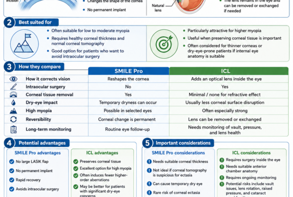
An Implantable Collamer Lens may correct remaining regular myopia or astigmatism without removing more corneal tissue.
However, ICL does not correct substantial irregular corneal optics and does not stop ectasia progression.
It may be considered only when:
- The cornea is stable
- Cross-linking has been completed when necessary
- A meaningful regular refractive component remains
- Internal eye anatomy is suitable
- The expected benefit is realistic
Patients may still require rigid or scleral lenses for irregular astigmatism.
When Is Corneal Transplantation Needed?
Most patients with ectasia do not immediately require a transplant.
Transplantation may be considered when there is:
- Severe corneal scarring
- Advanced irregularity
- Contact lens intolerance
- Inadequate vision despite other treatment
- Recurrent hydrops
- Extreme thinning
- Failure of less invasive rehabilitation
Possible procedures include:
- Deep anterior lamellar keratoplasty
- Penetrating keratoplasty
Transplantation can restore corneal structure and visual potential, but it involves:
- A long recovery
- Astigmatism
- Suture management
- Graft rejection risk
- Infection risk
- Possible future contact lens use
- Limited graft lifespan
The aim of earlier diagnosis, cross-linking and contact lens rehabilitation is partly to reduce the likelihood that transplantation will be required.
Can Ectasia Be Cured?
There is currently no treatment that reliably returns an ectatic cornea completely to its original natural structure.
Management can often:
- Stop or slow progression
- Improve the corneal shape
- Correct irregular optics
- Preserve functional vision
The condition may therefore be stabilised and rehabilitated rather than completely erased.
Can Ectasia Come Back After Cross-Linking?
Progression after cross-linking is possible.
Risk may be greater with:
- Advanced initial disease
- Very young age
- Severe corneal steepness
- Continued vigorous eye rubbing
- Inadequate treatment effect
- Long-term biological progression
Patients require continued serial tomography even when the early post-cross-linking result is reassuring.
Can Patients With Ectasia Still Drive?
Possibly.
Driving suitability depends on:
- Best-corrected visual acuity
- Contrast sensitivity
- Glare
- Night vision
- Visual fields
- Local licensing requirements
A patient may meet daytime visual-acuity requirements but remain uncomfortable or unsafe at night because of:
- Ghosting
- Halos
- Reduced contrast
- Contact lens intolerance
Driving should only continue when vision is legally adequate and functionally safe.
Can Patients With Ectasia Exercise?
Ordinary exercise does not usually worsen a stable ectatic cornea.
However:
- Eye rubbing should be avoided
- Protective eyewear should be used for impact sports
- Contact lenses must be handled hygienically
- Activities should be restricted during recovery from cross-linking or other surgery
The treating surgeon should advise when contact sport and swimming can resume after a procedure.
Can Ectasia Be Prevented?
Not every case can be prevented, but risk can be substantially reduced.
Careful Preoperative Screening
Screening may include:
- Stable refraction
- Corneal topography
- Corneal tomography
- Pachymetry distribution
- Epithelial thickness mapping
- Tissue calculations
- Corneal biomechanical assessment
- Family history
- Eye-rubbing history
- Allergy assessment
No one scan or index should be used alone.
Conservative Tissue Planning
The surgeon should assess:
- Flap thickness
- Ablation depth
- Residual stromal bed
- Percentage of tissue altered
- Optical-zone requirements
- Corneal shape
- Age and prescription
A technically achievable treatment is not necessarily the safest treatment.
Selecting Another Procedure
When corneal laser surgery would create an inadequate safety margin, alternatives may include:
- ICL
- Spectacles
- Contact lenses
- No surgery
PRK or SMILE should not be used merely to bypass suspicious tomography.
Avoiding Vigorous Eye Rubbing
Patients should treat allergy and avoid habitual rubbing before and after refractive surgery.
Long-Term Follow-Up
Patients with:
- Borderline preoperative findings
- High myopia
- Family history of keratoconus
- Significant eye rubbing
- Progressive astigmatism
may benefit from periodic corneal imaging even when their vision remains good.
Should Cross-Linking Be Performed Routinely With LASIK?
Some surgeons have investigated prophylactic cross-linking combined with LASIK in selected higher-risk eyes.
This is sometimes called LASIK Xtra.
Potential aims include:
- Increasing postoperative stiffness
- Reducing regression
- Reducing ectasia risk
However:
- Long-term evidence is still evolving
- Protocols vary
- Cross-linking introduces its own risks
- It does not make a clearly abnormal cornea suitable for LASIK
- It should not replace careful screening or conservative tissue planning
Prophylactic cross-linking should not be marketed as a guarantee against ectasia.
What Symptoms Require Prompt Review?
Arrange a corneal assessment for:
- Increasing myopia after previous stability
- Increasing or changing astigmatism
- Frequent spectacle-prescription changes
- Progressive ghosting
- Increasing glare or starbursts
- One eye becoming progressively worse
- Reduced vision despite updated glasses
- New contact lens intolerance
- Worsening corneal-map results
When Is Urgent Assessment Needed?
Seek urgent review for:
- Sudden marked loss of vision
- Significant eye pain
- Sudden corneal clouding
- A new white area on the cornea
- Severe light sensitivity
- Increasing redness
- Discharge
- Contact lens-related pain
- Eye trauma
These symptoms may indicate:
- Acute hydrops
- Corneal infection
- Abrasion
- Contact lens complication
- Another urgent eye condition
Frequently Asked Questions About Corneal Ectasia
Is Corneal Ectasia Rare?
Post-refractive ectasia is uncommon.
Naturally occurring keratoconus is more common, but prevalence varies among populations.
Is Corneal Ectasia the Same as Keratoconus?
Keratoconus is one form of corneal ectasia.
Post-LASIK ectasia is an ectatic condition developing after refractive surgery.
Does LASIK Cause Ectasia?
LASIK alters corneal biomechanics.
Ectasia may occur when surgical tissue alteration interacts with underlying susceptibility or other risk factors.
Most appropriately screened LASIK patients do not develop ectasia.
Can Ectasia Occur After a Normal LASIK Screening?
Yes, rarely.
Current screening greatly reduces risk but cannot detect every susceptible cornea.
Can Ectasia Develop Years After LASIK?
Yes.
It may present months or years after surgery.
Can PRK Cause Ectasia?
Yes, although published reports are less frequent than after LASIK.
PRK still removes stromal tissue.
Can SMILE Cause Ectasia?
Yes.
SMILE avoids a flap but still removes a stromal lenticule.
Does a Thin Cornea Mean I Have Ectasia?
No.
Some thin corneas are regular and stable.
Diagnosis depends on shape, thickness distribution, biomechanics and progression.
Does a Thick Cornea Mean LASIK Is Safe?
No.
A thick cornea may still have keratoconus susceptibility or abnormal tomography.
Can Ectasia Be Corrected With Glasses?
Early regular refractive error may improve with glasses.
Advanced irregular astigmatism often requires rigid or scleral contact lenses.
Can Soft Contact Lenses Correct Ectasia?
Custom soft lenses may help mild disease.
Rigid or scleral lenses generally provide better correction of significant irregular astigmatism.
Do Contact Lenses Stop Progression?
No.
They improve the optical surface but do not strengthen the cornea.
Does Cross-Linking Improve Vision?
It may produce some visual or topographic improvement, but its principal aim is to stop progression.
Does Cross-Linking Cure Ectasia?
No.
It strengthens and stabilises the cornea but does not restore the original untreated structure.
Can Ectasia Progress After Cross-Linking?
Yes.
Continued follow-up is necessary.
Can Cross-Linking Be Repeated?
Repeat treatment may be considered in selected cases with documented progression, but this requires individual assessment.
Is Cross-Linking Painful?
Epithelium-off cross-linking can cause several days of pain, watering and light sensitivity while the surface heals.
Can LASIK Be Repeated in an Ectatic Eye?
Routine LASIK enhancement should not be performed because additional tissue removal may worsen instability.
Can PRK Be Performed on Ectasia?
Only in highly selected cases, usually as a limited customised treatment combined with cross-linking.
Can Ring Segments Cure Ectasia?
No.
They reshape the cornea and may improve vision, but progression may still require cross-linking.
Can ICL Cure Ectasia?
No.
ICL can correct a regular refractive component but does not correct corneal weakness or substantial irregularity.
Will I Need a Corneal Transplant?
Most patients can be managed without transplantation.
Transplantation is generally reserved for advanced scarring, severe irregularity or contact lens failure.
Can Eye Rubbing Worsen Ectasia?
Yes.
Vigorous habitual eye rubbing should be stopped and underlying allergy treated.
Does Pregnancy Worsen Ectasia?
Progression has been reported in susceptible patients, although most pregnancies do not cause ectasia.
A change in vision during pregnancy should be assessed.
Can Ectasia Cause Blindness?
It does not usually cause total blindness.
It can cause severe visual impairment if progression, scarring or irregularity becomes advanced.
Can Ectasia Affect Only One Eye?
It may be much more severe in one eye.
The fellow eye should still be examined carefully for subtle abnormalities.
How Often Should Ectasia Be Monitored?
The interval depends on:
- Age
- Severity
- Evidence of progression
- Previous cross-linking
- Contact lens use
- Pregnancy or other risk factors
Active disease may require reviews several months apart, while stable treated disease may be monitored less frequently.
Key Takeaway
Corneal ectasia is progressive weakening and distortion of the cornea.
It may occur naturally as:
- Keratoconus
- Pellucid marginal degeneration
- Another ectatic disorder
It may also occur after:
- LASIK
- PRK or TransPRK
- SMILE
- Other corneal surgery
Possible symptoms include:
- Increasing myopia
- Increasing astigmatism
- Ghosting
- Halos and starbursts
- Reduced night vision
- Frequent spectacle changes
- Vision that glasses no longer correct fully
Important risk factors include:
- Keratoconus susceptibility
- Abnormal corneal topography or tomography
- Excessive tissue alteration
- Low residual stromal tissue
- High myopia
- Younger age
- Family history
- Vigorous eye rubbing
Diagnosis may require:
- Refraction
- Best-corrected visual acuity
- Corneal topography
- Corneal tomography
- Pachymetry
- Epithelial thickness mapping
- Corneal biomechanical testing
- Serial comparison over time
Treatment has two different goals.
To stabilise the cornea:
- Stop eye rubbing
- Treat allergy
- Perform corneal cross-linking when progression is present
To improve vision:
- Spectacles
- Rigid gas-permeable lenses
- Hybrid lenses
- Scleral lenses
- Intracorneal ring segments
- Selected customised surface treatment
- Corneal transplantation in advanced cases
Cross-linking is intended mainly to stop deterioration. It does not automatically eliminate irregular astigmatism or remove the need for contact lenses.
Further routine LASIK should not be performed until ectasia has been excluded.
Early diagnosis offers the best opportunity to preserve corneal structure, stabilise the condition and achieve useful long-term vision.
References
- Moshirfar M, et al. Ectasia After Corneal Refractive Surgery: A Systematic Review. 2021. PMID: 34417707.
- Zhao L, et al. Comprehensive Management of Post-LASIK Ectasia: From Prevention to Treatment. Acta Ophthalmologica. 2023. PMID: 36774646.
- Bohac M, et al. Incidence and Clinical Characteristics of Post-LASIK Ectasia: A Review of Over 30,000 LASIK Cases. 2018. PMID: 30359155.
- Randleman JB, Trattler WB, Stulting RD. Validation of the Ectasia Risk Score System for Preoperative LASIK Screening. 2008. PMID: 18328998.
- Santhiago MR, et al. Association Between the Percent Tissue Altered and Post-LASIK Ectasia in Eyes With Normal Preoperative Topography. 2014. PMID: 24727263.
- Santhiago MR, et al. Ectasia Risk Factors in Refractive Surgery. 2016. PMID: 27143849.
- Ambrósio R Jr, et al. Corneal Ectasia After LASIK Despite Low Preoperative Risk: Tomographic and Biomechanical Findings. 2010. PMID: 20481412.
- Wolle MA, Randleman JB, Woodward MA. Complications of Refractive Surgery: Ectasia After Refractive Surgery. 2016.
- Reinstein DZ, et al. Epithelial Thickness Mapping for Corneal Refractive Surgery. 2022. PMID: 35779050.
- Esporcatte LPG, et al. Corneal Biomechanics for Corneal Ectasia: Update. 2022.
- Sharif W, et al. Long-Term Efficacy and Stability of Corneal Collagen Cross-Linking for Post-LASIK Ectasia. 2019.
- Greenstein SA, et al. Long-Term Outcomes After Corneal Cross-Linking for Keratoconus and Corneal Ectasia. 2023. PMID: 37565471.
- Murchison CE, et al. Infectious Keratitis After Corneal Cross-Linking: A Systematic Review. 2021. PMID: 33769765.
- Woodward MA, et al. Visual Rehabilitation and Outcomes for Ectasia After Corneal Refractive Surgery.
- Tunc Z, et al. Evaluation of Intrastromal Corneal Ring Segments for Treatment of Post-LASIK Ectasia. 2011. PMID: 22011487.
- Peris-Martínez C, et al. Effect of Intracorneal Ring Segments in Post-LASIK Corneal Ectasia. 2020. PMID: 30384777.
- Kramer EG, Boshnick EL. Scleral Lenses in the Treatment of Post-LASIK Ectasia and Superficial Neovascularisation. 2015. PMID: 25746152.