Author: Dr Val Phua
Estimated reading time: 24 minutes
SMILE Pro can be an excellent form of laser vision correction—but being short-sighted does not automatically make someone suitable.
Before recommending treatment, I am not simply asking:
“Is your degree within the laser’s treatment range?”
I am asking a much broader question:
“Can I correct this prescription while leaving this particular cornea structurally healthy, optically good and appropriate for the patient’s long-term needs?”
That distinction matters.
Two patients may both have:
- -5.00 D of myopia
- 6/6 corrected vision
- Similar ages
yet I may recommend:
- SMILE Pro for one
- PRK for another
- ICL for another
- No refractive surgery at all for another
because their:
- Corneal shape
- Corneal thickness
- Amount of tissue required
- Ocular surface
- Astigmatism
- Pupil size
- Retinal health
- Prescription stability
- Age
- Lifestyle
are different.
Singapore National Eye Centre currently lists SMILE Pro for short-sightedness approximately -1.00 to -10.00 D and astigmatism up to -5.00 D, provided that the cornea meets the necessary thickness and health requirements. However, these numerical treatment ranges describe what the technology can treat—not who should necessarily be treated.
This is one of the most important concepts in refractive surgery:
Technical treatability and clinical suitability are not the same thing.
When I assess someone for SMILE Pro, the measurements I pay particular attention to include:
- Manifest and cycloplegic refraction
- Refractive stability
- Best-corrected visual acuity
- Corneal topography
- Corneal tomography
- Corneal thickness and thickness distribution
- Posterior corneal elevation
- Predicted lenticule thickness
- Predicted residual stromal thickness
- Overall percentage of corneal tissue altered
- Epithelial thickness profile where indicated
- Ocular-surface and tear-film health
- Pupil size and night-vision requirements
- Astigmatism magnitude and axis
- Treatment centration
- Intraocular pressure
- Natural-lens clarity
- Dilated retinal examination
The final recommendation comes from putting all these measurements together, rather than applying one universal cut-off.
The Quick Answer
Who Is Usually a Good Candidate for SMILE Pro?
A good candidate typically has:
- Myopia with or without regular astigmatism
- A stable prescription
- Good best-corrected vision
- Normal corneal topography and tomography
- Adequate corneal tissue for the intended correction
- A healthy ocular surface
- No significant cataract, glaucoma or retinal disease that would limit the benefit of surgery
- Realistic expectations
Singapore’s current SNEC guidance similarly describes suitable SMILE Pro patients as those with an appropriate prescription, stable refraction and relatively healthy eyes, while excluding pregnancy and nursing at the time of treatment.
Is There a Minimum Age?
Patients should be adults and, more importantly, have a stable prescription.
Age alone does not establish stability.
A 19- or 20-year-old whose myopia is still increasing may be less suitable than an older patient whose prescription has been unchanged for several years.
ZEISS’s current global patient guidance uses a useful practical benchmark of no more than approximately 0.50 D change during the preceding year, although specific regulatory age and treatment criteria differ between countries.
The US SMILE labelling, for example, uses a different regulatory age threshold and also requires documented refractive stability.
For clinical decision-making, I care more about the pattern:
Is this eye still becoming more myopic?
If it is, surgery can accurately correct today’s prescription while the eye subsequently continues changing.
Is There a Minimum Corneal Thickness?
There is no single corneal-thickness number that automatically determines suitability.
This is important.
A patient may ask:
“My cornea is 510 microns. Is that thick enough?”
The answer cannot be determined from 510 µm alone.
I need to know:
- What does the tomography look like?
- Where is the thinnest point?
- Is the thickness distribution normal?
- Is there inferior steepening?
- Is the posterior surface normal?
- How much tissue must be removed?
- How thick will the SMILE cap be?
- How much untouched stromal tissue will remain?
- How young is the patient?
- How high is the myopia?
- Is there a history of eye rubbing or keratoconus?
Abnormal corneal topography and tomography remain among the most important recognised risk factors for post-refractive corneal ectasia.
A thicker abnormal cornea may therefore worry me more than a somewhat thinner but completely regular cornea.
Is SMILE Pro Suitable for High Myopia?
Sometimes.
The technology can treat substantial myopia in appropriately selected patients.
However, increasing myopic correction requires a thicker lenticule and therefore alters more stromal tissue.
Recent SMILE Pro data show excellent outcomes overall, but high-myopic eyes have somewhat less predictable early unaided vision than low- and moderate-myopic eyes. A 2025 VISUMAX 800 study found one-month 20/20 vision in approximately:
- 99% of low-myopic eyes
- 97% of moderate-myopic eyes
- 81% of high-myopic eyes
in that particular cohort.
High myopia should therefore trigger a second question:
Would ICL produce a better long-term anatomical and optical solution than removing this amount of corneal tissue?
Being able to perform SMILE Pro does not automatically mean it is the best procedure.
Measurement 1: Your Prescription
The assessment begins with refraction.
I want to know:
- How much myopia is present?
- How much astigmatism?
- What is the astigmatic axis?
- Is the refraction consistent?
- Is it different from previous years?
- Does cycloplegia reveal hidden accommodative influence?
Manifest Refraction
Manifest refraction is the familiar:
“Which is clearer—one or two?”
test.
It determines the prescription the patient accepts under normal viewing conditions.
For refractive surgery, I pay attention to more than the final numbers.
I am looking for:
- Reproducibility
- Consistency between eyes
- Differences from the current spectacles
- Differences from automated refraction
- Unusual cylinder changes
- Signs that accommodation may be affecting the result
Cycloplegic Refraction
Cycloplegic drops temporarily relax focusing.
This can be particularly useful in:
- Younger patients
- Hyperopic patients
- Patients with inconsistent refraction
- Suspected accommodative spasm
- Unexpected differences between measurements
Formal SMILE patient-selection guidance includes cycloplegic evaluation as part of a complete preoperative examination.
Why Refractive Stability Matters
Suppose a patient was:
- -4.00 D two years ago
- -4.75 D last year
- -5.50 D today
The laser may accurately correct -5.50 D.
But the important question is whether the eye is still progressing.
If axial myopia continues, the patient may later become:
- -0.50 D
- -1.00 D
- or more
again.
This is not necessarily the laser “wearing off”.
It may be the eye continuing its natural biological progression.
Measurement 2: Best-Corrected Visual Acuity
Before discussing how well someone may see without glasses after surgery, I first determine how well the eye sees with its best glasses correction.
This establishes the visual potential of the eye.
For example, if an eye has always achieved only:
6/9
because of amblyopia, I would not promise that SMILE Pro will somehow produce:
6/6
Laser surgery corrects refractive error.
It does not cure:
- Amblyopia
- Retinal damage
- Optic-nerve disease
- Corneal scars
The preoperative corrected vision therefore helps set realistic expectations.
Measurement 3: Corneal Topography
Corneal topography creates a detailed map of the front surface of the cornea.
It can reveal:
- Steepening
- Flattening
- Astigmatism
- Asymmetry
- Irregularity
- Patterns suspicious for keratoconus
Why Is This So Important?
SMILE Pro permanently removes stromal tissue.
If the cornea already has an underlying structural tendency to weaken or bulge, refractive surgery may increase the risk of corneal ectasia.
Post-refractive ectasia can cause:
- Progressive myopia
- Increasing astigmatism
- Ghosting
- Distorted vision
- Loss of corrected visual acuity
Abnormal corneal shape is one of the strongest identifiable risk factors for ectasia after laser refractive surgery.
Measurement 4: Corneal Tomography
Topography and tomography are related but not identical.
Topography mainly describes the anterior corneal surface.
Tomography reconstructs the cornea in three dimensions.
I assess:
- Anterior curvature
- Posterior elevation
- Total corneal thickness
- Location of the thinnest point
- Pachymetric progression
- Relationship between the front and back surfaces
Modern tomography can detect abnormalities that may not be obvious from:
- Vision
- Slit-lamp examination
- Simple keratometry
- Central thickness alone
Tomographic screening is therefore central to identifying patients at risk of corneal ectasia before refractive surgery.
What Am I Looking for on Tomography?
Features that may concern me include:
- Inferior or inferotemporal steepening
- Abnormal posterior elevation
- Abnormal thickness progression
- An unusually displaced thinnest point
- Excessive asymmetry
- Patterns suggestive of keratoconus
- Pellucid-type changes
No single colour on one map should be interpreted in isolation.
The pattern across multiple maps matters.
The Belin/Ambrósio Display
Where available, Scheimpflug tomography may provide composite ectasia-screening parameters such as the Belin/Ambrósio Enhanced Ectasia Display.
These combine information about:
- Elevation
- Pachymetry
- Corneal shape
into screening indices.
They are useful tools.
They do not replace clinical judgement.
Measurement 5: Corneal Thickness
Pachymetry measures corneal thickness.
For refractive surgery I am interested in:
- Central corneal thickness
- Minimum corneal thickness
- Where the minimum occurs
- How rapidly the cornea thickens towards the periphery
Why a Single Thickness Cut-Off Is Misleading
Historically, patients often heard rules such as:
“Above 500 microns is safe.”
That is too simplistic.
A 495-µm cornea with:
- Completely normal tomography
- Low myopia
- Small required tissue removal
may have a more reassuring structural profile than a 530-µm cornea with:
- Abnormal posterior elevation
- Inferior steepening
- Suspicious pachymetric progression
Current ectasia research consistently emphasises the interaction between:
- Corneal shape
- Patient characteristics
- Tissue removal
- Residual tissue
rather than central thickness alone.
Measurement 6: Predicted Lenticule Thickness
The higher the myopic correction, the more tissue generally needs to be incorporated into the lenticule.
Before surgery, the laser software predicts the tissue geometry required for:
- Sphere
- Cylinder
- Optical zone
- Treatment parameters
I therefore examine not only the prescription but also:
How much stromal tissue will correcting that prescription actually cost?
A -2.00 D treatment and a -9.00 D treatment may both be technically possible, but their effects on the cornea are not equivalent.
Measurement 7: Residual Stromal Thickness
This is one of the most important concepts in corneal refractive surgery.
After SMILE Pro:
- The corneal cap remains.
- The lenticule has been removed.
- Stromal tissue remains beneath the treated plane.
I calculate the predicted residual stromal architecture before recommending surgery.
Why Does Residual Tissue Matter?
The cornea needs sufficient structural reserve to maintain its shape over decades.
Lower residual stromal thickness has been associated with greater biomechanical change after corneal refractive surgery.
For historical US SMILE labelling, residual stromal thickness below 250 µm was listed as a contraindication.
However, I would not regard 250 µm as a desirable target simply because it appears as a regulatory lower boundary.
A 2026 systematic review of 25 SMILE studies involving 3,247 eyes found more pronounced biomechanical weakening when residual stromal thickness fell below approximately 280 µm, and the authors proposed 280 µm as a useful provisional benchmark pending better validated metrics.
The correct interpretation is not:
“281 µm is safe and 279 µm is dangerous.”
Biology does not work in a binary fashion.
It means I become increasingly cautious as the planned surgery consumes more of the cornea’s structural reserve.
Measurement 8: Percentage of Tissue Altered
Rather than looking only at the number of microns remaining, another approach is to consider how large the treatment is relative to the patient’s original cornea.
This concept is often described as the percentage of tissue altered.
For LASIK, percentage tissue altered became an important ectasia-risk concept because it incorporated:
- Flap thickness
- Ablation depth
- Original corneal thickness
rather than relying on residual stromal thickness alone.
The exact threshold developed for LASIK should not simply be copied across to SMILE.
SMILE has different anatomy.
Research specifically examining SMILE has therefore proposed SMILE-specific measures.
What Does the Newer SMILE Evidence Suggest?
A 2026 systematic review found more pronounced biomechanical changes when the SMILE tissue-alteration proportion exceeded approximately 27–28%.
The authors suggested:
- Residual stromal thickness around 280 µm
- SMILE tissue-alteration proportion around 28%
as potential benchmarks, while explicitly recognising that more specific validated safety metrics are still needed.
I regard these as additional information rather than rigid universal pass/fail numbers.
The patient’s:
- Tomography
- Age
- Prescription
- Family history
- Eye rubbing
- Corneal biomechanics
still matter.
Measurement 9: Posterior Corneal Surface
The back surface of the cornea is important because early ectatic change may sometimes be detected there before obvious abnormalities develop on the front surface.
Tomography allows assessment of:
- Posterior elevation
- Best-fit reference surfaces
- Localised protrusion
A suspicious posterior surface may make me reluctant to remove corneal tissue even when:
- Vision is excellent
- Central thickness looks adequate
- The anterior map seems only mildly abnormal
Measurement 10: Epithelial Thickness Mapping
The corneal epithelium is not uniformly thick.
It can remodel to partially mask irregular stromal shape underneath.
This is sometimes described as the epithelium acting like biological masking tape.
For example, over an early cone the epithelium may become locally thinner, while adjacent regions become thicker.
The front surface may therefore look more regular than the underlying stromal anatomy actually is.
Why Might I Measure the Epithelium?
OCT epithelial mapping can provide another layer of information when the tomography is:
- Borderline
- Asymmetric
- Unexpected
- Difficult to interpret
A refractive-surgery screening study found that adding epithelial thickness maps changed:
- Whether surgery was offered in 16% of patients
- The preferred procedure in 16%
- The ranking of procedures in 25%
of the screened patients.
Epithelial mapping is therefore particularly useful as an additional safety test, not simply another colourful scan.
Does Everyone Need Epithelial Mapping?
Not necessarily.
The core assessment remains:
- History
- Refraction
- Slit lamp
- Corneal topography/tomography
- Pachymetry
- Dilated examination
Epithelial mapping becomes especially valuable when something does not fit neatly.
Measurement 11: Corneal Biomechanics
Some clinics use devices designed to assess how the cornea deforms when exposed to a controlled air pulse.
These measurements may include parameters derived from:
- Corvis ST
- Ocular Response Analyzer
- Other biomechanical models
They attempt to provide information beyond geometry alone.
Can a Biomechanical Test Prove That a Cornea Is Safe?
No.
Biomechanical assessment may add useful information, particularly for borderline cases.
But it does not override:
- Abnormal tomography
- Keratoconus
- Inadequate tissue
The 2026 tissue-safety review identified several biomechanical parameters as promising, but also concluded that more specific validated metrics are still required.
Measurement 12: Corneal Curvature
I assess:
- K readings
- Overall corneal steepness
- Regularity
- Asphericity
Extremely:
- Flat
- Steep
- Irregular
corneas may influence both:
- Suitability
- Treatment planning
The postoperative cornea should remain optically sensible rather than merely technically treatable.
Measurement 13: Astigmatism
Astigmatism is more than the cylinder number.
I evaluate:
- Magnitude
- Axis
- Regularity
- Reproducibility
- Whether refractive cylinder matches corneal measurements
What If the Spectacle Astigmatism and Corneal Astigmatism Do Not Match?
That may suggest contribution from:
- The natural lens
- Tear-film instability
- Measurement variability
This matters because surgery is being planned on the cornea.
Before treating a significant cylinder, I want to understand where it comes from.
Cyclotorsion
When a patient moves from sitting upright to lying beneath the laser, the eye may rotate slightly.
This matters particularly for higher astigmatism.
The newer VISUMAX 800 used for SMILE Pro includes OcuLign, which allows treatment-pattern rotation to compensate for cyclotorsional alignment, together with CentraLign for centration assistance.
Recent SMILE Pro studies demonstrate favourable results even in higher astigmatism.
Measurement 14: Treatment Centration
Where the optical treatment is centred can influence postoperative:
- Coma
- Glare
- Halos
- Night vision
- Optical quality
Earlier SMILE relied heavily on surgeon-controlled centration during docking.
SMILE Pro adds computer-assisted positioning.
This does not eliminate the need for surgical judgement.
I still assess:
- Pupil centre
- Corneal vertex
- Visual axis
- Angle kappa
- Fixation
Research predating SMILE Pro already showed that attention to angle-kappa alignment could reduce postoperative vertical coma, particularly in patients with larger angle kappa.
Measurement 15: Pupil Size
Pupil size does not determine candidacy by itself.
However, I pay more attention to it when a patient:
- Drives extensively at night
- Has large dark-adapted pupils
- Is especially concerned about halos
- Has high myopia
- Has demanding night-vision requirements
Why Does Pupil Size Matter?
Under dim conditions the pupil enlarges.
Light then passes through a larger area of the optical system.
This may make:
- Residual refractive error
- Decentration
- Higher-order aberrations
more noticeable.
Studies of SMILE show that higher preoperative myopia and postoperative coma can be associated with poorer mesopic contrast and greater night-vision complaints.
Pupil size should therefore contribute to counselling rather than act as a stand-alone exclusion criterion.
Measurement 16: Optical Zone
The optical zone is the central treatment region designed to provide the desired refractive correction.
Larger optical zones may be desirable for certain visual requirements but generally require:
- Different lenticule geometry
- More tissue
This creates a trade-off between:
- Optical performance
- Tissue preservation
A 2025 SMILE Pro study found that smaller optical zones and lower residual stromal thickness were associated with a lower likelihood of achieving 20/20 unaided vision at one month.
This is another reason I do not plan surgery using prescription alone.
Measurement 17: Ocular-Surface Health
Before refractive surgery, I examine:
- Tear film
- Corneal staining
- Tear break-up
- Eyelids
- Meibomian glands
- Blepharitis
- Allergy
Why Does Dry Eye Matter Before SMILE?
SMILE generally disrupts fewer anterior corneal nerves than LASIK, but dry-eye symptoms can still occur.
Studies demonstrate measurable temporary changes in:
- Tear-film stability
- Corneal sensation
- Ocular-surface regularity
after SMILE.
Severe dry eye is also listed as a contraindication in formal SMILE labelling.
Why Treat Dry Eye Before Surgery?
An unhealthy tear film can make measurements less reliable.
It may affect:
- Refraction
- Corneal topography
- Visual quality
It also increases the likelihood of postoperative discomfort.
I therefore prefer to optimise the surface first and repeat measurements when necessary.
What About Contact-Lens Intolerance?
Contact-lens intolerance is one reason patients consider refractive surgery.
SNEC specifically lists susceptibility to dry eye or contact-lens intolerance among the situations in which SMILE Pro may be attractive.
However, contact-lens intolerance caused by significant ocular-surface disease should still be treated rather than assumed to disappear automatically after surgery.
Measurement 18: Intraocular Pressure
I measure eye pressure before refractive surgery.
Why?
Because a patient may have:
- Previously undiagnosed ocular hypertension
- Glaucoma
- Steroid responsiveness
- Suspicious optic nerves
SMILE is not appropriate for uncontrolled glaucoma according to established labelling.
Why Does Preoperative Pressure Become Even More Useful Later?
After SMILE, the cornea becomes:
- Thinner
- Flatter
- Biomechanically different
Standard tonometry may therefore give a lower reading than it did before surgery.
Knowing the patient’s:
- Preoperative pressure
- Corneal thickness
- Optic-disc appearance
provides useful context in future glaucoma assessment.
Measurement 19: Optic-Nerve Health
I examine the optic nerves.
A young patient may have:
- Large physiological cups
- Glaucoma suspect discs
- Raised pressure
that deserves investigation before elective refractive surgery.
Additional tests may include:
- OCT retinal nerve-fibre layer
- Ganglion-cell analysis
- Visual field
- Gonioscopy
where indicated.
The goal is not merely to make someone spectacle-free.
It is to avoid performing elective surgery while an important eye disease remains undiagnosed.
Measurement 20: Natural-Lens Clarity
This becomes increasingly important with age.
SMILE Pro changes the cornea.
It does not treat the natural lens.
If the patient already has:
- Early cataract
- Significant nuclear sclerosis
- Lens-induced refractive change
a corneal laser treatment may provide only temporary benefit.
Formal SMILE guidance specifically recommends evaluating the crystalline lens, particularly in older patients, to exclude lens opacity before surgery.
Why a Changing Prescription in Someone Aged 45–55 Concerns Me
A new increase in:
- Myopia
- Astigmatism
at this age may not represent ordinary refractive instability.
It may be caused by early cataract.
Correcting the cornea while the lens continues changing may lead to rapid recurrence of refractive error.
Measurement 21: Dilated Retinal Examination
I consider retinal assessment an important part of refractive-surgery evaluation, particularly in:
- Moderate myopia
- High myopia
- Patients with flashes or floaters
- Previous retinal tears
- Lattice degeneration
- Family history of retinal detachment
Official SMILE preoperative guidance specifically states that dilated indirect ophthalmoscopy is essential to rule out retinal pathology.
Why Does the Retina Matter if SMILE Treats the Cornea?
Because laser surgery does not shorten a myopic eyeball.
A patient may have:
-8.00 D before SMILE Pro
and become:
plano with 6/6 unaided vision
afterwards.
The retina may still belong to an anatomically long, highly myopic eye.
The lifetime risks of:
- Retinal tear
- Retinal detachment
- Myopic macular degeneration
- Myopic choroidal neovascularisation
do not disappear.
What If I Find Lattice Degeneration?
Lattice degeneration alone does not automatically mean refractive surgery cannot be performed.
The retinal findings must be assessed according to:
- Presence of holes
- Previous symptoms
- Fellow-eye history
- Personal retinal-detachment risk
Selected lesions may require treatment or observation.
The decision should be made independently of the desire to proceed quickly with refractive surgery.
Pregnancy and Breastfeeding
Elective refractive surgery is generally deferred during pregnancy.
Singapore’s current SMILE Pro guidance lists pregnancy and nursing among reasons not to proceed at that time.
Pregnancy and the postpartum period may be associated with:
- Refractive change
- Hormonal changes
- Corneal changes
- Dry-eye changes
The aim is to perform surgery once the patient’s refraction and physiology have stabilised.
Diabetes
Diabetes is not simply a yes/no question.
I consider:
- Glycaemic control
- Refractive stability
- Retinopathy
- Corneal sensation
- Ocular-surface health
- Healing risk
Uncontrolled diabetes is a contraindication in established SMILE labelling, while controlled diabetes requires individualised risk assessment.
Fluctuating glucose may also produce fluctuating refraction.
I would not want to permanently reshape the cornea based on a prescription that is temporarily altered by unstable glucose.
Autoimmune and Connective-Tissue Disease
Conditions such as:
- Rheumatoid arthritis
- Lupus
- Sjögren syndrome
may influence:
- Ocular surface
- Wound healing
- Inflammation
- Infection risk
Active autoimmune or connective-tissue disease is listed as a contraindication in formal SMILE guidance, while controlled disease requires individual clinical judgement.
This should be discussed with both the treating physician and refractive surgeon where necessary.
Eye Rubbing and Allergy
I specifically ask about:
- Habitual eye rubbing
- Itchy eyes
- Atopy
- Allergic conjunctivitis
Why?
Chronic vigorous eye rubbing is strongly associated with keratoconus and corneal ectatic disease.
A patient who rubs the eyes aggressively should not simply undergo surgery and continue the same behaviour.
The allergy should be treated.
Previous Eye Surgery
Previous surgery changes the assessment.
Examples include:
- Previous LASIK
- Previous PRK
- Previous SMILE
- Retinal surgery
- ICL
A standard primary SMILE Pro plan may no longer apply.
I need to understand:
- What tissue has already been altered?
- Where is the previous treatment plane?
- What is the current corneal thickness?
- Is an enhancement safer using PRK?
- Would a lens-based correction be preferable?
Corneal Scars
A corneal scar may influence:
- Laser interface formation
- Visual potential
- Lenticule dissection
- Optical quality
The depth and location therefore matter.
Anterior-segment OCT may help determine how deep the opacity extends.
Previous Herpes Eye Disease
A history of:
- Herpes simplex keratitis
- Herpes zoster ophthalmicus
requires caution because corneal surgery may potentially trigger reactivation.
Established SMILE labelling identifies active or recent herpetic eye disease as an important contraindication or warning.
Keratoconus
A patient with established keratoconus is generally not an ordinary SMILE Pro candidate.
SMILE Pro is flapless, but it still removes stromal tissue.
Post-SMILE ectasia has been reported.
Reviews of such cases emphasise that the same careful tomographic screening principles used for other forms of laser refractive surgery remain essential before SMILE.
What About “Forme Fruste” Keratoconus?
This refers to subtle or incomplete keratoconus where the cornea may:
- Look normal clinically
- Have good spectacle-corrected vision
- Show only subtle topographic or tomographic abnormalities
This is precisely why refractive assessment requires more than:
- Spectacle prescription
- Corneal thickness
- Slit-lamp examination
Is a Normal Topography Enough?
Not always.
This is where I may add:
- Tomography
- Epithelial mapping
- Biomechanical assessment
particularly when risk is borderline.
The aim is to detect a vulnerable cornea before removing tissue rather than diagnose ectasia afterwards.
Family History of Keratoconus
A family history does not automatically exclude surgery.
It makes me more cautious.
I pay particular attention to:
- Age
- Tomography
- Epithelial profile
- Eye rubbing
- Allergy
- Biomechanics
because keratoconus has recognised genetic and environmental contributions.
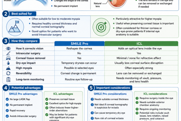
High Myopia: When I Start Thinking More Seriously About ICL
The higher the myopia, the more I ask:
Why remove a large amount of corneal tissue if the eye can instead be corrected with an implantable lens?
ICL may become attractive when:
- Myopia is high
- The cornea is relatively thin
- Predicted residual stroma is becoming limited
- Optical quality is particularly important
- Dry eye is significant
This does not mean there is one universal prescription at which I switch from SMILE Pro to ICL.
The boundary depends on anatomy.
Example 1: -3.00 D with a Healthy Cornea
Imagine a patient with:
- -3.00 D myopia
- Stable refraction
- Thick, regular cornea
- Normal tomography
- Healthy ocular surface
- Normal retina
SMILE Pro may be an extremely straightforward option.
Example 2: -8.00 D with a Thick, Normal Cornea
SMILE Pro may still be possible.
But I would examine:
- Required lenticule thickness
- Residual tissue
- Optical zone
- Pupil size
- Night-vision needs
and actively compare the result with ICL.
Example 3: -4.00 D with a 485-µm Cornea
The answer is not automatically:
“Too thin.”
Nor is it:
“PRK will definitely be fine.”
I would examine the complete tomography and the tissue requirements of each procedure.
Example 4: -2.50 D with a 550-µm Cornea but Suspicious Tomography
The prescription is easy.
The thickness appears generous.
But the corneal shape may make elective tissue-removing surgery inappropriate.
A thick cornea does not neutralise abnormal tomography.
Example 5: -6.00 D with Severe Dry Eye
The cornea may be structurally suitable but the ocular surface may not be.
I may first:
- Treat meibomian-gland dysfunction
- Control allergy
- Improve tear-film stability
- Repeat measurements
If significant dry-eye disease remains, ICL may deserve consideration if the internal anatomy is appropriate.
Example 6: -5.00 D in a 48-Year-Old with Early Nuclear Sclerosis
The problem is no longer simply corneal myopia.
The natural lens has begun changing.
Performing SMILE Pro may give only temporary refractive benefit before cataract progression alters the prescription again.
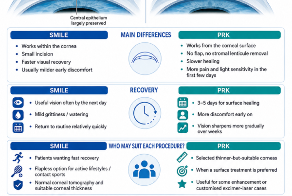
When I Prefer PRK Instead
PRK may be preferable when:
- A more superficial tissue-removal strategy makes greater anatomical sense
- Topography-guided treatment is desirable
- Wavefront-guided surface treatment is desirable
- Previous refractive surgery requires a surface enhancement
- SMILE tissue geometry is less favourable
PRK has slower recovery and a recognised haze risk, but those disadvantages may be acceptable when the corneal architecture favours surface treatment.
When I Prefer ICL Instead
ICL may be preferable when:
- Myopia is high
- Corneal tissue would be stretched close to my preferred safety margin
- Corneal optical quality is a priority
- Dry-eye concerns are substantial
- The internal anatomy is suitable
ICL does not remove refractive stromal tissue but introduces the separate risks of intraocular surgery.
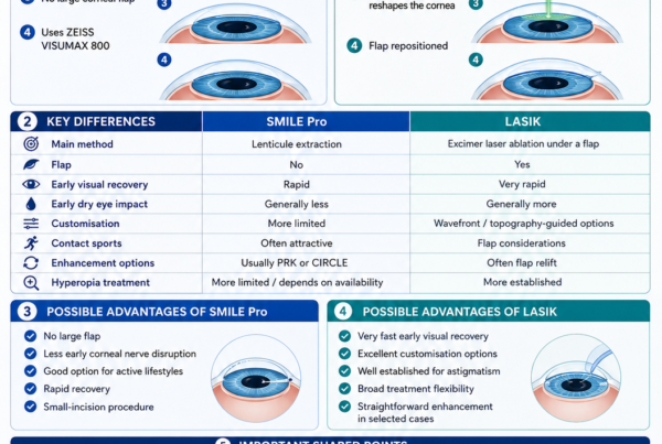
When LASIK May Be Preferable
LASIK may be selected when:
- Highly customised excimer ablation is desirable
- Hyperopic treatment is required
- Topographic or wavefront treatment characteristics favour LASIK
- A particular enhancement strategy is important
The decision should be based on the eye rather than assuming SMILE Pro is automatically better because it is newer.
What Does Not Automatically Disqualify You?
Some findings require judgement rather than automatic rejection.
Examples include:
- Mild dry eye
- Moderate myopia
- High astigmatism
- Relatively thin but normal cornea
- Contact-lens intolerance
- Large pupils
These may influence:
- Procedure selection
- Preoperative treatment
- Surgical parameters
- Counselling
rather than making surgery impossible.
What Makes Me Much More Cautious?
I become especially cautious when several risk factors occur together, such as:
- Young age
- Increasing myopia
- High correction
- Thin cornea
- Suspicious tomography
- Low predicted residual stroma
- Strong family history of keratoconus
- Vigorous eye rubbing
Ectasia risk is multifactorial.
Risk rises through the interaction between the patient’s native cornea and the amount of surgery planned.
Measurements Are Only Half the Assessment
A technically perfect cornea does not automatically make someone a good refractive-surgery candidate.
I also want to understand:
- Why does the patient want surgery?
- What does their work involve?
- Do they drive extensively at night?
- Do they play contact sports?
- Are they comfortable with occasional spectacles?
- Are expectations realistic?
- Do they understand presbyopia?
- Would they accept an enhancement if necessary?
Expectations Matter
SMILE Pro is intended to reduce dependence on optical correction.
It does not guarantee:
- Perfect vision
- Zero prescription
- No glare
- No halos
- No dry eye
- Permanent freedom from glasses
Even a technically excellent outcome may leave:
- -0.25 D
- +0.25 D
- small residual cylinder
without any meaningful functional problem.
Presbyopia
A patient approaching their 40s needs specific counselling.
If both eyes are corrected perfectly for distance, reading vision will progressively become more difficult as presbyopia develops.
Options may include:
- Distance correction in both eyes
- Monovision
- Continued reading glasses
Suitability therefore includes lifestyle and future visual needs, not just corneal measurements.
Common Myths
“My Prescription Is Within -10.00 D, So I Qualify”
False.
Treatment range is not the same as suitability.
“My Cornea Is Above 500 Microns, So SMILE Pro Is Safe”
False.
Shape and tissue distribution matter.
“My Cornea Is Below 500 Microns, So I Cannot Have SMILE Pro”
Also false as an absolute statement.
Some thinner corneas may be suitable when all other parameters are reassuring.
“There Is One Safe Residual-Stroma Number”
Too simplistic.
Modern evidence provides useful benchmarks, but ectasia risk is continuous and multifactorial.
“250 Microns Is Safe Because It Is in the FDA Label”
No.
That is an older regulatory lower boundary, not a target I would aim to approach routinely.
“SMILE Pro Cannot Cause Ectasia Because There Is No Flap”
False.
SMILE still removes stromal tissue.
“A Normal Thickness Means I Cannot Have Keratoconus”
False.
Tomographic shape is critical.
“If My Topography Looks Normal, No Other Testing Matters”
Not always.
Tomography and sometimes epithelial mapping can reveal additional risk.
“Higher Myopia Just Means the Laser Runs Longer”
False.
Higher myopia usually requires a larger volume of corneal tissue alteration.
“SMILE Pro Is Always Better Than ICL Because It Avoids Going Inside the Eye”
False.
For high myopia, the tissue and optical trade-off may favour ICL.
“ICL Is Only for People Who Fail SMILE Screening”
False.
ICL may be the preferred primary procedure in selected high-myopic patients.
“Dry Eye Means I Cannot Have SMILE”
Not necessarily.
Mild or moderate ocular-surface disease may first be treated and reassessed.
Severe uncontrolled dry eye is a different situation.
“Contact-Lens Intolerance Means SMILE Is Perfect for Me”
Not automatically.
The cause of the intolerance should first be understood.
“6/6 Vision Means My Eyes Are Healthy”
False.
Patients can have:
- Retinal disease
- Glaucoma
- Suspicious corneas
despite excellent central visual acuity.
“Once My Retina Is Checked Before Surgery, I No Longer Need Retinal Reviews”
False.
High-myopia retinal risks remain after refractive correction.
“SMILE Pro Stops My Myopia”
False.
It corrects the current refractive error.
“A Stable Glasses Prescription Means My Eye Has Stopped Ageing”
False.
Future:
- Presbyopia
- Cataract
- Retinal disease
- Glaucoma
can still occur.
Frequently Asked Questions
How Long Should My Prescription Be Stable?
A useful benchmark is no more than approximately 0.50 D of meaningful change over the preceding year, although individual surgeons and regulatory systems may use different criteria.
I also consider the longer-term trend.
What if My Prescription Changed by 0.25 D?
A change this small may represent:
- Measurement variability
- Tear-film variation
rather than true progression.
The entire history matters.
What if My Prescription Changed by 1.00 D Last Year?
I would generally want to understand why and demonstrate stability before elective surgery.
How Thick Does My Cornea Need to Be?
There is no universal number.
The answer depends on:
- Tomography
- Prescription
- Lenticule thickness
- Cap parameters
- Residual stroma
- Age
- Other ectasia risk factors
Is 500 Microns Enough?
Sometimes.
Sometimes not.
The number cannot be interpreted alone.
Is 480 Microns Too Thin?
Not automatically, but it deserves careful evaluation.
For some patients, PRK or ICL may provide a better tissue strategy.
What Residual Stromal Thickness Do You Look For?
I calculate the projected residual stromal tissue for every treatment.
Current research suggests increased biomechanical concern as SMILE residual stromal thickness falls below approximately 280 µm, but this is a risk benchmark rather than a universal threshold.
I prefer to evaluate the entire structural profile rather than operate to the edge of a number.
Do You Measure Percentage Tissue Altered?
Tissue-alteration metrics can add useful information.
Recent SMILE-specific research suggests that a tissue-alteration proportion around 27–28% may represent an important biomechanical region, but the authors themselves emphasise that more validated SMILE-specific metrics are needed.
Do You Check the Back of the Cornea?
Yes.
Posterior elevation is an important component of tomography.
Do You Perform Epithelial Mapping?
It can be particularly helpful for:
- Borderline tomography
- Unusual asymmetry
- Keratoconus screening
- Conflicting measurements
Studies show that epithelial mapping can materially change refractive-surgery candidacy decisions.
Do You Check Corneal Biomechanics?
Biomechanical measurements may be useful in selected cases as an additional layer of risk assessment.
They do not replace tomography.
Do You Check Pupil Size?
Yes, particularly when night vision is important or the pupil is relatively large.
Does a Large Pupil Mean I Cannot Have SMILE Pro?
No.
It influences:
- Optical planning
- Tissue requirements
- Night-vision counselling
rather than automatically excluding surgery.
Do You Dilate the Eyes?
A dilated examination is particularly important in myopic patients to assess the retina and natural lens.
Formal SMILE patient-selection guidance includes dilated retinal examination.
Do I Need Retinal Laser Before SMILE Pro?
Not routinely.
Retinal laser is performed only when the retinal specialist or ophthalmologist believes a particular lesion warrants treatment.
Can I Have SMILE Pro with Lattice Degeneration?
Possibly.
The retina must first be assessed appropriately.
Can I Have SMILE Pro After Retinal Laser?
Often yes once the retina is stable, but suitability depends on:
- Underlying retinal condition
- Visual potential
- Myopia
- Timing
Can I Have SMILE Pro if I Have Dry Eyes?
Potentially after appropriate treatment.
Severe uncontrolled dry eye is a reason not to proceed.
Can I Have SMILE Pro if I Have Allergic Eyes?
Possibly after controlling:
- Inflammation
- Itching
- Eye rubbing
Can I Have SMILE Pro if I Have Glaucoma?
This requires individual assessment.
Uncontrolled glaucoma is inappropriate for elective SMILE.
A stable glaucoma suspect requires consideration of:
- Optic nerve
- Visual field
- OCT
- Pressure
- Future interpretation of IOP
Can I Have SMILE Pro if I Have Diabetes?
Well-controlled diabetes may be considered in selected patients after evaluating:
- Retinopathy
- Glucose stability
- Ocular surface
- Corneal health
Uncontrolled diabetes is a contraindication.
Can I Have SMILE Pro During Pregnancy?
Elective treatment should be deferred.
Can I Have SMILE Pro While Breastfeeding?
It is generally postponed until postpartum hormonal and refractive stability is established.
Can I Have SMILE Pro if I Am 45?
Possibly.
But the consultation must include:
- Presbyopia
- Monovision
- Natural-lens status
- Cataract risk
Can I Have SMILE Pro at 55?
Sometimes, but developing lens changes increasingly make lens-based solutions more relevant.
Can I Have SMILE Pro if I Have a Cataract?
Generally the cataract should drive the treatment plan rather than permanently reshaping the cornea for a prescription being caused by an ageing lens.
Can I Have SMILE Pro if I Have Keratoconus?
Routine refractive SMILE Pro is generally inappropriate.
Can I Have SMILE Pro with a Family History of Keratoconus?
Possibly, but I would assess the cornea especially carefully.
Can I Have SMILE Pro if I Rub My Eyes?
The cause of rubbing should be addressed.
Persistent vigorous rubbing makes me more cautious about corneal structural risk.
Can I Have SMILE Pro if My Corneas Are Steep?
Possibly.
The whole tomographic pattern matters more than one keratometry reading.
Can I Have SMILE Pro if My Corneas Are Flat?
Possibly.
Again, the expected postoperative shape and optical result need to be assessed.
Can SMILE Pro Treat High Astigmatism?
Current VISUMAX 800 treatment capability extends to substantial astigmatism in appropriate patients, and the platform includes cyclotorsional alignment assistance.
What if My Astigmatism Axis Keeps Changing?
I would want to understand why before surgery.
Possible explanations include:
- Tear-film instability
- Contact-lens warpage
- Measurement inconsistency
- Corneal disease
- Lens-related astigmatism
What if Only One Eye Is Suitable?
Refractive surgery does not have to be symmetrical.
In selected cases, different eyes may require:
- Different procedures
- Treatment in only one eye
- No treatment
The goal is not procedural symmetry.
It is ocular safety.
My SMILE Pro Suitability Assessment: A Practical Checklist
1. Confirm the Visual Problem
I establish:
- Unaided vision
- Best-corrected vision
- Manifest refraction
- Cycloplegic refraction where appropriate
- Refractive stability
2. Analyse the Cornea
I review:
- Topography
- Tomography
- Pachymetry
- Posterior elevation
- Thickness distribution
- Keratoconus indices
- Epithelial map where indicated
- Biomechanics where useful
3. Model the Proposed Surgery
I consider:
- Sphere
- Cylinder
- Optical zone
- Cap thickness
- Lenticule thickness
- Predicted residual stroma
- Overall proportion of tissue altered
4. Assess Optical Quality
I consider:
- Pupil size
- Astigmatism
- Treatment centration
- Night-driving requirements
- Higher-order aberrations where relevant
5. Assess the Ocular Surface
I look for:
- Dry eye
- Meibomian-gland dysfunction
- Allergy
- Blepharitis
- Corneal staining
- Eye rubbing
6. Examine the Rest of the Eye
I assess:
- Eye pressure
- Optic nerve
- Natural lens
- Macula
- Peripheral retina
7. Compare Other Procedures
I ask whether:
- LASIK
- PRK
- ICL
- Continued spectacles or contact lenses
would provide a better overall risk-benefit balance.
8. Match the Surgery to the Patient
I consider:
- Occupation
- Contact sports
- Night driving
- Recovery time
- Age
- Presbyopia
- Expectations
The Bottom Line
Suitability for SMILE Pro cannot be determined by spectacle power alone.
Although current Singapore guidance lists a SMILE Pro treatment range of approximately -1.00 to -10.00 D of myopia and astigmatism up to -5.00 D, eligibility still depends on corneal thickness and the overall health of the eye.
The measurements I consider most important include:
- Prescription and refractive stability
- Best-corrected vision
- Corneal topography
- Corneal tomography
- Corneal-thickness distribution
- Posterior elevation
- Predicted lenticule thickness
- Predicted residual stromal tissue
- Tissue-alteration proportion
- Epithelial thickness where indicated
- Ocular-surface health
- Astigmatism and treatment alignment
- Pupil size
- Eye pressure
- Natural-lens clarity
- Retinal health
The most important safety principle is:
A normal-looking central corneal thickness does not prove that a cornea is safe for surgery.
Tomographic abnormalities, age, high myopia, tissue removal and residual stromal architecture all influence ectasia risk.
Newer evidence also reinforces that SMILE-specific tissue planning should go beyond an old minimum residual-stroma number. A 2026 systematic review found greater biomechanical change below approximately 280 µm of residual stromal thickness and around 27–28% SMILE tissue alteration, while emphasising that these remain benchmarks rather than absolute universal safety boundaries.
For borderline corneas, epithelial thickness mapping can provide useful additional information and has been shown to change surgical-candidacy decisions in a meaningful proportion of patients.
High myopia also deserves special consideration.
SMILE Pro can successfully treat substantial prescriptions, but as the correction increases:
- More corneal tissue is altered
- Early predictability may decrease
- Optical considerations become more important
Recent VISUMAX 800 data confirm excellent overall results but also show somewhat less predictable early 20/20 vision in high-myopic eyes.
That is when I increasingly ask whether an ICL would preserve more corneal tissue and provide a better overall optical strategy.
Finally, I examine the entire eye.
Formal preoperative SMILE guidance recommends evaluation of:
- The natural crystalline lens
- Corneal mapping
- A dilated retina
rather than treating refractive error in isolation.
The most important message is:
My aim in a SMILE Pro assessment is not to find a way to make the laser fit the prescription. It is to decide whether SMILE Pro is the safest and most sensible refractive strategy for that particular eye—and to recommend PRK, ICL, another option or no surgery when it is not.
References
- Singapore National Eye Centre. SMILE Pro: suitability, treatment range and preoperative considerations. Current information accessed August 2026.
- Singapore National Eye Centre. Refractive surgery suitability assessment and preoperative testing. Current information accessed August 2026.
- Carl Zeiss Vision Care. ZEISS SMILE Pro suitability criteria and refractive treatment ranges. Current information accessed August 2026.
- US Food and Drug Administration. VisuMax SMILE indications, contraindications and patient-selection precautions.
- Jin SX, Dackowski E, Chuck RS. Risk factors for post-laser refractive surgery corneal ectasia. Curr Opin Ophthalmol. 2020;31:288–292. PMID: 32398416.
- Santhiago MR, et al. Ectasia risk factors in refractive surgery. PMID: 27143849.
- Luz A, et al. Application of corneal tomography before keratorefractive procedures for laser vision correction. PMID: 27079610.
- Randleman JB, et al. Determining the utility of epithelial thickness mapping in refractive-surgery evaluations. Am J Ophthalmol. 2022. PMID: 35247335.
- Safety thresholds for removing and preserving corneal tissue in small-incision lenticule extraction: a PRISMA-compliant systematic review. 2026. PMID: 42180670.
- Santhiago MR, et al. Percent tissue altered and corneal ectasia. PMID: 27096376.
- Corneal ectasia risk and percentage tissue altered in myopic patients presenting for refractive surgery. PMID: 31686775.
- Factors affecting visual outcome after SMILE Pro using VISUMAX 800. 2025. PMID: 40976615.
- Corneal collagen cross-linking for ectasia after SMILE: review of risk and screening principles. PMID: 35790199.
- Effect of wavefront aberrations on night-vision problems and mesopic contrast after SMILE. PMID: 34236902.
- Effect of angle-kappa adjustment on higher-order aberrations after SMILE. PMID: 32040010.
- Dry-eye evaluation and relationship between tear-film stability and corneal-surface regularity after SMILE. PMID: 28940146.
- Corneal epithelial remodelling and higher-order aberrations after SMILE. PMID: 36630428.