Author: Dr Val Phua
Estimated reading time: 16 minutes
Implantable Collamer Lens surgery—commonly known as ICL surgery—does not appear to cause a substantial increase in long-term glaucoma risk in appropriately selected patients receiving a modern central-port ICL.
However, ICL surgery can occasionally cause a temporary or persistent rise in eye pressure. If a significant pressure increase is not recognised and treated, it can damage the optic nerve and lead to secondary glaucoma.
The risk is therefore best understood in three parts:
- Temporary eye-pressure elevation can occur during the hours or weeks after surgery.
- Implant-related glaucoma is uncommon but can occur because of angle narrowing, excessive vault, pupillary block or pigment dispersion.
- High myopia itself increases glaucoma risk, whether or not the patient undergoes ICL surgery.
Modern EVO ICL studies are reassuring. In the three-year United States FDA study, no eyes developed elevated pressure from angle narrowing or pigment dispersion, and no eyes experienced pupillary block requiring a peripheral iridotomy. Nevertheless, the FDA states that the longer-term risks of glaucoma, pigment dispersion and permanent drainage-angle adhesions are not yet fully established. [1–4]
What Is Glaucoma?
Glaucoma is a group of diseases that cause progressive damage to the optic nerve.
The optic nerve transmits visual information from the eye to the brain. If glaucoma is not detected and controlled, it can cause:
- Loss of peripheral vision
- Difficulty seeing in dim lighting
- Reduced contrast sensitivity
- Advanced visual-field loss
- Permanent blindness in severe cases
Raised intraocular pressure is an important glaucoma risk factor, but glaucoma is not defined by pressure alone.
A person may have:
- High eye pressure without optic nerve damage
- Glaucoma despite statistically normal eye pressure
- Temporary postoperative pressure elevation without developing glaucoma
What Is the Difference Between Raised Eye Pressure and Glaucoma?
These terms should not be used interchangeably.
Raised Eye Pressure
Raised intraocular pressure means that the fluid pressure inside the eye is above the desired level.
It may be:
- Temporary
- Medication-related
- Caused by retained surgical material
- Related to ICL vault or position
- Unassociated with permanent optic nerve damage
Ocular Hypertension
Ocular hypertension usually refers to persistently raised eye pressure without detectable glaucomatous optic nerve or visual-field damage.
Glaucoma
Glaucoma means that the optic nerve has developed characteristic damage, often accompanied by corresponding visual-field loss.
A temporary pressure spike after surgery is therefore not necessarily glaucoma. It becomes dangerous when the pressure is very high, lasts too long, repeatedly recurs or occurs in an optic nerve that is already vulnerable.
Does ICL Surgery Cause Glaucoma?
For most suitable patients, the answer is no.
Modern studies of central-port ICLs generally report stable average eye pressure and a low incidence of clinically significant glaucoma-related complications. Eight- and ten-year studies have shown favourable long-term safety in appropriately selected eyes. [5,6]
However, ICL surgery can contribute to secondary glaucoma through several uncommon mechanisms:
- Retained surgical viscoelastic
- Steroid response
- Pupillary block
- Excessively high vault
- Drainage-angle narrowing
- Pigment dispersion
- Abnormal ICL orientation or position
- Persistent inflammation
The risk is reduced substantially by careful patient selection, correct ICL sizing, meticulous surgery and appropriate follow-up.
Why Can Eye Pressure Rise After ICL Surgery?
The eye continuously produces a clear fluid called aqueous humour.
This fluid normally:
- Forms behind the iris.
- Moves through the pupil.
- Enters the anterior chamber.
- Leaves through the drainage angle.
Eye pressure rises when fluid production exceeds drainage.
The ICL sits behind the iris and in front of the natural lens. If the implant, surgical material, inflammation or medication interferes with aqueous-fluid flow, pressure may increase.
When Can Eye Pressure Rise?
Pressure problems may occur at different stages.
Within the First Few Hours
The most likely causes are:
- Retained ophthalmic viscoelastic
- Pupillary block
- Incorrect lens orientation
- Very high vault
- Acute inflammation
This is why FDA professional guidance recommends checking eye pressure approximately one to six hours after EVO ICL implantation. [1,2]
During the First One to Four Weeks
The commonest cause during this period is often a response to postoperative steroid eyedrops.
Other possible causes include:
- Persistent inflammation
- Incomplete removal of viscoelastic
- Angle narrowing
- ICL-related iris contact
A clinical series examining raised pressure after posterior-chamber ICL implantation found steroid response to be the most frequent cause, followed by retained viscoelastic and pupillary block. Most affected eyes were controlled without permanent glaucomatous damage when treated promptly. [3,7]
Months or Years Later
Later pressure elevation may occur because of:
- Excessive vault
- Progressive drainage-angle narrowing
- Pigment dispersion
- Peripheral anterior synechiae
- Natural development of open-angle glaucoma
- High-myopia-related glaucoma susceptibility
- Age-related anatomical changes
Late implant-related glaucoma appears rare, but published case reports show that it can occur many years after an initially uncomplicated procedure. [8,9]
What Is Retained Viscoelastic?
During ICL surgery, the surgeon may place a transparent gel-like material called ophthalmic viscosurgical device, or viscoelastic, inside the eye.
It helps:
- Protect the corneal endothelium
- Maintain space during surgery
- Reduce contact with the natural lens
- Allow the ICL to be positioned safely
If excessive viscoelastic remains after surgery, it may temporarily block the drainage system and cause a rapid pressure rise.
How Is It Managed?
Treatment may include:
- Pressure-lowering eyedrops
- Oral pressure-lowering medication
- Observation with repeated pressure measurements
- Releasing fluid through the surgical incision
- Washing retained viscoelastic out of the anterior chamber
When treated promptly, pressure usually returns to normal without permanent damage.
Can Steroid Eyedrops Raise Eye Pressure?
Yes.
Steroid eyedrops are prescribed after ICL surgery to control inflammation. Some people are steroid responders, meaning their pressure rises after steroid exposure.
The pressure increase commonly becomes apparent during the first few postoperative weeks.
Risk may be greater in patients with:
- A previous steroid response
- A family history of glaucoma
- Existing ocular hypertension
- High myopia
- Diabetes
- A suspicious optic nerve
- Prolonged or intensive steroid use
Management may involve:
- Reducing the steroid dose
- Switching to a less pressure-raising anti-inflammatory medication
- Adding pressure-lowering eyedrops
- Closer monitoring
Patients should not stop steroid treatment independently, because inadequately treated inflammation can also harm the eye.
What Is Pupillary Block Glaucoma?
Pupillary block occurs when aqueous fluid cannot move normally from behind the iris into the anterior chamber.
Fluid accumulates behind the iris, pushing it forwards and narrowing or closing the drainage angle. This can cause a rapid pressure increase.
Possible symptoms include:
- Severe eye pain
- Headache
- Redness
- Cloudy vision
- Coloured rings around lights
- Nausea
- Vomiting
Has the Central Port Reduced Pupillary Block Risk?
Yes.
Earlier ICL models without a central port generally required peripheral iridotomies to create alternative pathways for aqueous fluid.
Modern EVO ICLs contain a 0.36 mm central port that allows fluid to flow through the lens. Routine preoperative peripheral iridotomy is therefore generally unnecessary. [1,2]
In the three-year FDA clinical study, no eyes developed pupillary block requiring peripheral iridotomy or iridectomy. [4]
Pupillary block is nevertheless not theoretically impossible. It may occur if:
- The central port is obstructed
- The ICL is implanted upside down
- Residual viscoelastic blocks aqueous flow
- Severe inflammation develops
- The anatomy is unusually crowded
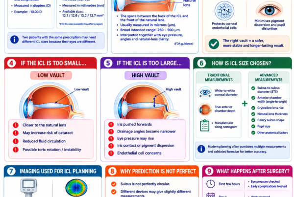
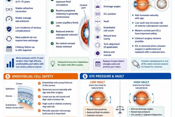
What Is ICL Vault?
Vault is the distance between the back surface of the ICL and the front surface of the natural crystalline lens.
An appropriate vault maintains separation from the natural lens without excessively crowding the front of the eye.
Low Vault
A very low vault is mainly associated with:
- Natural-lens proximity
- Anterior subcapsular cataract risk
- Reduced aqueous circulation
Low vault is not usually the main mechanism for glaucoma unless other anatomical factors are present.
High Vault
An excessively high vault may:
- Push the iris forwards
- Narrow the drainage angles
- Reduce aqueous outflow
- Raise eye pressure
- Increase iris contact
- Cause pigment dispersion
- Distort the pupil
The significance of high vault depends on more than the measurement alone.
The surgeon also considers:
- Eye pressure
- Gonioscopic angle appearance
- Anterior-segment OCT
- Iris configuration
- Corneal endothelial cell count
- Symptoms
- Change over time
In the FDA three-year study, two eyes required ICL exchange because of high vault, but neither had developed raised pressure. This demonstrates that clinically important vault abnormalities can sometimes be corrected before glaucoma occurs. [4]
Does an ICL Narrow the Drainage Angle?
Placing an ICL behind the iris generally causes some reduction in the available anterior-chamber angle width.
In a properly selected eye, the remaining angle should still allow adequate aqueous drainage.
The risk becomes more relevant when:
- The anterior chamber was shallow before surgery
- The drainage angles were already narrow
- The ICL is too large
- Vault is excessive
- The natural lens thickens with age
- Peripheral anterior synechiae develop
For this reason, FDA criteria contraindicate EVO ICL implantation in eyes with a true anterior chamber depth below 3.0 mm, an anterior chamber angle below Grade III or moderate-to-severe glaucoma. [2]
Local regulatory criteria and clinical practice may differ, but the underlying principle remains the same: there must be enough internal space for the ICL without compromising the drainage angle.
What Is Pigment Dispersion?
The back surface of the iris contains pigment.
If the ICL repeatedly contacts or rubs against the iris, pigment particles may be released into the eye.
The pigment may deposit on:
- The ICL
- The corneal endothelium
- The iris
- The drainage angle
- The trabecular meshwork
When pigment is present without optic nerve damage, the condition may be called pigment dispersion syndrome.
When pigment obstructs drainage sufficiently to cause raised pressure and glaucomatous damage, it is called pigmentary glaucoma.
How Common Is Pigmentary Glaucoma After ICL?
Clinically significant pigmentary glaucoma appears uncommon with modern ICLs.
The FDA three-year EVO study reported no elevated pressure attributed to pigment dispersion. [4]
However, rare late cases have been reported. A 2024 case described bilateral pigment dispersion syndrome 13 years after ICL implantation. A 2025 report described late-onset pigmentary glaucoma with visual-field loss after phakic ICL implantation. Case reports cannot establish how frequently this occurs, but they demonstrate why lifelong pressure and drainage-angle monitoring remain sensible. [8,9]
Can ICL Be Implanted Upside Down?
Incorrect orientation is rare but possible.
An upside-down ICL may produce:
- Abnormal vault
- Pupillary block
- Iris contact
- Raised eye pressure
- Pupil distortion
- Cataract risk
The ICL contains orientation markings to help the surgeon confirm that it is correctly positioned. [2]
Significant malposition usually requires prompt surgical correction.
Does High Myopia Itself Increase Glaucoma Risk?
Yes.
Most ICL patients have moderate or high myopia. High myopia is independently associated with a greater prevalence and incidence of open-angle glaucoma.
This means that a patient who develops glaucoma years after ICL surgery may have developed it because of:
- The underlying highly myopic eye
- Family history
- Age
- Optic nerve susceptibility
- Another glaucoma risk factor
- An ICL-related mechanism
- A combination of these factors
Recent reviews have reaffirmed the association between axial myopia and open-angle glaucoma while emphasising that diagnosing glaucoma in highly myopic eyes can be difficult. [10–12]
ICL surgery corrects the refractive blur but does not shorten the elongated eye or remove the glaucoma susceptibility associated with high myopia.
Why Is Glaucoma Harder to Diagnose in Highly Myopic Eyes?
High myopia can change the appearance of the optic nerve and surrounding retina.
Possible features include:
- Tilted optic discs
- Large or shallow optic cups
- Peripapillary atrophy
- Thin retinal nerve-fibre measurements
- Segmentation errors on OCT
- Macular ganglion-cell abnormalities
- Visual-field defects unrelated to glaucoma
These changes may imitate glaucoma or make true glaucoma more difficult to detect.
For this reason, glaucoma assessment should not rely on a single test.
Doctors may need to compare:
- Serial optic-disc photographs
- OCT retinal nerve-fibre layer scans
- Macular ganglion-cell analysis
- Visual fields
- Eye pressure
- Corneal thickness
- Gonioscopy
- Changes over time
Does ICL Make Eye-Pressure Measurement Less Accurate?
ICL surgery does not intentionally reshape the central cornea in the way that LASIK, PRK or SMILE do.
This means that corneal curvature and thickness changes caused by laser ablation are generally not introduced by ICL surgery.
However, every eye-pressure measurement still has limitations and may be influenced by:
- Corneal thickness
- Corneal biomechanics
- Measurement device
- Time of day
- Patient positioning
- Eyelid squeezing
Pressure should therefore be interpreted together with optic nerve, OCT, visual-field and drainage-angle findings.
Who Should Not Have ICL Surgery Because of Glaucoma Risk?
Under current FDA professional labelling, moderate-to-severe glaucoma is a contraindication to EVO ICL implantation. The safety and effectiveness of EVO ICL have also not been established in all patients with glaucoma or ocular hypertension. [1,2]
ICL may be unsuitable or require extreme caution when there is:
- Moderate or advanced glaucoma
- Progressive visual-field loss
- Uncontrolled eye pressure
- Narrow drainage angles
- A shallow anterior chamber
- Pigment dispersion syndrome
- Pseudoexfoliation
- Peripheral anterior synechiae
- Previous angle-closure attacks
- A marked steroid response
- Significant optic nerve damage
- Inability to attend long-term reviews
Can Someone With Mild or Suspected Glaucoma Have ICL?
Possibly in exceptional selected circumstances, but this is not a routine decision.
The surgeon must consider:
- Whether glaucoma is confirmed or only suspected
- The degree of optic nerve damage
- Visual-field status
- Eye-pressure control
- Drainage-angle anatomy
- Anterior chamber depth
- Expected postoperative vault
- Age
- Alternative methods of vision correction
- Whether implant-related monitoring would complicate glaucoma care
In many cases, glasses, contact lenses or another refractive approach may provide a safer balance of benefits and risks.
A glaucoma specialist’s assessment may be appropriate before surgery.
What Tests Are Needed Before ICL Surgery?
A comprehensive preoperative assessment should include the following.
Eye-Pressure Measurement
Pressure should be measured before surgery and interpreted in the context of the patient’s cornea and optic nerve.
Repeated measurements may be needed when:
- Pressure is borderline
- The readings vary
- There is a family history of glaucoma
- The optic nerve appears suspicious
Gonioscopy
Gonioscopy uses a specialised lens to examine the drainage angle directly.
It can identify:
- Narrow angles
- Pigment
- Peripheral anterior synechiae
- Abnormal angle anatomy
- Previous angle closure
Anterior Chamber Depth
The space between the corneal endothelium and natural lens must be sufficient to accommodate the ICL safely.
Anterior-Segment Imaging
Anterior-segment OCT or ultrasound biomicroscopy may assess:
- Angle width
- Internal eye dimensions
- Sulcus anatomy
- Iris configuration
- Crystalline lens rise
- Expected ICL sizing
Optic Nerve Examination
The surgeon examines:
- Cup-to-disc ratio
- Neuroretinal rim
- Disc haemorrhage
- Myopic disc changes
- Asymmetry between the eyes
OCT
OCT may assess:
- Retinal nerve-fibre layer
- Macular ganglion-cell complex
- Optic nerve anatomy
High-myopia-related artefacts must be interpreted carefully.
Visual-Field Testing
A visual field may be performed when:
- Glaucoma is suspected
- The optic nerve is abnormal
- Eye pressure is raised
- There is a strong family history
- A baseline is desirable for long-term comparison
Corneal Thickness
Central corneal thickness helps place the pressure measurement into context.
What Follow-Up Is Needed After ICL Surgery?
A typical glaucoma-related follow-up programme may include:
- Eye-pressure measurement within one to six hours
- Review the following day
- Review during the first week
- Review after several weeks while steroid drops are being used
- Further review at one to three months
- Regular long-term examinations
The exact schedule should be individualised.
Patients with borderline pressure, unusual vault or glaucoma risk may require more frequent reviews.
What Should Be Checked Long Term?
Long-term follow-up may include:
- Eye pressure
- ICL position
- Vault
- Drainage-angle width
- Angle pigmentation
- Peripheral anterior synechiae
- Optic nerve appearance
- OCT retinal nerve-fibre layer
- Macular ganglion-cell analysis
- Visual-field testing
- Natural-lens clarity
- Corneal endothelial cell count
Even patients with excellent vision should continue follow-up because glaucoma and angle changes may initially cause no symptoms.
What Symptoms Can Raised Eye Pressure Cause?
Possible symptoms include:
- Eye pain
- Headache
- Redness
- Cloudy or blurred vision
- Halos around lights
- Nausea
- Vomiting
- A fixed or distorted pupil
However, moderately raised pressure and early open-angle glaucoma may cause no symptoms.
The absence of pain does not guarantee that the pressure is normal.
When Should You Seek Urgent Care?
Seek urgent assessment after ICL surgery for:
- Severe or increasing eye pain
- Rapidly worsening vision
- Marked redness
- A severe headache
- Nausea or vomiting
- Cloudy vision
- Coloured rings around lights
- A distorted or poorly reactive pupil
- Increasing light sensitivity
- Sudden loss of peripheral vision
These symptoms may indicate a severe pressure rise, pupillary block, angle closure, inflammation or another urgent complication.
How Is Raised Pressure Treated?
Treatment depends on the cause.
Retained Viscoelastic
Possible treatment includes:
- Pressure-lowering medication
- Releasing fluid from the incision
- Anterior-chamber washout
Steroid Response
Management may include:
- Reducing or changing the steroid
- Adding pressure-lowering eyedrops
- Closer monitoring
Pupillary Block
Treatment may include:
- Pressure-lowering medication
- Pupil-modifying medication where appropriate
- Clearing an obstructed central port
- Peripheral iridotomy
- Repositioning or removing the ICL
Excessive Vault or Angle Narrowing
Options may include:
- Observation when pressure and angles remain safe
- Pressure-lowering medication
- Repositioning
- Exchange for a smaller ICL
- ICL removal
- Cataract surgery in selected older patients
Pigmentary Glaucoma
Treatment may include:
- Pressure-lowering eyedrops
- Laser treatment where appropriate
- ICL repositioning or removal
- Glaucoma surgery in advanced or uncontrolled cases
Promptly identifying the cause is essential because medication alone may not solve a mechanical problem caused by the implant. [3]
If the ICL Is Removed, Will the Pressure Return to Normal?
Pressure may improve when the ICL is causing:
- Angle narrowing
- Pupillary block
- Excessive vault
- Iris rubbing
- Pigment dispersion
However, pressure may remain elevated if the patient also has:
- Primary open-angle glaucoma
- Permanent drainage-angle scarring
- Peripheral anterior synechiae
- Significant pigment accumulation
- Steroid-related pressure elevation
- Another glaucoma mechanism
Removing the ICL cannot restore optic nerve tissue that has already been damaged.
This is why prevention and early detection are preferable to waiting for established visual-field loss.
Is EVO ICL Safer Than Older ICL Models for Glaucoma Risk?
The central-port EVO design appears to have improved the pressure-related safety profile.
Potential advantages include:
- More physiological aqueous flow
- No routine requirement for peripheral iridotomy
- Lower pupillary-block risk
- Reduced concern about aqueous stagnation in front of the natural lens
The FDA three-year study showed no pupillary block requiring iridotomy, no pressure elevation caused by angle narrowing and no pressure elevation caused by pigment dispersion. [4]
However, EVO ICL does not eliminate:
- Steroid responses
- Retained-viscoelastic pressure spikes
- High-vault angle narrowing
- Rare pigment dispersion
- Natural glaucoma risk from high myopia
- The need for lifelong monitoring
Does ICL Have a Higher Glaucoma Risk Than LASIK or SMILE?
The procedures have different pressure-related considerations.
ICL
ICL introduces possible intraocular mechanisms such as:
- Retained viscoelastic
- Pupillary block
- High vault
- Angle narrowing
- Pigment dispersion
LASIK and SMILE
LASIK and SMILE do not place a lens inside the eye, but they alter the cornea.
After corneal laser surgery:
- Standard eye-pressure readings may underestimate the true pressure
- Glaucoma detection may become more complicated
- Steroid responses can still occur
- High-myopia-related glaucoma risk remains
The safer procedure depends on the patient’s corneal anatomy, internal eye anatomy, prescription and glaucoma risk.
Does ICL Protect Against Glaucoma?
No.
ICL surgery corrects myopia but does not protect the optic nerve.
It does not reverse:
- Axial elongation
- Myopic optic-disc changes
- Family or genetic risk
- Age-related glaucoma risk
- Existing optic nerve damage
Regular glaucoma screening remains important after surgery.
Frequently Asked Questions
Does ICL Commonly Cause Glaucoma?
No.
Most appropriately selected patients do not develop glaucoma because of ICL surgery.
Temporary pressure elevation is more common than true glaucomatous optic nerve damage.
How Common Is Raised Eye Pressure After ICL?
Published rates vary because studies use different lens models, definitions and follow-up periods.
An older mixed-model clinical series reported postoperative ocular hypertension in approximately 5% of eyes, most commonly because of steroid response. Modern EVO studies have generally reported fewer implant-related pressure complications. [4,7]
Can Eye Pressure Rise on the Day of Surgery?
Yes.
Retained viscoelastic is a common early cause.
This is why a pressure check within the first several hours is important.
Can Pressure Rise Weeks Later?
Yes.
A steroid response commonly develops during the first one to four weeks.
Can Glaucoma Develop Years After ICL?
It can, although implant-related late glaucoma appears rare.
Possible causes include pigment dispersion, progressive angle narrowing or glaucoma developing independently in a highly myopic eye.
Does High Vault Always Cause Glaucoma?
No.
Some eyes with high vault maintain normal pressure and adequate drainage angles.
High vault becomes clinically important when it causes angle narrowing, pigment dispersion, pressure elevation or other anatomical concerns.
Does Low Vault Cause Glaucoma?
Low vault is more strongly associated with natural-lens proximity and cataract risk than glaucoma.
Can the Central Hole Become Blocked?
Rarely, it may be obstructed by viscoelastic, inflammatory material or abnormal lens orientation.
This can interfere with aqueous flow and contribute to pressure elevation.
Can ICL Cause Pigmentary Glaucoma?
Rarely, yes.
Chronic iris contact can release pigment that obstructs the drainage system.
Can I Have ICL if My Eye Pressure Is Slightly High?
Possibly, but the cause of the pressure elevation must be established first.
Additional glaucoma testing and specialist assessment may be required.
Can I Have ICL if I Have a Family History of Glaucoma?
A family history is not automatically a contraindication.
It does justify careful baseline assessment and long-term monitoring.
Can I Have ICL if I Am a Glaucoma Suspect?
This requires individual assessment.
The potential benefit of elective refractive surgery must be weighed against the uncertainty and need for lifelong glaucoma surveillance.
Can I Have ICL if I Already Have Glaucoma?
Moderate-to-severe glaucoma is contraindicated under current FDA labelling.
Mild or suspected disease requires highly individual specialist assessment, and another form of correction may be preferable. [1,2]
Will I Know if My Pressure Is High?
Not always.
Some pressure spikes cause pain or cloudy vision, but moderate elevation can be asymptomatic.
Does Normal Eye Pressure Mean I Cannot Have Glaucoma?
No.
Normal-tension glaucoma can occur, and pressure varies throughout the day.
The optic nerve and visual field must also be assessed.
Do I Need Yearly Eye Checks After ICL?
Yes.
Long-term reviews should generally assess eye pressure, angles, vault, optic nerve and other ocular structures.
Key Takeaway
Modern ICL surgery does not appear to cause a major increase in glaucoma risk in properly selected patients.
The available evidence is reassuring:
- Average long-term eye pressure is generally stable
- Pupillary block is less common with the EVO central port
- Modern clinical studies report very low rates of angle- or pigment-related pressure problems
- True secondary glaucoma is uncommon
However, ICL surgery can cause eye-pressure elevation through:
- Retained viscoelastic
- Steroid response
- Pupillary block
- Excessive vault
- Drainage-angle narrowing
- Pigment dispersion
- Abnormal lens position
High myopia itself also increases the lifetime risk of open-angle glaucoma and makes optic nerve and OCT interpretation more challenging.
Safety depends on:
- Adequate anterior chamber depth
- Open drainage angles
- Healthy preoperative eye pressure
- Careful glaucoma assessment
- Correct ICL sizing
- Early postoperative pressure checks
- Monitoring during steroid treatment
- Lifelong eye examinations
Seek urgent care for severe eye pain, cloudy or rapidly worsening vision, marked redness, headache, nausea, vomiting or a distorted pupil.
Even when vision after ICL surgery is excellent, regular monitoring of eye pressure, drainage angles and optic nerve health remains essential.
References
- US Food and Drug Administration. EVO and EVO+ Visian Implantable Collamer Lens: Patient Information Booklet. 2022.
- US Food and Drug Administration. EVO and EVO+ Visian Implantable Collamer Lens: Instructions for Use. 2022.
- Gong D, Deng S, Dang K, Yan Z, Wang J. Causes and Management Strategies for Elevated Intraocular Pressure After Implantable Collamer Lens Implantation. Frontiers in Medicine. 2024. PMID: 38384405.
- Parkhurst G, et al. Three-Year Results From the United States FDA Prospective Multicentre Clinical Study of the EVO Implantable Collamer Lens. 2025. PMID: 40937096.
- Kamiya K, et al. Eight-Year Outcomes of Implantation of Posterior Chamber Phakic Intraocular Lens With a Central Port for Moderate-to-High Ametropia. 2021. PMID: 34977099.
- Alfonso-Bartolozzi B, et al. Ten-Year Follow-up of Posterior Chamber Phakic Intraocular Lenses With Central-Port Design. 2024. PMID: 38085219.
- Senthil S, Choudhari NS, Vaddavalli PK, Murthy S, Reddy JC, Garudadri CS. Etiology and Management of Raised Intraocular Pressure Following Posterior Chamber Phakic Intraocular Lens Implantation in Myopic Eyes. PLOS ONE. 2016. PMID: 27855172.
- Katsimpris A, Kumarasamy M. Bilateral Late-Onset Pigment Dispersion Syndrome Following Implantable Collamer Lens Surgery: A Case Report. 2024. PMID: 38682039.
- Li Z, et al. Late-Onset Pigment Dispersion Glaucoma After Phakic Implantable Collamer Lens Implantation: A Case Report. 2025. PMID: 40660586.
- Qiu M, et al. Comprehensive Assessment of Glaucoma in Patients With High Myopia: A Systematic Review. 2024. PMID: 39392516.
- Jiravarnsirikul A, et al. Evaluating Glaucoma in Myopic Eyes: Challenges and Advances. 2025. PMID: 39701308.
- Jonas JB, et al. Clinical and Anatomical Features of Myopia and Their Association With Open-Angle Glaucoma. 2024. PMID: 39622437.
- Passaro ML, et al. Potential Harms of Posterior Chamber Phakic Intraocular Lenses: A Systematic Review and Meta-analysis of Complication Incidence. 2026. PMID: 41611001.
- Larivoir NB, et al. Efficacy, Predictability and Safety of Phakic Implantable Collamer Lenses With a Central Port: A Meta-analysis. 2025. PMID: 41369665.
- Zhang H, et al. Analysis of Perioperative Problems Related to Implantable Collamer Lens Implantation and Their Management. 2022. PMID: 35731355.