Author: Dr Val Phua
Estimated reading time: 15 minutes
ICL and LASIK are two established surgical options for correcting short-sightedness and astigmatism. Both aim to reduce dependence on spectacles or contact lenses, and both can provide excellent vision in appropriately selected patients.
However, they correct vision in fundamentally different ways.
LASIK reshapes the cornea using a laser.
ICL surgery places an additional corrective lens inside the eye without removing central corneal tissue.
This difference affects who may be suitable, the range of prescriptions that can be treated, the recovery process, the possible side effects and the type of long-term monitoring required.
ICL is often considered for patients with high myopia, thin corneas or other factors that make corneal laser surgery less suitable. LASIK is frequently suitable for patients with low-to-moderate myopia, healthy corneas and a stable tear film who prefer to avoid an operation inside the eye.
Neither procedure is universally better. The more appropriate option depends on:
- Your spectacle prescription
- Corneal shape and thickness
- The health of the eye surface
- The amount of space inside the front of the eye
- Your age
- Your lifestyle and occupation
- Your tolerance for the different risks of corneal and intraocular surgery
Systematic reviews comparing phakic intraocular lenses such as ICL with excimer-laser surgery have generally found both approaches effective. Some evidence favours phakic lenses for refractive accuracy, contrast sensitivity or preservation of corrected vision in moderate-to-high myopia, but the quality and age of the comparative studies vary.
What Is ICL Surgery?
ICL stands for Implantable Collamer Lens.
During ICL surgery, a soft, foldable corrective lens is inserted through a small corneal incision and positioned:
- Behind the iris
- In front of the natural crystalline lens
The eye’s natural lens is not removed.
The ICL works together with the natural lens to focus light more accurately on the retina. A toric ICL can correct myopia together with astigmatism.
Modern EVO ICL models contain a small central port that permits the natural circulation of aqueous fluid through the lens. This design generally avoids the need for the peripheral iridotomy that was commonly performed before implantation of earlier ICL models.
What Is LASIK?
LASIK stands for Laser-Assisted In Situ Keratomileusis.
During LASIK:
- A thin flap is created within the cornea, usually with a femtosecond laser.
- The flap is lifted.
- An excimer laser removes a precisely calculated amount of corneal tissue.
- The underlying cornea is reshaped.
- The flap is repositioned.
For myopia, the laser generally flattens the central cornea so that light focuses farther back and more accurately on the retina.
LASIK can correct myopia, hyperopia and astigmatism within the approved range of the particular laser platform. The precise treatment limits vary according to the machine, corneal measurements and local regulatory approval.
ICL vs LASIK at a Glance
ICL
- Places a corrective lens inside the eye
- Does not remove central corneal tissue
- Does not create a corneal flap
- Often considered for moderate-to-very-high myopia
- Can correct astigmatism with a toric lens
- Requires adequate internal eye dimensions
- Requires endothelial cell assessment
- Carries intraocular surgical risks
- Requires long-term monitoring of the lens, vault, eye pressure and natural lens
LASIK
- Reshapes the cornea with a laser
- Removes a calculated amount of corneal tissue
- Creates a corneal flap
- Is commonly used for low-to-moderate myopia and selected higher prescriptions
- Can correct astigmatism
- Requires adequate corneal thickness and normal corneal tomography
- Does not leave an implant inside the eye
- Carries corneal, flap and eye-surface risks
- Generally requires less implant-specific long-term surveillance
What Is the Main Difference Between ICL and LASIK?
The main difference is the part of the eye used to correct the prescription.
ICL Is an Additive Procedure
The ICL adds optical power inside the eye. Central corneal tissue is not removed.
This can be advantageous when a large myopic correction would otherwise require substantial corneal tissue removal.
LASIK Is a Corneal Reshaping Procedure
LASIK permanently changes the curvature and thickness profile of the cornea.
There is no permanent lens implant, but the removed corneal tissue does not grow back.
Which Procedure Can Correct Higher Myopia?
ICL generally has an advantage when the degree of myopia is high.
As the LASIK prescription increases:
- More corneal tissue must be removed
- The optical treatment zone may need to be modified
- The risk of regression may increase
- Visual quality may be affected
- The remaining corneal tissue must still meet safety requirements
ICL can correct high and very high myopia without removing a correspondingly large amount of corneal tissue.
Long-term LASIK studies involving very high myopia have reported greater regression and higher retreatment rates than studies involving lower prescriptions. These older studies used previous-generation laser technology, so their numerical results should not be applied directly to every modern LASIK patient. They nevertheless demonstrate why the amount of correction matters when choosing a procedure.
Central-port ICL studies have reported favourable efficacy and predictability across low-to-moderate and high-myopia groups. ICL should not therefore be viewed exclusively as an option for extreme myopia, although its relative advantages often become more apparent as the prescription increases.
Is ICL Better Than LASIK for High Myopia?
ICL is frequently preferred when myopia is high, but the decision remains individual.
Potential reasons to favour ICL include:
- No removal of central corneal tissue
- A wider range of myopic correction
- Less induction of certain corneal higher-order aberrations
- Potentially better contrast sensitivity in high myopia
- Less concern about leaving an excessively thin residual corneal bed
- No LASIK flap
A comparative study in high myopia found that ICL implantation induced fewer ocular higher-order aberrations than wavefront-guided LASIK and produced more favourable contrast-sensitivity results. A subsequent systematic review of LASIK and toric ICL studies similarly found evidence favouring ICL for several visual-quality outcomes in moderate-to-high myopia.
LASIK may still be reasonable for selected patients with relatively high myopia when:
- The cornea is thick and regularly shaped
- The proposed tissue removal remains within safe limits
- The treatment is expected to remain optically acceptable
- The patient wishes to avoid intraocular surgery
- The surgeon considers the expected benefits greater than the corneal risks
Which Is Better for Low or Moderate Myopia?
For lower prescriptions, both procedures may provide excellent results.
LASIK may be preferred because it:
- Avoids entering the eye
- Does not leave an implant
- Is usually a shorter and less expensive procedure
- Has a long clinical track record
- Does not require ICL sizing or vault monitoring
- Usually provides rapid recovery
ICL may still be considered when:
- The cornea is relatively thin
- Dry eye is a significant concern
- Corneal laser surgery is anatomically unsuitable
- Preserving the corneal structure is a priority
- The patient prefers an additive rather than an ablative correction
- Lifestyle considerations make a LASIK flap less desirable
Studies of central-port ICL implantation have reported good outcomes even for low-to-moderate myopia, but the presence of a technically treatable prescription does not automatically mean that intraocular surgery is the preferred choice.
Which Is Better if You Have Thin Corneas?
ICL may be preferable when the cornea is too thin for the proposed LASIK correction.
LASIK suitability does not depend on corneal thickness alone. The assessment also considers:
- Corneal topography and tomography
- The degree of myopia and astigmatism
- Planned flap thickness
- Planned tissue removal
- Residual stromal bed
- Percentage of tissue altered
- Signs of keratoconus or corneal instability
- Age and refractive history
A thin but otherwise normal cornea may be unsuitable for LASIK if the proposed correction would leave insufficient structural reserve.
ICL does not require central corneal tissue removal, but it is not automatically safe merely because the cornea is thin. Corneal tomography remains necessary to identify keratoconus or other corneal disease.
ICL corrects the spectacle prescription but does not treat progressive keratoconus. A patient with suspected corneal instability may require monitoring, corneal cross-linking or another management plan.
Which Is Better for Dry Eyes?
ICL may have an advantage in patients with significant pre-existing dry eye because it does not require a corneal flap or laser ablation across the central corneal nerves.
LASIK can temporarily reduce corneal sensation and disrupt the normal feedback mechanisms involved in blinking and tear production. Dryness, burning, grittiness and fluctuating vision are therefore common during the early postoperative period. Most patients improve as the corneal nerves recover, but symptoms can be prolonged or severe in a minority of patients.
The risk of post-LASIK dry eye may be greater in patients with:
- Existing dry eye disease
- Meibomian gland dysfunction
- Blepharitis
- Contact lens intolerance
- Autoimmune disease
- Certain medications
- A higher refractive correction
- Reduced corneal sensitivity
- Hormonal or age-related tear-film changes
ICL surgery generally causes less direct disruption to the central corneal nerves. However, ICL does not guarantee that dry-eye symptoms will disappear. Temporary irritation can still occur from:
- The surgical incision
- Eyedrops and their preservatives
- Eyelid inflammation
- Reduced blinking
- Pre-existing tear-film disease
Dry eye and eyelid disease should therefore be evaluated and treated before either operation.
Which Produces Better Quality of Vision?
Both procedures can provide sharp unaided vision. Visual quality involves more than the smallest line that can be read on an eye chart.
It also includes:
- Contrast sensitivity
- Night vision
- Glare
- Halos
- Starbursts
- Ghosting
- Stability of vision
- Performance under low-light conditions
In moderate-to-high myopia, ICL may have an optical advantage because it avoids substantial reshaping of the central cornea. Studies have reported fewer induced higher-order aberrations and favourable contrast sensitivity after ICL compared with LASIK in high myopia.
This does not mean that every ICL patient will see better than every LASIK patient.
Visual quality after either procedure depends on:
- The degree of correction
- Pupil size
- Residual refractive error
- Astigmatism alignment
- Tear-film stability
- Corneal optical quality
- Retinal health
- The accuracy of treatment planning
- Healing and neural adaptation
Can Both Procedures Cause Glare and Halos?
Yes.
After LASIK
Glare, halos, starbursts and double images may occur because of:
- Tear-film instability
- Healing of the corneal flap
- Residual refractive error
- Induced higher-order aberrations
- A pupil that extends beyond the effective optical zone
- Decentration or irregular healing
FDA-supported patient-reported outcome studies found high overall satisfaction after LASIK, but some participants developed new visual symptoms or dry-eye symptoms after surgery. Many symptoms improved with time, although they did not disappear in every patient.
After ICL
Glare, halos or starbursts may occur because of:
- The edge or optical design of the lens
- The central port
- Residual myopia or astigmatism
- Toric lens rotation
- Pupil size
- Incorrect vault or lens position
- Normal early postoperative adaptation
The visual system often adapts, and many symptoms become less noticeable over time. Persistent or functionally significant night-vision symptoms require assessment.
Which Has Faster Visual Recovery?
Both LASIK and ICL can provide rapid visual recovery.
LASIK Recovery
Many patients notice substantially clearer vision within the first day.
Vision can initially fluctuate because of:
- Dryness
- Tear-film disturbance
- Mild corneal swelling
- Flap healing
- Residual refractive error
ICL Recovery
Many ICL patients also notice major improvement by the following day.
Early visual fluctuations may result from:
- Dilated pupils
- Mild inflammation
- Temporary pressure changes
- Corneal incision swelling
- Residual surgical medication
- Adjustment to the implanted optics
The speed of visual improvement should not be confused with complete healing. Both procedures require postoperative eyedrops, activity restrictions and follow-up examinations.
Which Procedure Is More Painful?
Both operations are usually performed using topical anaesthetic eyedrops.
LASIK
Patients may notice:
- Pressure while the flap is created
- Brief dimming of vision
- Mild burning or watering after surgery
- Grittiness for several hours
Significant pain is unusual after uncomplicated LASIK.
ICL
Patients may notice:
- Bright lights
- Movement
- Mild pressure
- Water around the eye
- Temporary blurring
Mild discomfort or a foreign-body sensation may occur afterwards. Severe or increasing pain after either procedure is not normal and requires urgent review.
Which Procedure Has the Easier Recovery?
LASIK generally involves fewer restrictions related to an intraocular wound, but it requires protection of the corneal flap.
ICL recovery involves precautions associated with an operation inside the eye and monitoring for early pressure elevation or inflammation.
After either procedure, patients are usually advised to:
- Avoid rubbing the eyes
- Use prescribed eyedrops correctly
- Avoid swimming initially
- Keep soap and contaminated water out of the eyes
- Avoid eye makeup for the advised period
- Avoid strenuous activity until permitted
- Attend all follow-up appointments
The precise restrictions differ between surgeons and should follow the postoperative instructions provided.
ICL vs LASIK for Contact Sports
ICL does not create a corneal flap, which can be an advantage for people involved in:
- Martial arts
- Boxing
- Rugby
- Contact sports
- Military or tactical activities
- Occupations with a significant risk of facial impact
A LASIK flap heals strongly at its edges but does not completely regenerate the original uninterrupted corneal architecture. Significant trauma can rarely displace or wrinkle a flap even years after surgery. FDA patient information therefore advises patients involved in activities that could damage the flap to discuss this risk with their surgeon.
ICL eliminates the flap-related risk but not the risk of eye trauma itself. A sufficiently strong impact can still damage:
- The ICL
- The iris
- The natural lens
- The retina
- Other internal structures of the eye
Protective eyewear remains important after either procedure.
Which Procedure Is More Reversible?
ICL is often described as reversible because the implanted lens can usually be removed or exchanged.
This may be necessary because of:
- Incorrect vault
- Incorrect power
- Toric lens rotation
- Cataract formation
- Raised eye pressure
- Patient intolerance
- Another intraocular complication
However, ICL should more accurately be described as removable, rather than completely reversible.
Removing it requires another intraocular operation, and no surgeon can guarantee that the eye will return to precisely its preoperative state. FDA information similarly cautions that the residual effects after ICL removal are not fully known.
LASIK is not reversible because corneal tissue has been permanently removed. However, a residual prescription may sometimes be corrected with an enhancement, provided the cornea remains suitable.
What Are the Main Risks of LASIK?
Dry Eye Disease
LASIK can worsen existing dryness or cause new postoperative dry-eye symptoms.
Symptoms may include:
- Burning
- Grittiness
- Fluctuating vision
- Light sensitivity
- Watering
- Contact lens intolerance
Flap Complications
Possible flap-related problems include:
- Flap displacement
- Flap folds or striae
- Epithelial ingrowth
- Inflammation beneath the flap
- Irregular healing
- Traumatic flap injury
Corneal Ectasia
Ectasia is progressive weakening and bulging of the cornea after refractive surgery.
It can cause:
- Increasing myopia
- Increasing astigmatism
- Ghosting
- Distorted vision
- Reduced corrected visual acuity
Careful corneal tomography, pachymetry and surgical planning substantially reduce the risk but cannot eliminate it completely. A systematic review found ectasia to be uncommon after modern corneal refractive surgery but more frequently reported after LASIK than after PRK in eyes without identifiable preoperative risk factors.
Under-Correction, Overcorrection or Regression
Some patients retain a spectacle prescription or experience a gradual return of myopia.
Management may include:
- Glasses
- Contact lenses
- Observation
- Laser enhancement
- Another refractive procedure
Glare and Night-Vision Symptoms
Halos, glare, starbursts and reduced low-contrast vision can occur, particularly during early healing or when the correction is high.
Infection or Inflammation
Infection after LASIK is uncommon but potentially serious. Inflammation can also occur beneath the flap.
Reduced Best-Corrected Vision
Rarely, irregular astigmatism, infection, scarring, ectasia or another complication can reduce vision even with glasses.
What Are the Main Risks of ICL Surgery?
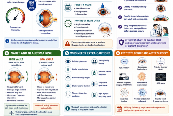
Raised Eye Pressure
Eye pressure may rise because of:
- Retained surgical viscoelastic
- Postoperative inflammation
- A steroid response
- Excessive vault
- Narrowing of the drainage angle
- Pupillary block
- Pre-existing glaucoma susceptibility
Eye pressure is therefore checked during the early postoperative period.
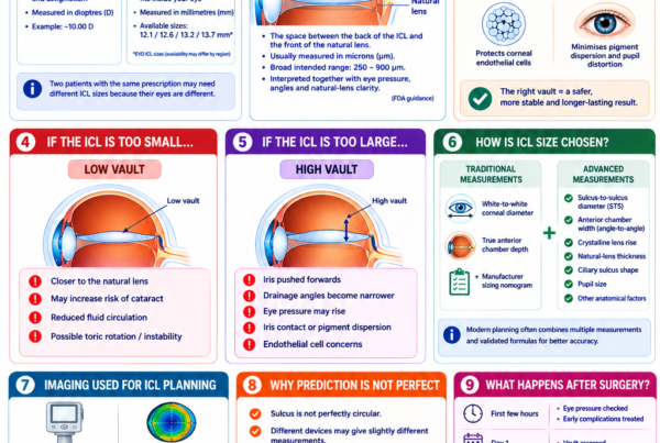
Incorrect Vault
Vault is the distance between the ICL and the natural lens.
A very low vault may increase concern about contact with the natural lens and cataract formation.
A very high vault can crowd the front of the eye and contribute to:
- Narrow drainage angles
- Raised eye pressure
- Iris contact
- Inflammation
Incorrect vault is an important reason for lens exchange in published ICL series.
Cataract
The ICL sits in front of the natural lens. Cataract may develop because of:
- Normal ageing
- Low vault
- Proximity to the natural lens
- Surgical trauma
- Other individual risk factors
Modern central-port ICL studies have reported favourable long-term cataract outcomes, but cataract risk cannot be eliminated.
Endothelial Cell Loss
The endothelial cells line the inner surface of the cornea and help keep it clear.
Long-term monitoring may include endothelial cell counts because excessive cell loss could eventually affect corneal clarity. Ten-year data for a central-port model showed a modest mean endothelial cell reduction in the studied cohort, but results from one series should not be applied identically to every patient.
Toric ICL Rotation
A toric ICL must remain aligned with the intended axis.
Significant rotation can produce:
- Residual astigmatism
- Blurred vision
- Ghosting
- Reduced visual quality
Surgical repositioning or lens exchange may occasionally be required.
Infection Inside the Eye
Endophthalmitis is a rare but potentially sight-threatening intraocular infection.
Symptoms may include:
- Increasing pain
- Increasing redness
- Rapidly worsening vision
- Marked light sensitivity
- Increasing discharge or swelling
Urgent treatment is required.
Inflammation
Mild inflammation is expected after surgery and usually responds to prescribed anti-inflammatory drops.
Persistent or severe inflammation requires investigation and additional treatment.
Need for Lens Exchange or Removal
Published series have reported lens exchange or removal for inappropriate vault, cataract, raised pressure, refractive error or dissatisfaction. One retrospective series reported exchange or explantation in approximately 2% of implanted lenses over seven years, but this is a study-specific figure and not a universal complication rate.
Which Procedure Has More Serious Risks?
LASIK risks mainly involve the:
- Cornea
- Corneal flap
- Tear film
- Quality and stability of the corneal optics
ICL risks mainly involve:
- Eye pressure
- The natural lens
- Corneal endothelial cells
- Lens sizing and vault
- Intraocular inflammation or infection
LASIK avoids entering the eye, which avoids the risks unique to intraocular surgery.
ICL avoids corneal ablation and flap creation, which avoids the structural and nerve-related risks unique to LASIK.
The probability and potential severity of complications depend on the patient’s anatomy and the quality of screening, planning, surgery and follow-up. Comparing procedures based only on the total number of listed complications is therefore misleading.
Which Procedure Is Safer?
Both can be safe when performed on appropriately selected patients.
A procedure may become less safe when used outside its ideal setting.
LASIK Becomes Less Attractive When:
- The cornea is thin or irregular
- Tomography suggests ectasia risk
- A high correction would require excessive tissue removal
- Significant dry eye remains uncontrolled
- The patient faces a high risk of direct eye trauma
- Corneal epithelial disease is present
ICL Becomes Less Attractive When:
- The anterior chamber is too shallow
- The drainage angle is narrow
- The endothelial cell count is inadequate
- The natural lens is already developing cataract
- Glaucoma or significant optic nerve damage is present
- The internal dimensions are unsuitable
- The patient is unwilling to attend long-term reviews
Reviews of moderate-to-high myopia have found phakic lenses at least comparable with excimer-laser correction and sometimes superior for certain safety, accuracy or visual-quality outcomes. However, much of the comparative evidence includes older lens and laser platforms, limited sample sizes and relatively short follow-up.
Is LASIK Safer Because It Is Not Inside the Eye?
LASIK avoids the risks of intraocular surgery, including:
- Endophthalmitis
- ICL-related cataract
- Incorrect vault
- ICL-related pressure elevation
- Endothelial-cell concerns related to the implant
- Lens rotation or exchange
However, LASIK introduces risks that ICL does not, including:
- Flap complications
- Corneal ectasia
- Permanent removal of corneal tissue
- Greater corneal nerve disruption
- Post-LASIK dry eye
The safest procedure is therefore the one whose risk profile best matches the patient’s eyes.
Which Procedure Is More Accurate?
Both procedures are generally predictable in appropriately selected patients.
Accuracy depends on:
- Measurement quality
- Prescription stability
- Surgeon planning
- Corneal shape
- Astigmatism
- Healing
- Lens sizing
- Toric alignment
- The degree of myopia
In moderate-to-high myopia, reviews have found phakic lenses at least as predictable as excimer-laser treatment and sometimes more accurate.
For low-to-moderate prescriptions with favourable corneas, modern LASIK can also provide highly accurate outcomes.
No calculation can guarantee an exact zero prescription. A small residual refractive error can remain after either procedure.
Which Is More Stable Over Time?
Both ICL and LASIK have demonstrated long-term stability, but the eye itself can continue to change.
After LASIK
Potential changes include:
- Myopic regression
- Age-related prescription changes
- Cataract development
- Presbyopia
- Changes in corneal shape or tear-film quality
Older ten-year LASIK studies reported mild regression, with greater change and more retreatment in patients treated for very high myopia.
After ICL
Potential changes include:
- Natural progression of myopia
- Vault changes
- Toric rotation
- Cataract development
- Presbyopia
- Retinal or macular changes related to high myopia
Central-port ICL studies have demonstrated favourable stability at eight and ten years, although some eyes still required lens exchange, laser enhancement or cataract treatment.
Does Either Procedure Stop Myopia From Progressing?
No.
ICL and LASIK correct the current refractive error. They do not stop the eye from elongating or prevent future prescription changes.
Surgery should generally be postponed until the prescription is reasonably stable.
Patients with high myopia remain at increased lifetime risk of:
- Retinal tears
- Retinal detachment
- Myopic macular degeneration
- Glaucoma
- Cataract
Correcting the spectacle prescription does not remove these structural risks.
Does Either Procedure Prevent Presbyopia?
No.
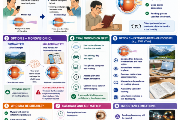
Presbyopia is the age-related loss of the natural lens’s ability to focus at near. It commonly becomes noticeable during the forties.
Both LASIK and standard ICL surgery can correct the eyes for clear distance vision, but reading glasses may eventually be required.
A myopic patient who can currently read by removing their glasses may lose that unaided near ability when both eyes are fully corrected for distance.
Possible strategies include:
- Reading glasses
- Monovision
- Leaving a small amount of myopia in one eye
- Other presbyopia-management options
A contact lens monovision trial is often useful before choosing permanent monovision.
Which Procedure Is Better After Age 40?
Age alone does not determine suitability, but the decision becomes more complex after 40.
The assessment should consider:
- Presbyopia
- Early cataract
- Natural-lens clarity
- Current unaided near vision
- Glare and night-driving symptoms
- Retinal health
- The expected years before cataract surgery may be required
ICL may be less attractive when the natural lens is already showing cataract changes, because the lens may need to be removed for cataract surgery within a relatively short period.
LASIK may correct distance vision but will not prevent reading-glasses dependence.
For some older patients with cataract or significant presbyopia, lens-replacement surgery may be more appropriate than either LASIK or ICL.
What Happens When Cataract Develops?
After ICL
The ICL is usually removed during or before cataract surgery. The cloudy natural lens is then removed and replaced with a cataract intraocular lens.
After LASIK
The patient can undergo cataract surgery, but previous LASIK changes the normal relationship between corneal curvature and focusing power.
Modern formulas and diagnostic devices have improved cataract-lens calculations after LASIK, but selecting the intraocular lens power can remain less straightforward than in an untreated cornea.
Patients should retain records of their pre-LASIK prescription and treatment where possible.
Which Procedure Requires More Long-Term Follow-Up?
ICL generally requires more implant-specific long-term surveillance.
Follow-up may assess:
- Vision and refraction
- Eye pressure
- ICL position
- Vault
- Drainage-angle anatomy
- Corneal endothelial cells
- The natural lens for cataract
- Retinal health
FDA patient information recommends ongoing monitoring of cataract formation, eye pressure and endothelial health while the ICL remains in the eye.
LASIK patients also require routine eye examinations, particularly for:
- Dry eye
- Refractive changes
- Corneal stability
- Cataract
- Glaucoma
- Retinal disease
However, there is no implanted lens requiring vault or endothelial surveillance.
Which Procedure Costs More?
ICL surgery is generally more expensive than LASIK because it involves:
- A customised implantable lens
- More detailed internal eye measurements
- Intraocular surgery
- Operating-theatre resources
- More extensive implant-related follow-up
A toric ICL may cost more than a spherical ICL.
Actual fees vary according to:
- The clinic and surgeon
- The lens model
- Whether astigmatism correction is required
- Whether one or both eyes are treated
- The investigations included
- Postoperative care
- Whether an enhancement is required
Cost should not be the sole factor when the procedures have substantially different suitability and risk profiles.
What Tests Are Needed Before Choosing?
A complete refractive surgery assessment may include:
Refraction
To determine:
- Myopia
- Astigmatism
- Best-corrected vision
- Prescription stability
Corneal Topography and Tomography
To assess:
- Corneal shape
- Corneal thickness
- Astigmatism
- Keratoconus risk
- Suitability for laser reshaping
Tear-Film and Eyelid Assessment
To identify:
- Dry eye disease
- Meibomian gland dysfunction
- Blepharitis
- Allergy
- Contact lens-related inflammation
Anterior Chamber Measurements
Particularly important for ICL planning:
- Anterior chamber depth
- White-to-white distance
- Sulcus or other internal dimensions
- Drainage-angle anatomy
- Lens sizing
Endothelial Cell Count
Required when considering ICL to ensure adequate corneal endothelial reserve.
Eye Pressure and Optic Nerve Assessment
To detect glaucoma or pressure-related risk.
Dilated Retinal Examination
Particularly important in moderate and high myopia to identify:
- Retinal holes
- Retinal tears
- Lattice degeneration
- Macular disease
- Other myopia-related changes
Pupil Size
Large pupils may influence the risk of night-vision symptoms after either procedure.
Who May Be Better Suited to LASIK?
LASIK may be more suitable when the patient:
- Has low-to-moderate myopia or astigmatism
- Has a thick, regularly shaped cornea
- Has normal corneal tomography
- Has a healthy and stable tear film
- Has a stable prescription
- Wishes to avoid an intraocular implant
- Prefers a lower-cost procedure
- Is unlikely to experience direct eye trauma
- Accepts the possibility of dry eye and flap-related complications
Who May Be Better Suited to ICL?
ICL may be more suitable when the patient:
- Has moderate, high or very high myopia
- Has myopia with astigmatism
- Has insufficient corneal tissue for the proposed laser correction
- Has corneal anatomy that makes LASIK less desirable
- Has significant concern about post-laser dry eye
- Wants to avoid removal of central corneal tissue
- Participates in activities where a corneal flap is undesirable
- Has sufficient internal eye dimensions
- Has an adequate endothelial cell count
- Has a clear natural lens
- Accepts intraocular surgical risks and long-term follow-up
Who May Be Unsuitable for Both?
Neither procedure may be appropriate when there is:
- An unstable prescription
- Active eye infection or inflammation
- Untreated severe dry eye
- Progressive keratoconus
- Significant cataract
- Retinal or optic nerve disease limiting vision
- Unrealistic expectations
- Pregnancy or breastfeeding with unstable measurements
- A systemic condition affecting healing
- Inadequate understanding of the risks and alternatives
Glasses and contact lenses remain valid alternatives. Choosing not to undergo elective surgery is also a reasonable decision.
Can You Have LASIK After ICL?
Possibly.
A small residual refractive error after ICL can sometimes be corrected with corneal laser surgery. This combined approach is sometimes called bioptics.
Suitability depends on:
- Corneal thickness and shape
- Residual prescription
- Tear-film health
- ICL position and vault
- Stability of the refraction
- The expected benefit
Laser enhancement should not be assumed before the ICL procedure. A separate assessment is required.
Can You Have ICL After LASIK?
In selected cases, yes.
A patient who has undergone LASIK may later develop a prescription that is not suitable for further corneal laser treatment. ICL may be considered if:
- The internal eye anatomy is suitable
- The natural lens is clear
- The endothelial cell count is adequate
- The residual prescription falls within the available ICL range
- Further corneal ablation is undesirable
Previous LASIK does not automatically prevent ICL implantation, but treatment planning becomes more individualised.
What Questions Should You Ask Your Surgeon?
Useful questions include:
- Why do you recommend ICL or LASIK for my eyes?
- What features of my cornea or internal eye anatomy influence the choice?
- How much corneal tissue would LASIK remove?
- Is my tomography completely normal?
- What is my expected ICL vault?
- Is my endothelial cell count adequate?
- What is my risk of dry eye?
- What visual result is realistic?
- What are the likely night-vision symptoms?
- What happens if a prescription remains?
- Would I be suitable for an enhancement?
- How will presbyopia affect me?
- What long-term follow-up will I require?
- How would future cataract surgery be managed?
- What symptoms require urgent review?
When Should You Seek Urgent Care After Surgery?
After either procedure, seek urgent assessment for:
- Severe or increasing pain
- Rapidly worsening vision
- Marked redness
- Significant light sensitivity
- Increasing discharge
- Difficulty opening the eye
- Nausea or vomiting with eye pain
- Severe headache
- A new white spot on the cornea
- Trauma to the operated eye
- New flashes or floaters
- A curtain-like shadow across the vision
Do not wait for the next routine appointment when symptoms are rapidly worsening.
Frequently Asked Questions About ICL vs LASIK
Is ICL Better Than LASIK?
Not for everyone.
ICL may be preferable for high myopia, thin corneas or when preservation of central corneal tissue is important. LASIK may be preferable for lower prescriptions with healthy corneas when the patient wishes to avoid intraocular surgery.
Is ICL Safer Than LASIK?
The procedures have different risks rather than a simple safer-versus-less-safe relationship.
ICL avoids flap complications, corneal ectasia from tissue removal and much of the corneal nerve disruption associated with LASIK.
LASIK avoids cataract, vault, endothelial and intraocular-infection risks related to an implant inside the eye.
Does ICL Give Sharper Vision Than LASIK?
Both can provide excellent vision.
ICL may provide advantages in contrast sensitivity and higher-order aberrations for higher myopic prescriptions, but individual results vary.
Is LASIK More Accurate Than ICL?
Both are highly predictable in appropriately selected patients.
ICL may have an accuracy advantage in moderate-to-high myopia, while LASIK is highly accurate for many low-to-moderate corrections.
Does ICL Cause Less Dry Eye?
ICL generally disrupts fewer central corneal nerves and may be preferable for some patients with dry-eye risk.
However, it does not cure dry eye, and pre-existing tear-film or eyelid disease should still be treated.
Is LASIK Recovery Faster?
Both procedures can provide useful vision within the first day. LASIK may involve less intraocular monitoring, while ICL may require closer early checks of eye pressure, lens position and inflammation.
Is ICL Truly Reversible?
The lens can usually be removed or exchanged, but removal requires another intraocular operation. The procedure should therefore be described as removable rather than completely reversible.
Can ICL Cause Cataract?
Yes, although modern central-port lenses have improved the safety profile.
Cataract risk is influenced by age, vault, anatomy, surgical factors and the duration of follow-up.
Can LASIK Cause Keratoconus?
LASIK does not usually cause true inherited keratoconus, but it can contribute to progressive corneal ectasia in susceptible or inadequately screened eyes.
Careful tomography and tissue planning reduce this risk.
Will Either Procedure Last Forever?
The physical LASIK reshaping and the implanted ICL are intended to provide long-term correction.
However, the eye can continue to change because of:
- Myopia progression
- Presbyopia
- Cataract
- Retinal disease
- Other age-related changes
Future glasses or further treatment may still be required.
Which Is Better for Night Driving?
This depends on prescription, pupil size, optical quality, residual refractive error and healing.
ICL may preserve contrast sensitivity better in high myopia, while both procedures can cause temporary or persistent halos and glare.
Which Is Better for Athletes?
ICL avoids a corneal flap and may be advantageous for contact-sport athletes.
LASIK may still be suitable for many non-contact athletes. Protective eyewear and adequate healing time remain important after either procedure.
Can I Choose Based Only on My Prescription?
No.
Two patients with the same prescription may require different procedures because of differences in:
- Corneal shape
- Corneal thickness
- Tear-film health
- Anterior chamber dimensions
- Endothelial cell count
- Pupil size
- Retinal health
- Age and lifestyle
Key Takeaway
ICL and LASIK are both effective forms of vision-correction surgery, but they use different parts of the eye.
LASIK reshapes the cornea and avoids placing a lens inside the eye. It is often well suited to patients with low-to-moderate prescriptions, healthy corneas and a stable eye surface.
ICL places a corrective lens behind the iris without removing central corneal tissue. It is often advantageous for patients with moderate-to-very-high myopia, thin or laser-unsuitable corneas, significant dry-eye concerns or lifestyles in which a corneal flap is undesirable.
ICL avoids flap, corneal-ablation and ectasia-related concerns but introduces risks involving vault, eye pressure, cataract, endothelial cells and intraocular infection.
LASIK avoids implant-related and intraocular risks but introduces risks involving dry eye, the corneal flap, corneal stability and permanent tissue removal.
The best procedure is not determined by which technology appears more advanced. It is the procedure with the most favourable balance of benefits and risks for the individual eye after comprehensive assessment.
References
- Goes S, et al. Posterior Chamber Toric Implantable Collamer Lenses Versus LASIK for Myopic Astigmatism: A Systematic Review. 2022. PubMed.
- Barsam A, Allan BDS. Excimer Laser Refractive Surgery Versus Phakic Intraocular Lenses for the Correction of Moderate to High Myopia. Cochrane systematic review. PubMed.
- Chen H, et al. Excimer Laser Versus Phakic Intraocular Lenses for Myopia and Astigmatism: A Meta-analysis of Randomised Controlled Trials. PubMed.
- Barsam A, Allan BDS. Meta-analysis of Randomised Controlled Trials Comparing Excimer Laser and Phakic Intraocular Lenses. PubMed.
- Igarashi A, et al. Visual Performance After Implantable Collamer Lens Implantation and Wavefront-Guided LASIK for High Myopia. PubMed.
- Kamiya K, et al. Posterior Chamber Phakic Intraocular Lens Implantation for Low-to-Moderate and High Myopia. PubMed.
- Alfonso-Bartolozzi B, et al. Ten-Year Follow-up of Posterior Chamber Phakic Intraocular Lenses With Central-Port Design. PubMed.
- Kamiya K, et al. Eight-Year Outcomes of Posterior Chamber Phakic Intraocular Lens Implantation With Central-Port Technology. PubMed.
- Fernández-Vega-Cueto L, et al. Seven-Year Follow-up of Posterior Chamber Phakic Intraocular Lens With Central Port. PubMed.
- Alhamzah A, et al. Indications for Exchange or Explantation of Phakic Implantable Collamer Lenses With a Central Port. PubMed.
- Alió JL, et al. Ten-Year Follow-up of LASIK for Myopia of Up to −10 Dioptres. PubMed.
- Alió JL, et al. Ten-Year Follow-up of LASIK for High Myopia. PubMed.
- Dossari SK, et al. Post-Refractive Surgery Dry Eye: A Systematic Review. 2024. PubMed.
- Moshirfar M, et al. Ectasia After Corneal Refractive Surgery: A Systematic Review. PubMed.
- Sahay P, et al. Complications of Laser-Assisted In Situ Keratomileusis. PubMed.
- Eydelman M, et al. Symptoms and Satisfaction of Patients in the Patient-Reported Outcomes With LASIK Studies. PubMed.
- Lee MD, et al. Patient-Reported Outcomes With Wavefront-Guided Laser Vision Correction. PubMed.
- US Food and Drug Administration. EVO/EVO+ Visian Implantable Collamer Lens Patient Information.
- US Food and Drug Administration. ZEISS MEL 90 LASIK Patient Information Booklet.
- US Food and Drug Administration. WaveLight EX500 LASIK Patient Information Booklet.