Author: Dr Val Phua
Estimated reading time: 14–16 minutes
What Is Glaucoma Surgery?
Glaucoma surgery lowers the pressure inside the eye to reduce the risk of further optic-nerve damage.
The operation may:
- Create a new drainage pathway
- Bypass a blocked part of the natural drainage system
- Open the existing drainage channel
- Insert a tube or microscopic stent
- Reduce the amount of aqueous fluid produced
- Combine glaucoma treatment with cataract surgery
Glaucoma surgery cannot normally:
- Restore optic-nerve fibres already lost
- Reverse an established visual-field defect
- Cure glaucoma permanently
- Guarantee that eye drops will never be required
- Remove the need for long-term monitoring
Its purpose is to protect the vision that remains.
The National Eye Institute advises that surgery cannot undo vision loss, but it can help protect vision and prevent further deterioration when medication or laser has not provided adequate control.
Why Does Glaucoma Damage Vision?
Glaucoma progressively damages the optic nerve, which carries visual information from the retina to the brain.
Eye pressure is the main modifiable risk factor.
The appropriate pressure differs between patients. A pressure that is acceptable for an eye with early, stable disease may be too high for an eye with:
- Advanced visual-field loss
- Damage close to central fixation
- Rapid OCT progression
- Previous loss in the other eye
- Normal-tension glaucoma
- A long remaining life expectancy
The ophthalmologist therefore establishes an individual target pressure.
Glaucoma surgery is considered when the pressure cannot be lowered sufficiently—or kept reliably low—using less invasive treatment.
When Is Glaucoma Surgery Needed?
Surgery may be recommended when:
- Glaucoma continues progressing despite eye drops
- Laser treatment has not achieved the target pressure
- The pressure remains dangerously high
- The glaucoma is advanced at diagnosis
- Visual-field loss is approaching central fixation
- Medication causes significant eye or systemic side effects
- The patient cannot use drops reliably
- Several medications are required but remain insufficient
- The drainage angle is permanently scarred or closed
- Previous glaucoma treatment has failed
- Secondary glaucoma is unlikely to respond adequately to drops alone
- A child has congenital or developmental glaucoma
Surgery should not always be viewed as a treatment of last resort.
The five-year Treatment of Advanced Glaucoma Study found that primary trabeculectomy produced lower pressure and better protection against disease progression than beginning with medication in patients who presented with advanced open-angle glaucoma, with a similar overall safety profile.
Does Surgery Mean the Glaucoma Is Very Severe?
Not necessarily.
Some operations are offered for mild or moderate glaucoma to:
- Reduce medication dependence
- Treat glaucoma during cataract surgery
- Avoid long-term preservative exposure
- Improve adherence
- Intervene before severe damage develops
Other operations, particularly trabeculectomy and glaucoma drainage implants, are more commonly used when:
- A low target pressure is required
- Disease is advanced
- Previous treatments have failed
- The glaucoma mechanism is complex
The type of surgery provides more information than the word “surgery” alone.
What Are the Main Types of Glaucoma Surgery?
The principal operations include:
- Trabeculectomy
- Glaucoma drainage implant surgery
- Minimally invasive glaucoma surgery
- Subconjunctival microshunt surgery
- Cataract or lens extraction for angle closure
- Cyclophotocoagulation
- Goniotomy or trabeculotomy for childhood glaucoma
These procedures lower pressure through different pathways and are not interchangeable.
Trabeculectomy
Trabeculectomy is a traditional filtering operation used to create a new drainage pathway from inside the eye to the space beneath the upper eyelid.
It remains one of the most effective operations for achieving a low eye pressure.
The National Eye Institute describes trabeculectomy as an operation in which a small opening is created beneath the upper eyelid so that excess aqueous fluid can drain away.
How Does Trabeculectomy Work?
During trabeculectomy, the surgeon creates:
- A small opening beneath the conjunctiva—the transparent tissue covering the white of the eye.
- A partial-thickness flap in the sclera.
- A controlled internal opening through which aqueous fluid can leave the anterior chamber.
- Sutures that regulate how much fluid flows beneath the flap.
The fluid collects in a small reservoir beneath the upper eyelid called a filtering bleb.
From the bleb, aqueous fluid is absorbed into surrounding tissues and blood vessels.
The bleb is usually hidden beneath the upper eyelid and is not normally noticeable during ordinary conversation.
What Is a Filtering Bleb?
A filtering bleb is an area where aqueous fluid collects beneath the conjunctiva after filtration surgery.
A healthy bleb allows controlled drainage while maintaining enough pressure to preserve the eye’s structure.
A bleb may appear:
- Slightly raised
- Pale or translucent
- Diffuse
- Located beneath the upper eyelid
A bleb is not a tumour or blister filled with pus.
It is the intended external drainage reservoir created by the surgery.
Why Is Mitomycin C Used?
The body naturally attempts to heal surgical wounds by producing scar tissue.
Excessive scarring may seal the new drainage channel and cause the pressure to rise again.
Antiscarring medication may therefore be applied during surgery.
The most commonly used agent is:
- Mitomycin C
Another agent used in selected situations is:
- 5-fluorouracil
These medications reduce fibroblast activity and improve the likelihood that the drainage pathway remains open.
The concentration and duration are selected according to factors including:
- Age
- Ethnicity
- Previous eye surgery
- Inflammation
- Conjunctival scarring
- Type of glaucoma
- The pressure target
A stronger antiscarring effect may improve filtration but may also increase the risk of:
- A thin bleb
- Leakage
- Excessively low pressure
- Late bleb infection
Who May Benefit from Trabeculectomy?
Trabeculectomy may be considered when:
- Open-angle glaucoma is advanced
- A low target pressure is required
- Visual-field damage is progressing
- Eye drops and laser are insufficient
- Medication cannot be tolerated
- The patient is young with severe disease
- Pressure remains high despite several treatments
- The conjunctiva remains suitable for filtration surgery
Trabeculectomy may also be used for selected:
- Angle-closure glaucoma
- Normal-tension glaucoma
- Uveitic glaucoma
- Juvenile glaucoma
- Secondary glaucoma
The suitability depends on the individual eye.
How Low Can Trabeculectomy Lower the Pressure?
Trabeculectomy often achieves lower pressures than angle-based minimally invasive procedures.
It can sometimes produce pressures in the low teens or even below, which may be required for:
- Advanced glaucoma
- Rapid progression
- Central-field involvement
- Normal-tension glaucoma progressing at low pressures
The final result is not completely predictable.
The eye must maintain a balance between:
- Sufficient drainage to protect the optic nerve
- Sufficient pressure to maintain normal eye anatomy and vision
How Effective Is Trabeculectomy?
Success depends on:
- Definition of success
- Target pressure
- Previous surgery
- Glaucoma type
- Scarring response
- Follow-up duration
- Need for additional medication
The National Eye Institute states that trabeculectomy lowers pressure successfully in approximately seven out of ten people, although outcomes vary according to the eye and the definition used.
The five-year TAGS trial found that trabeculectomy provided better pressure reduction and protection against progression than primary medication in patients presenting with advanced glaucoma.
What Happens After Trabeculectomy?
Postoperative care is a major part of the treatment.
The eye may require:
- Frequent steroid drops
- Antibiotic drops for a limited period
- Pressure checks
- Adjustment of glaucoma medication
- Removal or release of flap sutures
- Laser suture lysis
- Bleb massage
- Antiscarring injections
- Needling of scar tissue
- Additional sutures if pressure is too low
The final drainage level is actively adjusted during healing.
The operation should therefore not be thought of as completed the moment the patient leaves the operating theatre.
What Is Laser Suture Lysis?
Sutures in the scleral flap regulate aqueous drainage.
If pressure remains too high because the flap is too tightly closed, the surgeon may use a laser to cut one or more sutures through the conjunctiva.
This allows more fluid to pass beneath the flap.
Laser suture lysis is commonly performed in the clinic after numbing drops have been applied.
What Is Bleb Massage?
The surgeon may gently press the eye to encourage fluid to pass through the new drainage pathway.
Selected patients may be taught a specific massage technique.
Bleb massage should only be performed when instructed.
Excessive or incorrect pressure may cause:
- Excessive drainage
- Low pressure
- Bleeding
- Injury to the eye
What Is Bleb Needling?
Scar tissue may form around a trabeculectomy or microshunt bleb and restrict drainage.
Bleb needling uses a fine needle to release scar tissue and restore aqueous flow.
An antiscarring medication may be injected at the same time.
Needling may be performed:
- Soon after surgery
- Months later
- Years later
Moorfields Eye Hospital notes that needling may be required when scar tissue reduces drainage after trabeculectomy, microshunt or selected tube procedures.
What Are the Main Risks of Trabeculectomy?
Possible complications include:
- Temporary blurred vision
- Eye redness
- Grittiness
- Inflammation
- Pressure that remains too high
- Pressure that becomes too low
- Shallow anterior chamber
- Choroidal detachment
- Bleeding
- Bleb leakage
- Cataract progression
- Astigmatism or prescription change
- Corneal problems
- Infection
- Need for further surgery
- Permanent visual loss in rare cases
The National Eye Institute lists cataract, corneal problems, excessively low pressure and visual loss among recognised glaucoma-surgery risks.
What Is Hypotony?
Hypotony means that the eye pressure is too low.
This may occur when:
- Too much fluid drains
- The surgical wound leaks
- The ciliary body temporarily produces less fluid
- Inflammation is present
- The scleral flap is too loose
Mild early hypotony may resolve without permanent harm.
More significant hypotony can cause:
- Blurred or distorted vision
- A shallow anterior chamber
- Choroidal detachment
- Retinal or macular folds
- Hypotony maculopathy
- Cataract-related problems
- Permanent visual reduction
Treatment may include:
- Adjusting medication
- Pressure patching
- Additional sutures
- Injection of viscoelastic material
- Repair of a wound leak
- Surgical revision
Can the Pressure Remain Too High After Surgery?
Yes.
Possible causes include:
- Early inflammation
- Blood or debris blocking the opening
- Tight flap sutures
- Steroid response
- Rapid scar formation
- Encapsulation of the bleb
- Long-term closure of the drainage pathway
Management may include:
- Medication
- Bleb massage
- Laser suture lysis
- Antiscarring injection
- Needling
- Surgical revision
- Another glaucoma operation
Scarring is one of the principal reasons filtering surgery loses effectiveness.
What Is a Bleb Leak?
A bleb leak occurs when aqueous fluid escapes through a defect in the conjunctival surface.
A leak may occur:
- Soon after surgery
- Months or years later
- After trauma
- When a bleb becomes very thin
Possible symptoms include:
- Excessive watering
- Irritation
- Blurred vision
- Low pressure
- A foreign-body sensation
A leaking bleb increases the risk of infection and requires assessment.
Can a Bleb Become Infected?
Yes.
Bleb infection can occur months or years after filtration surgery.
The infection may remain limited to the bleb, called blebitis, or spread into the eye, causing bleb-related endophthalmitis.
Endophthalmitis can cause rapid and severe visual loss.
Warning symptoms include:
- Increasing redness
- Eye pain
- Light sensitivity
- Discharge
- Sudden blurred or reduced vision
- A white or yellow appearance within the bleb
- Eyelid swelling
These symptoms require immediate, preferably same-day, ophthalmic assessment.
Moorfields Eye Hospital emphasises that a bleb-related infection can spread into the eye and cause blindness unless treated promptly.
Is the Infection Risk Lifelong?
Yes.
A functioning filtering bleb remains present after the eye has healed.
Patients should remember that a painful red eye in an eye with a trabeculectomy bleb is never something to observe for several days without assessment.
Ordinary conjunctivitis can potentially spread into a vulnerable bleb.
Patients should tell any new eye-care provider that they have had filtering glaucoma surgery.
Can Contact Lenses Be Worn After Trabeculectomy?
Contact-lens wear over or near a bleb may cause:
- Friction
- Bleb breakdown
- Leakage
- Infection
- Discomfort
Conventional contact-lens wear is therefore often avoided or approached cautiously after bleb-forming surgery.
Specialist fitting may occasionally be considered when the visual need is substantial and the bleb position permits it.
The glaucoma and contact-lens teams should assess the risk together.
Glaucoma Drainage Implant Surgery
A glaucoma drainage implant creates a new pathway through a small tube attached to a plate positioned on the white of the eye.
These devices are also called:
- Tube shunts
- Aqueous shunts
- Glaucoma drainage devices
- Glaucoma valves
The tube carries aqueous fluid from inside the eye to a reservoir around the plate.
The fluid is then absorbed by surrounding tissues.
The National Eye Institute notes that tube implants are used for several glaucoma types, including congenital, neovascular, traumatic and other complex glaucomas.
What Are the Main Types of Tube Implant?
Common devices include:
- Ahmed glaucoma valve
- Baerveldt glaucoma implant
- Other non-valved or valved drainage implants
Valved Implant
A valved device is designed to restrict flow when pressure becomes low.
The Ahmed valve is a common example.
Non-Valved Implant
A non-valved implant does not contain a mechanical valve.
The tube may initially be tied closed with a dissolvable ligature to allow scar tissue to form around the plate before full flow begins.
The Baerveldt implant is a common example.
How Does Tube Surgery Differ from Trabeculectomy?
Both operations create an alternative drainage route.
Trabeculectomy
- Uses a scleral flap
- Creates a filtering bleb directly over the surgical opening
- Has no permanent tube or plate
- Often achieves very low pressures
- Requires intensive bleb management
Drainage Implant
- Uses a permanent tube and plate
- Directs fluid to a reservoir farther back under the eyelid
- May be preferred in scarred or complex eyes
- May have a delayed full effect with non-valved devices
- Carries device-specific risks
Neither operation is universally superior.
Who May Benefit from a Drainage Implant?
Tube surgery may be considered for:
- Previous failed trabeculectomy
- Previous conjunctival surgery
- Neovascular glaucoma
- Uveitic glaucoma
- Traumatic glaucoma
- Congenital or childhood glaucoma
- Aphakic glaucoma
- Glaucoma after corneal transplantation
- Eyes with extensive conjunctival scarring
- Selected primary open-angle glaucoma
- Complex secondary glaucoma
Tube implants are increasingly also used as primary incisional surgery in selected eyes without previous surgery.
What Is the Hypertensive Phase?
Some eyes—particularly after a valved implant—develop a period of increased pressure several weeks after surgery.
This may occur as the capsule around the drainage plate thickens during healing.
The hypertensive phase may require:
- Temporary glaucoma medication
- Continued observation
- Adjustment of steroid treatment
- Needling or revision in selected cases
The pressure may later improve as the capsule remodels.
Why Does a Non-Valved Tube Take Time to Work?
A non-valved tube may be tied closed temporarily so that a protective capsule develops around the plate before aqueous begins flowing freely.
During this period:
- Pressure may remain high.
- Glaucoma drops may still be needed.
- A small temporary opening or internal stent may allow limited early drainage.
When the ligature dissolves, pressure may fall relatively suddenly.
Close monitoring is required because the eye can become too soft when the tube opens.
What Are the Risks of Tube Surgery?
Possible complications include:
- Temporary blurred vision
- Inflammation
- Bleeding
- Pressure that remains too high
- Excessively low pressure
- Shallow anterior chamber
- Choroidal detachment
- Tube blockage
- Tube displacement
- Tube exposure through the conjunctiva
- Corneal endothelial-cell loss
- Corneal decompensation
- Double vision
- Cataract
- Infection
- Need for revision or further surgery
A tube that becomes exposed can allow microorganisms to enter the eye and requires urgent repair.
Can the Tube Damage the Cornea?
Yes.
A tube placed in the anterior chamber may come close to the corneal endothelium—the cells maintaining corneal clarity.
Long-term endothelial-cell loss may contribute to:
- Corneal swelling
- Reduced vision
- Need for corneal transplantation
Risk may be greater when:
- The tube sits close to the cornea
- The anterior chamber is shallow
- A corneal graft is present
- The endothelium is already weak
- The tube moves with eye rubbing or blinking
Alternative tube positions may be considered in selected eyes.
Can a Tube Cause Double Vision?
Yes, although this is uncommon.
The plate and surrounding scar tissue may affect the movement of an eye muscle.
Double vision may be:
- Temporary
- Position-dependent
- Persistent
Management may include:
- Observation
- Prism glasses
- Strabismus treatment
- Implant revision in selected cases
Trabeculectomy Versus Tube Implant
The optimal choice depends on:
- Previous eye surgery
- Conjunctival scarring
- Target pressure
- Corneal health
- Glaucoma type
- Age
- Risk of hypotony
- Surgeon experience
- Ability to attend postoperative follow-up
The five-year Primary Tube Versus Trabeculectomy Study found similar overall failure rates between primary tube implantation and trabeculectomy in eyes without previous incisional surgery. Trabeculectomy required fewer glaucoma medications but had more early postoperative complications.
The earlier Tube Versus Trabeculectomy Study, which involved eyes with previous cataract or glaucoma surgery, found a higher five-year success rate with tube implantation, although both operations achieved pressure in the low teens.
These studies do not establish one operation as best for every patient. They demonstrate that the eye’s previous surgical history matters.
Minimally Invasive Glaucoma Surgery
Minimally invasive glaucoma surgery, or MIGS, refers to a group of procedures designed to lower pressure with less tissue disruption and a faster recovery than traditional filtering surgery.
MIGS generally has:
- A favourable safety profile
- Small internal incisions
- Limited manipulation of the conjunctiva
- Faster visual recovery
- Moderate pressure-lowering ability
The 2026 American Academy of Ophthalmology guideline notes that MIGS usually provides less pressure reduction than trabeculectomy or aqueous-shunt surgery but has a more favourable safety profile.
Is MIGS One Operation?
No.
MIGS is a broad category.
Procedures may:
- Bypass the trabecular meshwork
- Remove or open trabecular tissue
- Dilate Schlemm’s canal
- Insert a trabecular microstent
- Access the suprachoroidal pathway
- Drain beneath the conjunctiva
Different procedures have different:
- Mechanisms
- Pressure-lowering effects
- Safety profiles
- Regulatory approvals
- Evidence bases
The name of the specific operation matters.
Trabecular Microstents
Trabecular stents create a small bypass through the trabecular meshwork into Schlemm’s canal.
Examples include:
- iStent devices
- Hydrus Microstent
These are commonly implanted during cataract surgery in patients with open-angle glaucoma.
They may:
- Reduce eye pressure modestly
- Reduce the number of glaucoma drops
- Improve access to the natural drainage system
- Preserve the conjunctiva for future surgery
Goniotomy and Trabeculotomy Procedures
These procedures open or remove part of the trabecular meshwork.
Examples include:
- Goniotomy
- Kahook Dual Blade goniotomy
- Gonioscopy-assisted transluminal trabeculotomy
- Ab interno trabeculotomy
They aim to improve access from the anterior chamber to Schlemm’s canal.
Possible temporary effects include:
- Blood reflux into the anterior chamber
- Blurred vision
- Pressure fluctuation
- Inflammation
Canaloplasty
Canaloplasty procedures dilate or tension Schlemm’s canal to improve drainage through the natural outflow pathway.
They may involve:
- A microcatheter
- Viscoelastic injection
- Internal suture placement
- An ab interno or external approach
The expected effect varies according to the procedure.
Who Is a Good Candidate for Angle-Based MIGS?
MIGS is commonly considered for patients with:
- Mild or moderate open-angle glaucoma
- A cataract requiring surgery
- Pressure controlled with several medications
- Medication intolerance
- Difficulty using drops
- A target pressure in the mid or upper teens
- Relatively preserved visual fields
It may also be considered as a standalone treatment in selected circumstances, depending on the device and local approval.
Who May Not Be Suitable for Angle-Based MIGS?
Angle-based MIGS may be insufficient when:
- Glaucoma is advanced
- A very low target pressure is required
- Pressure is extremely high
- The drainage angle is closed
- Extensive angle scarring is present
- Neovascular tissue obstructs the angle
- Previous angle surgery has failed
- Rapid progression requires more powerful pressure reduction
Trabecular MIGS relies on the downstream natural drainage system.
It cannot generally lower pressure below the level permitted by episcleral venous pressure and the remaining drainage channels.
How Effective Is MIGS?
The effect differs between devices.
In general, angle-based procedures:
- Produce a modest additional pressure reduction
- Reduce medication burden
- Have a lower risk of severe hypotony than filtering surgery
- Are often combined with cataract surgery
An American Academy of Ophthalmology report concluded that trabecular procedures combined with cataract surgery provide a mild additional pressure reduction over cataract surgery alone in hypertensive open-angle glaucoma.
The five-year HORIZON trial found that cataract surgery combined with a Schlemm’s canal microstent provided more durable pressure and medication control than cataract surgery alone in patients with mild-to-moderate open-angle glaucoma and cataract.
Does MIGS Replace Trabeculectomy?
No.
MIGS fills a treatment gap between:
- Eye drops or laser
- Traditional filtering surgery
MIGS is attractive when the goal is:
- Modest pressure reduction
- Fewer medications
- Faster recovery
- Lower surgical risk
Trabeculectomy or tube surgery remains more appropriate when:
- The target pressure is very low
- Glaucoma is advanced
- The visual field is deteriorating rapidly
- Previous lower-risk treatment has failed
Can MIGS Be Combined with Cataract Surgery?
Yes.
This is one of its most common uses.
The surgeon may:
- Remove the cataract.
- Implant an intraocular lens.
- Perform the glaucoma procedure through the same or an additional small incision.
Combining surgery may:
- Restore cataract-related vision
- Lower pressure
- Reduce medication use
- Avoid a second operation
However, the cataract and glaucoma parts have separate goals.
The patient should understand whether the glaucoma procedure is expected to:
- Reduce drops
- Lower pressure modestly
- Prevent future escalation
- Reach a specific target
Will Cataract Surgery Alone Lower Eye Pressure?
Cataract surgery often lowers pressure modestly, particularly when the starting pressure is elevated or the drainage angle is crowded.
The effect is not predictable enough to replace glaucoma surgery when:
- Very low pressure is required
- Glaucoma is advanced
- Progression is rapid
- Several medications are already insufficient
The additional benefit of MIGS should be assessed against cataract surgery alone.
Subconjunctival Microshunts
Subconjunctival microshunts create a pathway from the anterior chamber to a bleb beneath the conjunctiva.
Examples include:
- XEN Gel Stent
- PreserFlo MicroShunt
They are less invasive than conventional trabeculectomy in some respects but still create a filtering bleb.
They should therefore not be grouped uncritically with angle-based MIGS.
How Does a Microshunt Work?
A small tube is positioned so that:
- One end sits within the anterior chamber.
- The device passes through the eye wall.
- Aqueous fluid drains beneath the conjunctiva.
- A filtering bleb develops.
Antiscarring medication is usually required.
The operation may involve:
- An internal approach
- An external conjunctival approach
- Postoperative bleb needling
- Antiscarring injections
Who May Benefit from a Subconjunctival Microshunt?
A microshunt may be considered when:
- Greater pressure reduction is required than angle MIGS can provide
- The surgeon wishes to avoid a traditional scleral flap
- Open-angle glaucoma is inadequately controlled
- Medication reduction is important
- The conjunctiva is suitable
The exact role depends on:
- Device
- Regulatory availability
- Target pressure
- Glaucoma severity
- Surgeon experience
Are Microshunts as Effective as Trabeculectomy?
Not necessarily.
A five-year randomised comparison found that both PreserFlo MicroShunt implantation and trabeculectomy reduced pressure and medication use, but the probability of surgical success was lower with the microshunt. Microshunt surgery generally produced fewer or less severe early complications but did not achieve the same degree of pressure control as trabeculectomy in all patients.
Microshunts may be appropriate when the target is around the mid-teens, while trabeculectomy may remain preferable when very low pressure is required.
Can a Microshunt Bleb Scar?
Yes.
Like a trabeculectomy, a subconjunctival shunt depends on controlled wound healing.
Scarring may cause:
- Rising pressure
- Reduced drainage
- Bleb encapsulation
- Need for antiscarring injections
- Needling
- Revision
- Further surgery
Long-term follow-up remains necessary.
Cataract or Lens Surgery for Angle-Closure Glaucoma
The natural lens becomes thicker and moves forward with age.
In anatomically crowded eyes, the lens may push the iris towards the trabecular meshwork and narrow the drainage angle.
Removing the lens can:
- Deepen the anterior chamber
- Widen the angle
- Reduce pupillary-block crowding
- Lower pressure
- Treat cataract
- Reduce the risk of future angle closure
The 2026 AAO angle-closure guideline recommends considering lens extraction or incisional glaucoma surgery when the angle remains closed and pressure is not controlled adequately after laser or medication.
Does Lens Surgery Cure Angle-Closure Glaucoma?
No.
Lens removal improves the anatomy but does not reverse:
- Existing optic-nerve damage
- Permanent angle adhesions
- Trabecular-meshwork damage
- Visual-field loss
Some patients continue to require:
- Glaucoma drops
- Goniosynechialysis
- MIGS
- Trabeculectomy
- Tube surgery
- Continued OCT and visual-field monitoring
What Is Goniosynechialysis?
Peripheral anterior synechiae are permanent adhesions between the iris and the drainage angle.
During goniosynechialysis, the surgeon mechanically separates selected adhesions to expose the trabecular meshwork.
It is often combined with:
- Cataract surgery
- Angle surgery
- Other glaucoma procedures
The likelihood of success depends partly on how long the angle has been closed and how healthy the trabecular meshwork remains.
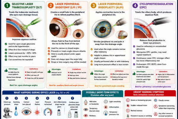
Cyclophotocoagulation
Cyclophotocoagulation treats the ciliary body to reduce aqueous-fluid production.
Forms include:
- Continuous-wave transscleral cyclophotocoagulation
- Slow-coagulation treatment
- Micropulse transscleral treatment
- Endoscopic cyclophotocoagulation
It may be considered for:
- Refractory glaucoma
- Painful high-pressure eyes
- Eyes with previous failed surgery
- Neovascular glaucoma
- Eyes unsuitable for conventional filtration
- Selected eyes with useful vision
Cyclophotocoagulation is discussed separately from drainage surgery because it reduces fluid production rather than creating a new outflow pathway.
Childhood Glaucoma Surgery
Medication usually provides only temporary support in primary congenital glaucoma.
Surgery is the main treatment.
Common childhood procedures include:
- Goniotomy
- Trabeculotomy
- Circumferential trabeculotomy
- Glaucoma drainage implants
- Trabeculectomy in selected cases
- Cyclophotocoagulation for refractory disease
Angle surgery is generally the preferred initial treatment for primary congenital glaucoma when the anatomy permits it.
Children require long-term monitoring because:
- The eye continues growing.
- Pressure may become difficult to measure.
- Amblyopia may develop.
- Corneal enlargement and scarring affect vision.
- Additional surgery may be required.
How Is the Type of Surgery Chosen?
The decision depends on:
- Glaucoma type
- Open or closed drainage angle
- Target pressure
- Disease severity
- Rate of progression
- Previous eye surgery
- Conjunctival scarring
- Cataract status
- Corneal health
- Age
- General health
- Medication tolerance
- Ability to attend frequent follow-up
- Patient preference
Mild or Moderate Open-Angle Glaucoma with Cataract
Possible options include:
- Cataract surgery alone
- Cataract surgery with trabecular stent
- Cataract surgery with goniotomy
- Cataract surgery with canaloplasty
- Cataract surgery with another MIGS procedure
Advanced Open-Angle Glaucoma
Possible options include:
- Trabeculectomy
- Glaucoma drainage implant
- Subconjunctival microshunt in selected cases
- Cyclophotocoagulation in selected eyes
Previous Failed Trabeculectomy or Scarred Conjunctiva
A glaucoma drainage implant may be favoured.
Neovascular or Complex Secondary Glaucoma
Possible treatments include:
- Drainage implant
- Cyclophotocoagulation
- Trabeculectomy in selected circumstances
- Treatment of the underlying retinal ischaemia or inflammation
Narrow-Angle or Angle-Closure Glaucoma
Possible treatments include:
- Laser iridotomy
- Cataract or lens extraction
- Goniosynechialysis
- Trabeculectomy
- Tube implant
- Combined procedures
What Happens Before Glaucoma Surgery?
The preoperative assessment may include:
- Visual acuity
- Eye-pressure measurement
- Gonioscopy
- Corneal examination
- Optic-nerve evaluation
- OCT
- Visual-field review
- Cataract assessment
- Conjunctival examination
- Review of previous surgery
- General medical history
- Medication review
- Anaesthetic assessment
Patients should disclose:
- Blood-thinning medication
- Asthma
- Cardiac disease
- Diabetes
- Kidney disease
- Steroid use
- Previous eye infection
- Previous LASIK, PRK or SMILE
- Allergy to medication
- Difficulty lying flat
- Pregnancy or breastfeeding
Should Blood Thinners Be Stopped?
Not automatically.
The risks of stopping anticoagulant or antiplatelet medication may be greater than the surgical bleeding risk.
The ophthalmologist and prescribing physician should consider:
- Reason for the blood thinner
- Previous stroke or heart attack
- Cardiac stents
- Atrial fibrillation
- Type of surgery
- Anaesthesia
- Individual bleeding risk
Patients should never stop these medications independently.
What Type of Anaesthesia Is Used?
Many adult glaucoma operations are performed using:
- Local anaesthesia around or within the eye
- Intravenous sedation
- Topical anaesthesia for selected MIGS procedures
General anaesthesia may be used for:
- Children
- Patients unable to remain still
- Severe anxiety
- Certain complex operations
- Particular medical or communication needs
The patient is usually awake but should not feel sharp pain.
How Long Does the Operation Take?
The duration depends on the procedure.
Approximate operating times vary widely:
- A simple MIGS procedure may add only a short time to cataract surgery.
- Trabeculectomy often takes less than an hour.
- Tube implantation may take one to two hours.
- Combined or complex surgery may take longer.
The patient may spend substantially more time in the facility for preparation, anaesthesia and postoperative observation.
Will Both Eyes Be Operated on Together?
Filtering glaucoma surgery is generally performed on one eye at a time.
This allows:
- Recovery of the first eye
- Adjustment of treatment
- Assessment of the surgical response
- Preservation of vision in the untreated eye during healing
The National Eye Institute advises that when both eyes require glaucoma surgery, they are normally operated on separately.
What Should I Expect Immediately After Surgery?
The eye may be:
- Patched or protected by a clear shield
- Red
- Watery
- Gritty
- Mildly sore
- Light-sensitive
- Blurred
Temporary blurred vision may result from:
- Inflammation
- Corneal swelling
- Changes in pressure
- Pupil-dilating medication
- Astigmatism from sutures
- Blood in the anterior chamber
- Ointment
- A change in spectacle prescription
Vision may fluctuate during the first several weeks.
Will Surgery Improve My Vision?
Glaucoma surgery is performed primarily to preserve vision, not improve it.
Vision may improve when surgery is combined with cataract removal.
Vision may remain unchanged when:
- The optic nerve is damaged
- No significant cataract was present
- The operation is purely pressure-lowering
Temporary reduction in vision during recovery does not automatically indicate permanent damage.
Sudden or progressive visual loss still requires urgent assessment.
What Eye Drops Are Used After Surgery?
Postoperative medication may include:
- Steroid drops
- Antibiotic drops
- Pupil-dilating or relaxing drops
- Lubricants
- Pressure-lowering medication when required
Steroid treatment is particularly important after filtering surgery because inflammation encourages scarring.
The dose may be frequent initially and gradually reduced over weeks or months.
Patients should not:
- Stop steroids suddenly
- Substitute another bottle
- Restart old glaucoma drops in the operated eye unless instructed
- Continue antibiotics beyond the prescribed period without advice
The National Eye Institute notes that postoperative anti-inflammatory and infection-preventing drops are used for several weeks after glaucoma surgery.
Should Previous Glaucoma Drops Be Continued?
This depends on the operation and the eye pressure.
After trabeculectomy or tube surgery, the old glaucoma drops in the operated eye are often stopped initially.
Drops should usually continue in the unoperated eye.
After MIGS, some or all medications may remain necessary until the response is measured.
Follow the written instructions rather than assuming that all previous medication should continue or stop.
How Frequent Are Follow-Up Visits?
Follow-up is usually more intensive after trabeculectomy or tube surgery than after ordinary cataract surgery.
Visits may occur:
- The first day
- Within the first week
- Weekly or more frequently during early healing
- At increasing intervals once the pressure stabilises
Additional visits may be needed for:
- Suture adjustment
- Bleb massage
- Antiscarring injections
- Needling
- Pressure spikes
- Hypotony
- Inflammation
- Tube opening
Frequent follow-up is part of achieving a successful result.
How Long Is Recovery?
Recovery depends on:
- Operation type
- Preoperative vision
- Pressure stability
- Inflammation
- Need for postoperative adjustments
- Work and lifestyle
Approximate expectations are:
MIGS
- Often relatively rapid visual recovery
- Frequently performed with cataract surgery
- Activity restrictions may resemble cataract-surgery precautions
Trabeculectomy or Tube Surgery
- Blurred vision may continue for several weeks
- The eye may take one to three months to feel fully settled
- Pressure may fluctuate during healing
- The spectacle prescription may change
The National Eye Institute advises avoiding heavy lifting and selected activities for approximately two to four weeks after glaucoma surgery, with the exact restriction determined by the surgeon.
When Can I Return to Work?
This depends on:
- Type of surgery
- Vision in the other eye
- Job demands
- Need to drive
- Exposure to dust or water
- Heavy lifting
- Frequency of follow-up
Office-based work may resume earlier than:
- Construction
- Heavy manual work
- Contact sport
- Swimming instruction
- Work in dusty or contaminated environments
Patients undergoing trabeculectomy or tube surgery should plan for repeated early appointments.
When Can I Drive?
Do not drive until:
- Vision is adequate
- The operated eye is comfortable
- The other eye provides sufficient vision
- The surgeon confirms that driving is appropriate
- Legal visual requirements are met
Advanced glaucoma may affect driving through visual-field loss even when visual acuity is clear.
A binocular driving-field test may be required in selected cases.
What Activities Should Be Avoided?
Follow the individual surgical instructions.
Common temporary restrictions include:
- Rubbing or pressing the eye
- Heavy lifting
- Straining
- Bending with the head far below the waist
- Contact sports
- Swimming
- Hot tubs
- Dusty environments
- Eye makeup
- Getting soap or contaminated water into the eye
Reading, watching television and ordinary walking are generally permitted according to comfort.
Why Must the Eye Be Protected at Night?
A clear shield may be worn while sleeping to prevent:
- Accidental rubbing
- Pressure from the pillow
- A finger entering the eye
- Trauma during sleep
The duration depends on the procedure and the surgeon’s instructions.
Warning Signs After Glaucoma Surgery
Seek urgent eye care for:
- Severe or increasing eye pain
- Sudden reduced vision
- Marked redness
- Thick discharge
- Increasing light sensitivity
- Nausea or vomiting with eye pain
- A new curtain or shadow
- Trauma to the operated eye
- Fluid leaking from the eye
- A visible exposed tube
- Pain or redness around a filtering bleb
- Symptoms that worsen rather than improve
A painful red eye after trabeculectomy remains an emergency even many years after surgery because of the lifetime risk of bleb infection.
Can Glaucoma Surgery Fail?
Yes.
Failure may occur because:
- Scar tissue closes the drainage pathway
- The implant becomes encapsulated
- The tube becomes blocked
- The tube or stent moves
- The operation does not lower pressure enough
- The effect reduces over time
- The glaucoma continues progressing at the achieved pressure
Failure does not necessarily mean the operation was performed incorrectly.
Wound healing differs substantially between patients.
What Happens If Surgery Fails?
Possible next steps include:
- Restarting or adding eye drops
- Laser treatment
- Bleb needling
- Antiscarring injections
- Surgical revision
- A second trabeculectomy
- Tube implantation
- A second tube
- Cyclophotocoagulation
- Another MIGS procedure in selected eyes
The next operation depends on:
- Why the first operation failed
- Conjunctival condition
- Corneal health
- Target pressure
- Remaining vision
- Glaucoma type
Will Eye Drops Still Be Needed?
Possibly.
The likelihood depends on the procedure.
Trabeculectomy
Many patients can stop or reduce medication, but drops may be restarted if pressure rises.
Tube Implant
Medication may still be needed, particularly during the early postoperative period or if the final pressure remains above target.
MIGS
Reducing the number of medications is often a primary goal, but complete medication independence is not guaranteed.
Microshunt
Some patients remain medication-free while others require additional drops.
Surgery can still be successful when medication remains necessary, provided the pressure and progression are better controlled.
Can Surgery Be Repeated?
Yes.
Repeated surgery may be necessary when:
- The original pathway scars
- Pressure later increases
- Glaucoma progresses
- The first procedure was intentionally conservative
- A different drainage mechanism is needed
Each additional operation may become more complex because of:
- Conjunctival scarring
- Altered anatomy
- Corneal vulnerability
- Previous implants
- Reduced tissue flexibility
This is one reason the sequence of glaucoma procedures is planned carefully.
Does Surgery Stop Glaucoma Permanently?
No.
Surgery lowers pressure.
The patient still has glaucoma and requires:
- Pressure measurement
- OCT
- Visual-field testing
- Optic-nerve examination
- Monitoring of the bleb or implant
- Review of medication
- Assessment of the cornea and cataract
A stable pressure does not prove that the optic nerve is stable.
Progression may require a lower target.
Can Lost Vision Be Restored?
Established glaucomatous optic-nerve damage is generally irreversible.
Surgery may improve vision when blur was partly caused by:
- Cataract
- Corneal swelling from very high pressure
- Medication-related ocular-surface disease
- Acute pressure elevation
This improvement does not mean that lost optic-nerve fibres have regenerated.
Can Glaucoma Suddenly Worsen After Surgery?
Pressure and vision may fluctuate during recovery.
Urgent problems include:
- Acute pressure elevation
- Hypotony
- Bleeding
- Infection
- Choroidal detachment
- Tube blockage
- Wound leakage
- Severe inflammation
Prompt follow-up allows many complications to be corrected before permanent harm occurs.
Can Glaucoma Surgery Cause Cataract?
Trabeculectomy and other intraocular operations may accelerate cataract formation in a natural-lens eye.
This risk may be greater when there is:
- Postoperative inflammation
- Shallow anterior chamber
- Very low pressure
- Older age
- Pre-existing lens opacity
Cataract surgery can usually be performed later, but operating after trabeculectomy may affect the bleb through inflammation and scarring.
The cataract and glaucoma plans should therefore be considered together.
Should Cataract and Trabeculectomy Be Performed Together?
Combined cataract and filtering surgery may be considered when:
- Cataract significantly impairs vision
- Glaucoma requires surgical pressure reduction
- Two separate operations are undesirable
Advantages include:
- One anaesthetic episode
- Cataract treatment
- Pressure lowering
Possible limitations include:
- Greater postoperative inflammation
- More scarring
- Less pressure lowering than trabeculectomy alone in some eyes
- More complex refractive planning
In selected advanced glaucoma, separate trabeculectomy followed by later cataract surgery may achieve a lower pressure.
Does Glaucoma Surgery Affect the Spectacle Prescription?
It can.
Trabeculectomy and tube surgery may temporarily or permanently alter:
- Corneal curvature
- Astigmatism
- Anterior-chamber depth
- Lens position
- Tear-film quality
A new spectacle prescription should usually wait until the eye and pressure have stabilised unless temporary correction is needed.
Can Glaucoma Surgery Be Performed After LASIK or PRK?
Yes.
Previous corneal refractive surgery may affect:
- Pressure measurement
- Corneal biomechanics
- Interpretation of the glaucoma risk
- Choice of tonometer
It does not usually prevent trabeculectomy, tube or MIGS surgery.
Pre-LASIK pressure and corneal records are useful when available.
Can Surgery Be Performed on a Patient with Only One Seeing Eye?
Yes, when the expected benefit outweighs the risk.
The decision requires careful consideration of:
- Current pressure
- Rate of progression
- Remaining visual field
- Consequences of doing nothing
- Surgical alternatives
- Ability to attend intensive follow-up
- Surgeon experience
Avoiding surgery is not necessarily the safest option when the remaining optic nerve is actively deteriorating.
Can Elderly Patients Undergo Glaucoma Surgery?
Yes.
Chronological age alone does not determine suitability.
The assessment includes:
- General health
- Ability to lie still
- Anaesthetic risk
- Life expectancy
- Rate of progression
- Severity of damage
- Treatment burden
- Patient goals
An older patient with rapidly progressing advanced glaucoma may benefit significantly from surgery.
Can Surgery Be Used in Normal-Tension Glaucoma?
Yes.
Normal-tension glaucoma may progress even when pressure is within the statistical normal range.
Surgery may be considered when:
- Progression is confirmed
- Eye drops and laser cannot achieve a sufficiently low target
- Medication causes unacceptable side effects
- Visual-field loss threatens fixation
These eyes require careful pressure control because the therapeutic range between the target pressure and hypotony may be relatively narrow.
Frequently Asked Questions
Is glaucoma surgery painful?
The eye is numbed during surgery.
Patients may feel pressure, movement or light but should not feel sharp pain.
Mild postoperative discomfort is common.
Will I be awake?
Many adults are awake with local anaesthesia and sedation.
General anaesthesia is used in selected circumstances.
Will the operation remove my glaucoma?
No.
It lowers pressure but does not eliminate the lifelong disease.
Does surgery improve vision?
It primarily preserves vision.
Vision may improve when cataract surgery is also performed.
Is trabeculectomy still used now that MIGS exists?
Yes.
Trabeculectomy remains one of the most effective ways to achieve a very low eye pressure and is particularly important in advanced or rapidly progressing glaucoma.
Is MIGS safer than trabeculectomy?
MIGS generally has a more favourable safety profile and faster recovery.
It usually provides less pressure reduction.
Is a tube safer than trabeculectomy?
The risks differ.
Tube surgery may be more successful in eyes with previous surgery or scarring, while trabeculectomy may require fewer postoperative medications and can achieve very low pressure.
Which operation has the highest success rate?
There is no universal answer.
Success depends on:
- Target pressure
- Glaucoma type
- Previous surgery
- Follow-up duration
- Definition of success
Is a microshunt the same as MIGS?
Subconjunctival microshunts are sometimes grouped under MIGS, but unlike ordinary angle-based MIGS, they create a bleb and share some filtration-surgery risks.
Can surgery be performed before trying every eye drop?
Yes.
Primary trabeculectomy may be appropriate for advanced glaucoma, while MIGS may be offered during cataract surgery before the patient requires multiple medications.
Can I stop my drops before surgery?
Only when instructed.
Continue the usual medication unless the surgical team provides a different plan.
Why do I need so many steroid drops after trabeculectomy?
Inflammation promotes scar formation, which can close the new drainage pathway.
Steroids help control inflammation and scarring.
Can steroid drops raise pressure after surgery?
Yes.
The surgical team balances the need to prevent scarring against the possibility of a steroid pressure response.
Why is my pressure high immediately after surgery?
Possible causes include:
- Inflammation
- Tight sutures
- Blood
- Viscoelastic material
- Tube ligation
- Early scarring
- Steroid response
Why is my pressure very low?
The eye may be draining too freely or producing less fluid temporarily.
The surgeon will assess whether treatment is needed.
Why is my vision blurry even though the pressure is lower?
Possible causes include:
- Corneal swelling
- Inflammation
- Hypotony
- Astigmatism
- Dilating drops
- Cataract
- Blood in the eye
- Ointment
Can the bleb burst?
A bleb can leak or become damaged, particularly if it is thin or injured.
Do not rub the eye.
Can I feel the tube implant?
The plate and tube are normally covered by the eyelid and conjunctiva.
Some patients notice mild awareness during early healing.
Can the tube be seen?
The external plate is hidden beneath the eyelid.
A small part of the tube may be visible to an eye doctor inside the eye.
Can airport security detect a glaucoma implant?
The implant is very small and does not normally trigger airport security systems.
Can I have an MRI with a glaucoma implant?
Most modern glaucoma drainage devices and stents are MRI compatible under specified conditions.
The patient should provide the implant name to the radiology team.
Can I fly after surgery?
Flying may be possible once the surgeon confirms that the eye is stable.
Different advice applies when a gas bubble has been placed inside the eye.
Can I swim after surgery?
Swimming is generally avoided during early healing because of infection risk.
Resume only after approval from the surgical team.
Can I bend down?
Brief gentle bending may be unavoidable, but prolonged deep bending, heavy lifting and straining are commonly avoided during the early recovery period.
Can I sleep on the operated side?
Follow the surgeon’s instructions.
Avoid direct pressure on the operated eye.
Can I read after surgery?
Yes, according to comfort.
Reading does not damage the operation, although vision may initially be blurred.
Can I use a phone or computer?
Yes, according to comfort.
Screen use does not cause surgical failure.
When can I wear eye makeup?
Wait until the eye has healed and the surgeon approves it.
Old eye makeup should be discarded when contamination is a concern.
Can I rub my eye after it has healed?
Forceful rubbing should remain avoided, particularly over a bleb or tube.
Can I have cataract surgery after trabeculectomy?
Yes.
The cataract surgeon must protect the filtering bleb and minimise postoperative inflammation.
Will cataract surgery damage the bleb?
It can increase inflammation and scarring, which may reduce bleb function.
The risk must be monitored.
Can a failed trabeculectomy be repaired?
Sometimes.
Options include:
- Needling
- Revision
- Antiscarring treatment
- A new filtration site
- Tube implantation
Will I still need visual-field tests after surgery?
Yes.
Pressure control is intended to prevent field progression, but fields must be repeated to confirm stability.
A Practical Comparison of Glaucoma Operations
| Procedure | Main mechanism | Typical role | Main limitation |
|---|---|---|---|
| Trabeculectomy | Creates a new filtering bleb | Advanced glaucoma or very low target pressure | Intensive follow-up, hypotony, scarring and bleb infection risk |
| Tube implant | Drains through a tube to a plate | Complex glaucoma, scarring or previous failed surgery | Device exposure, corneal damage, diplopia and possible ongoing drops |
| Trabecular MIGS | Improves the natural drainage pathway | Mild-to-moderate open-angle glaucoma, often with cataract surgery | Usually provides modest pressure lowering |
| Subconjunctival microshunt | Creates a controlled bleb through a small tube | Intermediate option between MIGS and trabeculectomy | Scarring, needling and bleb-related risks |
| Lens extraction | Deepens the anterior chamber and opens the angle | Angle closure with cataract or lens crowding | Does not reverse permanent angle or optic-nerve damage |
| Cyclophotocoagulation | Reduces aqueous-fluid production | Refractory or selected uncontrolled glaucoma | Inflammation, hypotony and possible retreatment |
| Goniotomy or trabeculotomy | Opens the developmental drainage system | Primary congenital and selected childhood glaucoma | May require repeat or additional surgery |
A Practical Surgical Pathway
Step 1: Confirm Progression and Target Pressure
Review:
- Eye-pressure trend
- Optic nerve
- OCT
- Visual field
- Treatment adherence
- Remaining lifetime risk
Step 2: Identify the Glaucoma Mechanism
Determine whether the angle is:
- Open
- Narrow
- Closed
- Scarred
- Abnormal from trauma
- Obstructed by new blood vessels
Step 3: Review Previous Treatment
Consider:
- Eye drops
- Laser
- Cataract surgery
- Previous glaucoma surgery
- Conjunctival condition
Step 4: Match the Operation to the Required Pressure
- Mild or moderate disease with cataract: consider MIGS.
- Very low target: consider trabeculectomy.
- Previous surgery or complex secondary glaucoma: consider tube implantation.
- Intermediate target with suitable conjunctiva: consider microshunt.
- Angle closure with lens crowding: consider lens extraction.
- Refractory disease: consider cyclophotocoagulation.
Step 5: Discuss Benefits, Risks and Alternatives
Alternatives may include:
- Additional medication
- Preservative-free drops
- Selective laser trabeculoplasty
- Repeat laser
- Observation in selected stable disease
- A different surgical procedure
Step 6: Plan Postoperative Care
Confirm:
- Drop schedule
- Follow-up visits
- Activity restrictions
- Transport
- Work leave
- Emergency contact instructions
Step 7: Monitor Long-Term Stability
Continue:
- Pressure measurement
- OCT
- Visual fields
- Bleb or implant examination
- Corneal assessment
- Review of medication
Common Myths About Glaucoma Surgery
“Surgery restores the vision glaucoma has taken.”
False.
Existing optic-nerve damage is usually permanent.
“Surgery means the glaucoma is cured.”
False.
Pressure can rise again and monitoring remains lifelong.
“MIGS is always better because it is newer.”
False.
MIGS is safer and less invasive but usually less powerful than trabeculectomy or tube surgery.
“Trabeculectomy is outdated.”
False.
It remains an important operation when a very low pressure is required.
“A tube is only used after trabeculectomy fails.”
False.
Tube implantation may be chosen as the first incisional operation in selected complex or scarred eyes.
“A normal pressure after surgery means I no longer need OCT or fields.”
False.
The optic nerve may progress despite a pressure that appears normal.
“The operation is finished once I leave the theatre.”
False.
Postoperative adjustment and scar control are essential parts of filtration surgery.
“A painful red eye years after trabeculectomy is probably ordinary conjunctivitis.”
False.
It may be a bleb infection and requires urgent assessment.
The Bottom Line
Glaucoma surgery lowers eye pressure to protect the optic nerve.
It does not normally restore vision already lost.
The principal operations include:
- Trabeculectomy: creates a new drainage pathway and filtering bleb. It is highly effective when a low target pressure is required.
- Glaucoma drainage implant: uses a tube and plate to divert aqueous fluid and is particularly useful in complex or previously operated eyes.
- MIGS: improves natural drainage with less tissue disruption and faster recovery, but generally provides more modest pressure reduction.
- Subconjunctival microshunts: create a filtering bleb through a small device and sit between angle MIGS and traditional trabeculectomy.
- Lens extraction: opens crowded drainage angles in selected angle-closure eyes.
- Cyclophotocoagulation: reduces aqueous-fluid production in refractory or selected uncontrolled glaucoma.
- Goniotomy and trabeculotomy: are central treatments for congenital and childhood glaucoma.
No operation is best for every patient.
The correct procedure depends on:
- Type of glaucoma
- Target pressure
- Severity and progression
- Previous surgery
- Corneal and conjunctival health
- Cataract status
- Ability to attend postoperative follow-up
- Individual priorities
Filtering surgery requires active postoperative management. Sutures may need adjustment, antiscarring treatment may be given and blebs may require needling.
Long-term monitoring remains essential because:
- Scar tissue may develop.
- Pressure may rise again.
- Additional medication may be required.
- The optic nerve may progress despite an apparently acceptable pressure.
The aim of glaucoma surgery is not simply to produce a lower pressure reading. It is to achieve a pressure low enough—and stable enough—to preserve useful vision for the rest of the patient’s life.
References
- Gedde SJ, et al. Primary Open-Angle Glaucoma Preferred Practice Pattern. Ophthalmology. 2026.
- National Eye Institute. Glaucoma Surgery. Updated December 2024.
- National Institute for Health and Care Excellence. Glaucoma: Diagnosis and Management. Guideline NG81. Reviewed March 2025.
- King AJ, et al. Five-Year Results of the Treatment of Advanced Glaucoma Study. Ophthalmology. 2024.
- Gedde SJ, et al. Treatment Outcomes in the Primary Tube Versus Trabeculectomy Study After Five Years. Ophthalmology. 2022.
- Gedde SJ, et al. Postoperative Complications in the Primary Tube Versus Trabeculectomy Study During Five Years of Follow-Up. Ophthalmology. 2022.
- Gedde SJ, et al. Treatment Outcomes in the Tube Versus Trabeculectomy Study After Five Years. American Journal of Ophthalmology. 2012.
- Ahmed IIK, et al. Long-Term Outcomes from the HORIZON Randomised Trial for a Schlemm’s Canal Microstent Combined with Cataract Surgery. Ophthalmology. 2022.
- Richter GM, et al. Trabecular Procedures Combined with Cataract Surgery for Open-Angle Glaucoma: An American Academy of Ophthalmology Report. Ophthalmology. 2024.
- Panarelli JF, et al. Ab Externo MicroShunt Versus Trabeculectomy in Primary Open-Angle Glaucoma: Five-Year Randomised Trial Results. Ophthalmology Glaucoma. 2025.
- Scheres LMJ, et al. Five-Year Follow-Up with the PreserFlo MicroShunt for Open-Angle Glaucoma. Eye. 2025.
- American Academy of Ophthalmology. Primary Angle-Closure Disease Preferred Practice Pattern. Ophthalmology. 2026.
- Moorfields Eye Hospital. Bleb Needling.
- Moorfields Eye Hospital. Bleb-Related Infection.
- Kaur K, et al. Primary Congenital Glaucoma. StatPearls. Updated 2024.