Author: Dr Val Phua
Estimated reading time: 20 minutes
A vitreous haemorrhage occurs when blood leaks into the clear gel filling the inside of the eye.
The vitreous normally allows light to pass unobstructed from the front of the eye to the retina. Even a relatively small amount of blood may scatter light and produce:
- New floaters
- Cobwebs
- Spots or shadows
- A red or smoky haze
- Blurred vision
- Sudden severe visual loss
A vitreous haemorrhage is not a diagnosis by itself. It is a sign that bleeding has occurred somewhere within or around the vitreous cavity.
Possible causes include:
- A retinal tear associated with posterior vitreous detachment
- Proliferative diabetic retinopathy
- Retinal vein occlusion
- Trauma
- Retinal neovascularisation
- Retinal arterial macroaneurysm
- Age-related macular degeneration or polypoidal choroidal vasculopathy
- Retinal vasculitis
- Sickle-cell retinopathy
- Terson syndrome
- Complications following eye surgery
Some causes can threaten vision urgently.
In particular, a sudden vitreous haemorrhage may hide a retinal tear or retinal detachment. Prompt dilated retinal examination—and ultrasound when the retina cannot be seen—is therefore essential.
Treatment depends on the cause, severity and condition of the retina.
Management may involve:
- Careful observation
- Repeated retinal examinations
- Ocular ultrasound
- Retinal laser treatment
- Anti-vascular endothelial growth factor injections
- Treatment of an underlying systemic disease
- Pars plana vitrectomy surgery
The most important first step is not simply to wait for the blood to clear. It is to identify why the bleeding occurred and determine whether the retina requires urgent treatment.
The Quick Answer
What Is a Vitreous Haemorrhage?
A vitreous haemorrhage is bleeding into the vitreous cavity—the large space behind the lens and in front of the retina.
Blood may remain:
- Suspended throughout the vitreous gel
- Layered inferiorly
- Trapped behind the vitreous
- Located immediately in front of the retina
- Mixed with inflammatory or fibrous tissue
The amount of bleeding can range from a few red blood cells causing mild floaters to a dense haemorrhage that prevents the retina from being seen.
What Does It Feel Like?
Possible symptoms include:
- A sudden shower of floaters
- Black or red spots
- Cobwebs
- Strings or wisps
- A smoky appearance
- A reddish or brown haze
- Shadows moving with the eye
- Blurred vision
- Marked loss of vision
- Vision reduced to recognising hand movements or light
Vitreous haemorrhage is usually painless.
Is It an Emergency?
A new vitreous haemorrhage should be assessed urgently.
The blood itself is not always immediately damaging. However, the cause may be urgent.
A retinal tear can progress to retinal detachment. Active abnormal blood vessels may continue bleeding. Trauma may have damaged the retina, lens or wall of the eye.
Seek same-day assessment for:
- Sudden new floaters or smoky vision
- A curtain or shadow
- Flashes of light
- Rapid loss of vision
- Recent eye trauma
- Eye pain or redness
- Symptoms after eye surgery or an injection
Will the Blood Clear Naturally?
Some vitreous haemorrhages clear gradually without surgery.
The process may take:
- Days for a very mild haemorrhage
- Several weeks for moderate bleeding
- Several months for a dense haemorrhage
Clearance may be slower when:
- The bleeding is dense
- New bleeding continues
- The vitreous remains firmly attached
- There is extensive diabetic fibrovascular tissue
- The patient has recurrent haemorrhage
- The eye has not previously undergone vitrectomy
Observation is appropriate only when the underlying retina has been assessed adequately and immediate surgery is not required.
What Is the Main Treatment?
There is no single treatment for every vitreous haemorrhage.
Possible approaches include:
- Laser treatment for a retinal tear or abnormal retinal vessels
- Anti-VEGF injections for proliferative vascular disease
- Observation for selected stable cases
- Vitrectomy to remove the blood and treat the retina
- Treatment of diabetes, hypertension or another systemic condition
Understanding the Vitreous
What Is the Vitreous?
The vitreous is a transparent gel occupying most of the inside of the eye.
It consists mainly of water together with:
- Collagen fibres
- Hyaluronic acid
- Small numbers of specialised cells
- A fine extracellular framework
It helps maintain the shape of the eye while allowing light to reach the retina.
Where Is It Located?
The vitreous lies:
- Behind the lens
- In front of the retina
- Within the central vitreous cavity
Its outer surface is called the posterior hyaloid.
In younger eyes, the posterior vitreous is generally attached to the retina. With age, the vitreous becomes more liquid and may separate from the retina.
Is the Vitreous Normally Supplied by Blood Vessels?
No.
The normal adult vitreous does not contain blood vessels.
Blood within the vitreous must therefore have entered from another structure, usually:
- The retina
- The optic disc
- Abnormal new vessels
- The ciliary body
- A damaged ocular structure
- A bleeding site beneath or in front of the retina
How Does Blood Affect Vision?
Light Scattering
Red blood cells prevent light from travelling cleanly through the eye.
A small amount of blood may cause:
- Specks
- Floaters
- Fine shadows
- Mild haze
A larger amount may block much of the incoming light.
Location Matters
Blood close to the visual axis may cause major symptoms even when the overall volume is small.
Blood settling outside the central visual axis may allow vision to improve temporarily.
This is why patients sometimes report:
- Clearer vision after sitting upright
- Worse vision on waking
- Blood or floaters shifting with eye movement
- Intermittent windows of clearer sight
Dense Vitreous Haemorrhage
A dense haemorrhage may reduce vision to:
- Counting fingers
- Hand movements
- Perception of light
The degree of visual loss does not, by itself, reveal whether the retina is attached or whether the macula is healthy.
What Are the Symptoms of Vitreous Haemorrhage?
Sudden Floaters
The patient may notice:
- Hundreds of small black dots
- A sudden shower of spots
- Moving strings
- Cobwebs
- Clumps
- Rings
- Clouds
New floaters may represent blood cells, condensed vitreous or both.
A Red or Brown Haze
Vision may appear:
- Red
- Brown
- Rust-coloured
- Grey
- Smoky
- Cloudy
The colour depends on the amount, age and distribution of the blood.
Blurred Vision
Blur may range from mild to profound.
Fine visual tasks may become difficult, including:
- Reading
- Recognising faces
- Driving
- Using a computer
- Judging steps
- Seeing in dim lighting
Shadows or Dark Areas
A dark area may be caused by:
- A clump of blood
- A retinal tear
- A retinal detachment
- A large preretinal haemorrhage
- An underlying retinal vascular event
A curtain-like shadow is particularly concerning for retinal detachment.
Flashes of Light
Flashes suggest mechanical traction on the retina.
When flashes occur together with vitreous haemorrhage, posterior vitreous detachment and a retinal tear must be considered urgently.
Painless Vision Loss
Most vitreous haemorrhages are painless.
Pain may indicate another problem, including:
- Eye trauma
- Acute glaucoma
- Neovascular glaucoma
- Severe inflammation
- Infection
- Recent surgery or injection complications
Is Vitreous Haemorrhage Dangerous?
The seriousness depends on the cause.
The Blood May Hide the Retina
A dense haemorrhage can prevent direct examination of:
- The macula
- The peripheral retina
- Retinal tears
- Retinal detachment
- Abnormal blood vessels
- Intraocular tumours
An apparently stable eye may therefore require repeated examinations or surgery to establish the diagnosis.
A Retinal Tear May Be Present
Vitreous haemorrhage occurring with an acute posterior vitreous detachment is strongly associated with retinal breaks.
Research involving symptomatic posterior vitreous detachment has consistently shown that the presence of vitreous or retinal haemorrhage substantially increases the likelihood of a retinal tear. Some tears or detachments are detected only during follow-up.
Abnormal Vessels May Continue Bleeding
Fragile new blood vessels may bleed repeatedly.
This may occur in:
- Proliferative diabetic retinopathy
- Retinal vein occlusion
- Retinal vasculitis
- Sickle-cell retinopathy
- Eales disease
- Ocular ischaemic syndrome
Scar Tissue May Pull on the Retina
In proliferative retinal disease, abnormal vessels often grow along fibrous tissue.
As this tissue contracts, it may cause:
- Recurrent bleeding
- Macular distortion
- Tractional retinal detachment
- Combined tractional and rhegmatogenous retinal detachment
The Main Causes of Vitreous Haemorrhage
The most likely cause varies according to:
- Age
- Diabetes status
- Recent symptoms
- Trauma
- Previous retinal disease
- Previous eye surgery
- Findings in the other eye
Historically, proliferative diabetic retinopathy, retinal tears and retinal vascular disease have accounted for a large proportion of spontaneous vitreous haemorrhage.
Posterior Vitreous Detachment and Retinal Tears
What Is a Posterior Vitreous Detachment?
A posterior vitreous detachment, or PVD, occurs when the vitreous gel separates from the retina.
This is common with increasing age.
During separation, the vitreous may pull firmly on a point of retinal attachment.
How Does a Retinal Tear Cause Bleeding?
The traction may:
- Tear the retina
- Damage a small retinal vessel
- Release blood into the vitreous
- Allow fluid to pass through the tear
- Lead to retinal detachment
The tear may be small and difficult to identify when blood obscures the retina.
Typical Symptoms
Possible symptoms include:
- Sudden floaters
- Flashes
- A shower of black spots
- A red haze
- A moving shadow
- A curtain or field defect
Why Is This Cause So Important?
A retinal tear can often be sealed with laser before detachment occurs.
Once retinal detachment develops, surgery may be required.
Dense fundus-obscuring haemorrhage associated with acute vitreous separation carries a substantial risk of retinal tears and detachment. Recent reviews increasingly support earlier vitrectomy in selected non-diabetic haemorrhages when the retina cannot be assessed reliably.
Proliferative Diabetic Retinopathy
Why Does Diabetes Cause Vitreous Haemorrhage?
Long-standing diabetes can damage retinal capillaries.
When areas of the retina receive insufficient oxygen, the eye releases vascular endothelial growth factor, or VEGF.
VEGF stimulates abnormal new blood vessels to grow on:
- The retinal surface
- The optic disc
- The posterior vitreous surface
These vessels are fragile and may rupture.
What May Trigger the Bleeding?
Bleeding may occur:
- Spontaneously
- During vitreous separation
- When fibrous tissue contracts
- After strenuous activity
- Following changes in traction
- Despite previous laser treatment
The activity itself is not usually the fundamental cause. The underlying problem is unstable neovascular tissue.
Associated Retinal Problems
Diabetic vitreous haemorrhage may coexist with:
- Active retinal neovascularisation
- Diabetic macular oedema
- Tractional retinal detachment
- Fibrovascular membranes
- Macular traction
- Previous panretinal photocoagulation
- Cataract
Can It Clear Without Surgery?
Yes, selected diabetic haemorrhages may clear sufficiently for retinal laser or observation.
However, persistent or recurrent haemorrhage may prevent:
- Completion of panretinal photocoagulation
- Monitoring of traction
- Assessment of the macula
- Safe daily activity
- Timely treatment of active proliferative disease
Modern small-gauge vitrectomy has led many retinal surgeons to consider surgery earlier than was customary in previous decades.
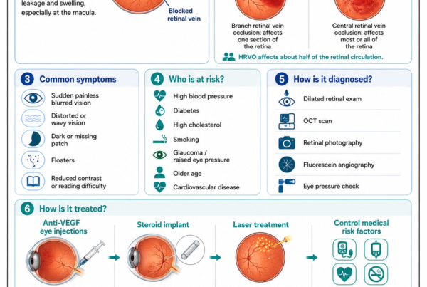
Retinal Vein Occlusion
A branch or central retinal vein occlusion may cause areas of retinal ischaemia.
The ischaemic retina can stimulate abnormal vessels to develop on:
- The retina
- The optic disc
- The iris
- The drainage angle
Retinal or disc new vessels may bleed into the vitreous.
Treatment may involve:
- Anti-VEGF injection
- Sector retinal laser
- Panretinal photocoagulation
- Vitrectomy
- Glaucoma treatment when required
The precise treatment depends on the location of neovascularisation and whether the retina can be visualised.
Retinal Arterial Macroaneurysm
What Is It?
A retinal arterial macroaneurysm is a localised dilatation of a retinal artery.
It is more frequently found in older adults and is often associated with hypertension.
How Can It Cause Vitreous Haemorrhage?
A macroaneurysm may rupture and produce bleeding:
- Within the retina
- Beneath the retina
- In front of the retina
- Beneath the internal limiting membrane
- Into the vitreous
The pattern may involve several retinal layers simultaneously.
Treatment
Management depends on:
- The position of the macroaneurysm
- Whether the macula is involved
- The amount of blood
- Continued leakage
- The presence of vitreous haemorrhage
Options may include observation, laser, anti-VEGF treatment, pneumatic displacement or vitrectomy.
Age-Related Macular Degeneration and Polypoidal Choroidal Vasculopathy
Breakthrough Vitreous Haemorrhage
Bleeding from abnormal vessels beneath the retina may sometimes break through:
- The retina
- The internal limiting membrane
- The posterior hyaloid
- Into the vitreous cavity
This may occur with:
- Neovascular age-related macular degeneration
- Polypoidal choroidal vasculopathy
- Large subretinal haemorrhage
Visual Prognosis
The visual outcome depends not only on the vitreous blood but also on:
- Macular damage
- Submacular haemorrhage
- Fibrosis
- Retinal pigment epithelial damage
- Duration of bleeding
- Activity of the underlying neovascular lesion
Vitrectomy can clear the media, but it cannot reverse established macular scarring. Studies of breakthrough haemorrhage from neovascular age-related macular degeneration and polypoidal choroidal vasculopathy report visual improvement after surgery in some eyes, although macular pathology commonly limits the final result.
Eye Trauma
Blunt Trauma
A direct blow may cause:
- Retinal tears
- Retinal dialysis
- Retinal detachment
- Choroidal rupture
- Traumatic retinal damage
- Damage to the ciliary body
- Lens dislocation
- Vitreous haemorrhage
Penetrating Trauma
A sharp object may injure:
- The cornea
- The sclera
- The lens
- The retina
- The choroid
- Intraocular blood vessels
Suspected open-globe injury is an emergency.
Pressure should not be applied to the eye.
Children
Trauma is a major cause of vitreous haemorrhage in children.
Other paediatric causes may include:
- Retinopathy of prematurity
- Persistent fetal vasculature
- Retinal vascular abnormalities
- Coagulation disorders
- Non-accidental injury
- Intraocular tumours
Children require prompt specialist assessment because prolonged visual obstruction may also interfere with visual development.
Retinal Vasculitis and Inflammatory Disease
Inflammation of retinal blood vessels may cause:
- Vessel-wall damage
- Retinal occlusion
- Retinal ischaemia
- Neovascularisation
- Vitreous haemorrhage
Possible underlying conditions include:
- Sarcoidosis
- Behçet disease
- Systemic lupus erythematosus
- Tuberculosis-associated retinal vasculitis
- Syphilis
- Other infectious or autoimmune disorders
Treatment may require coordinated management with:
- A uveitis specialist
- An infectious-disease physician
- A rheumatologist
- A general physician
Eales Disease
Eales disease is an idiopathic occlusive retinal vasculitis seen particularly in young adults.
It may progress through:
- Retinal periphlebitis
- Peripheral retinal non-perfusion
- Retinal neovascularisation
- Recurrent vitreous haemorrhage
- Traction or retinal detachment
Treatment may involve:
- Investigation for associated systemic or infectious disease
- Corticosteroid or anti-inflammatory treatment during active vasculitis
- Retinal laser for ischaemic retina
- Anti-VEGF injections
- Vitrectomy for persistent haemorrhage or traction
Recurrent vitreous haemorrhage is a recognised complication of its proliferative stage.
Sickle-Cell Retinopathy
Sickle-cell disease can obstruct peripheral retinal vessels.
The resulting ischaemia may produce peripheral neovascular formations sometimes described as sea-fan neovascularisation.
These abnormal vessels may cause:
- Vitreous haemorrhage
- Fibrous proliferation
- Tractional retinal detachment
- Rhegmatogenous retinal detachment
Management may include laser, anti-VEGF treatment or vitrectomy according to the findings.
Ocular Ischaemic Syndrome
Severe narrowing of the carotid circulation may reduce blood supply to the eye.
Possible ocular findings include:
- Mid-peripheral retinal haemorrhages
- Dilated retinal veins
- Retinal or iris neovascularisation
- Vitreous haemorrhage
- Neovascular glaucoma
- Ocular pain
This condition requires both ophthalmic and urgent systemic vascular assessment.
Terson Syndrome
What Is Terson Syndrome?
Terson syndrome refers to intraocular haemorrhage associated with acute intracranial bleeding or a sudden marked increase in intracranial pressure.
It may occur following:
- Subarachnoid haemorrhage
- Intracerebral haemorrhage
- Severe traumatic brain injury
- Other acute neurological events
Types of Ocular Bleeding
Bleeding may occur:
- Within the retina
- Beneath the internal limiting membrane
- Beneath the posterior hyaloid
- Within the vitreous cavity
Diagnosis and Treatment
Patients may be unable to report their visual symptoms because of impaired consciousness.
Diagnosis may require:
- Dilated examination
- Ocular ultrasound
- Neuroimaging
- Review after neurological stabilisation
Mild haemorrhage may be observed.
Vitrectomy may be considered for:
- Dense non-clearing haemorrhage
- Bilateral visual impairment
- Prolonged premacular haemorrhage
- Patients requiring faster visual rehabilitation
Terson syndrome is associated with more severe neurological disease and should be recognised as part of the patient’s overall assessment.
Blood Disorders and Abnormal Coagulation
Conditions that may contribute to intraocular bleeding include:
- Severe anaemia
- Thrombocytopenia
- Leukaemia
- Polycythaemia
- Haemophilia
- Disseminated intravascular coagulation
- Hyperviscosity syndromes
- Other haematological disease
The eye findings may be the first sign of a systemic disorder.
Testing should be guided by:
- Age
- Medical history
- Examination findings
- Recurrence
- Bilateral involvement
- Other bleeding symptoms
Can Blood Thinners Cause Vitreous Haemorrhage?
Antiplatelet and anticoagulant medicines may increase the extent or visibility of bleeding from an existing abnormal vessel.
Examples include:
- Aspirin
- Clopidogrel
- Warfarin
- Apixaban
- Rivaroxaban
- Dabigatran
- Edoxaban
However, these medicines do not usually create a retinal tear or proliferative retinopathy by themselves.
An underlying ocular source must still be identified.
Should Blood Thinners Be Stopped?
Do not stop prescribed antiplatelet or anticoagulant medication without consulting the doctor managing the cardiovascular or thrombotic condition.
Stopping treatment may increase the risk of:
- Stroke
- Heart attack
- Deep-vein thrombosis
- Pulmonary embolism
- Embolic complications from atrial fibrillation
The decision must balance ocular bleeding against systemic risk.
Vitreous Haemorrhage After Eye Surgery
Bleeding may occur after:
- Retinal surgery
- Diabetic vitrectomy
- Cataract surgery
- Glaucoma surgery
- Intravitreal injection
- Retinal laser
- Trauma to a surgical wound
Possible causes include:
- Residual abnormal vessels
- Inadequately treated retinal ischaemia
- A retinal tear
- Low eye pressure
- Iris or angle neovascularisation
- A vascular wound complication
- Recurrent diabetic fibrovascular bleeding
Postoperative bleeding ranges from mild and self-limiting to severe haemorrhage requiring additional surgery.
Recurrent Haemorrhage After Diabetic Vitrectomy
Vitreous cavity haemorrhage may recur after vitrectomy for proliferative diabetic retinopathy.
Possible sources include:
- Residual retinal neovascularisation
- Fibrovascular tissue at the retinal surface
- Anterior hyaloidal fibrovascular proliferation
- Entry-site neovascularisation
- Incomplete retinal laser treatment
- Recurrent traction
- New retinal breaks
Management may include:
- Observation
- Anti-VEGF injection
- Additional retinal laser
- Fluid-air exchange
- Repeat vitrectomy
- Investigation for retinal detachment
How Is Vitreous Haemorrhage Diagnosed?
Medical and Ocular History
Important questions include:
- When did the symptoms begin?
- Were the symptoms sudden or gradual?
- Are there flashes?
- Is there a curtain or shadow?
- Was there recent trauma?
- Does the patient have diabetes?
- Has retinal laser been performed?
- Is there a history of retinal tear or detachment?
- Has the eye undergone surgery?
- Is the patient taking blood-thinning medication?
- Is there hypertension or a blood disorder?
- Was there a recent severe headache or neurological event?
The history often narrows the likely cause before the retina is examined.
Visual-Acuity Testing
Visual acuity provides a baseline.
The result depends on:
- Density of the blood
- Location of the blood
- Macular health
- Lens opacity
- Optic-nerve function
- Retinal detachment
- Previous ocular disease
Very poor vision from dense haemorrhage does not automatically indicate permanent visual loss.
Pupil Examination
The pupil response may provide information about:
- Severe retinal dysfunction
- Optic-nerve disease
- Extensive retinal detachment
- Previous ocular damage
A relative afferent pupillary defect cannot be explained by uncomplicated vitreous haemorrhage alone and suggests significant underlying retinal or optic-nerve dysfunction.
Eye-Pressure Measurement
Eye pressure is measured because vitreous haemorrhage may coexist with:
- Glaucoma
- Neovascular glaucoma
- Trauma
- Postoperative pressure changes
- Ghost-cell glaucoma
- Haemolytic glaucoma
Both high and low pressure may be clinically significant.
Slit-Lamp Examination
The ophthalmologist examines the front of the eye for:
- Iris neovascularisation
- Inflammation
- Red blood cells in the anterior chamber
- Lens injury
- Cataract
- Surgical wounds
- Signs of trauma
- Abnormal eye pressure
Dilated Retinal Examination
When the view permits, the retina is examined with:
- Slit-lamp biomicroscopy
- Indirect ophthalmoscopy
- Scleral indentation when appropriate
- Wide-field retinal imaging
The examination looks for:
- Retinal tears
- Retinal holes
- Retinal detachment
- Diabetic neovascularisation
- Retinal vascular occlusion
- Macroaneurysm
- Retinal vasculitis
- Subretinal haemorrhage
- Choroidal lesions
- Trauma
A careful peripheral examination is essential.
Examination of the Other Eye
The fellow eye may reveal important clues.
Possible findings include:
- Proliferative diabetic retinopathy
- Previous retinal laser
- Retinal vein occlusion
- Peripheral retinal degeneration
- Sickle-cell retinopathy
- Hypertensive retinal changes
- Age-related macular degeneration
- Inflammatory disease
B-Scan Ocular Ultrasound
When Is Ultrasound Needed?
B-scan ultrasound is used when blood prevents a clear view of the retina.
It may help identify:
- Retinal detachment
- Posterior vitreous detachment
- Tractional membranes
- Intraocular foreign bodies
- Choroidal detachment
- Large retinal or choroidal lesions
- Abnormal tissue within the vitreous cavity
Can Ultrasound Detect Every Retinal Tear?
No.
Ultrasound is highly useful for detecting retinal detachment, but a small peripheral retinal tear may not be visible.
A normal ultrasound does not always eliminate the need for:
- Repeat examination
- Serial ultrasound
- Early vitrectomy in selected high-risk cases
Ultrasound is an adjunct rather than a replacement for a complete retinal examination.
Optical Coherence Tomography
OCT may be useful when the macula can be imaged.
It may detect:
- Diabetic macular oedema
- Epiretinal membrane
- Vitreomacular traction
- Macular detachment
- Subretinal fluid
- Submacular haemorrhage
- Photoreceptor damage
- Premacular haemorrhage
Dense vitreous blood may prevent a usable scan.
Fundus Photography
When sufficient retinal detail is visible, photography helps:
- Document the haemorrhage
- Record neovascularisation
- Compare clearing over time
- Monitor laser treatment
- Educate the patient
- Support long-term follow-up
Fluorescein Angiography
Fluorescein angiography may reveal:
- Retinal non-perfusion
- Active neovascularisation
- Retinal vasculitis
- Vascular leakage
- Macroaneurysm
- Retinal vein occlusion
It is often delayed until enough blood has cleared to permit useful imaging.
OCT Angiography
OCT angiography may demonstrate abnormal retinal or choroidal blood flow without dye injection.
Its usefulness may be limited by:
- Dense media opacity
- Motion artefact
- Segmentation error
- Inability to display leakage directly
Blood and Systemic Tests
Testing is individualised.
Possible investigations include:
- Blood pressure
- Fasting glucose
- HbA1c
- Full blood count
- Platelet count
- Lipid profile
- Kidney function
- Coagulation tests
- Sickle-cell testing
- Inflammatory or autoimmune testing
- Infectious-disease investigations
- Carotid vascular assessment
Not every patient requires an extensive blood panel.
Assessing the Severity
Mild Vitreous Haemorrhage
The retina remains substantially visible.
Symptoms may include:
- A few floaters
- Mild haze
- Small mobile shadows
The cause may be identifiable immediately.
Moderate Vitreous Haemorrhage
The retinal view is partly obscured.
Some areas can be examined, while others cannot.
Repeat examination may reveal pathology as the blood settles.
Dense Vitreous Haemorrhage
The fundus cannot be examined adequately.
Vision may be severely reduced.
Ultrasound and close retinal follow-up are required, and vitrectomy may be recommended depending on the likely cause.
Treatment Goals
Treatment aims to:
- Identify the source of bleeding
- Detect retinal tears or detachment
- Stop ongoing neovascular bleeding
- Treat retinal ischaemia
- Clear the visual axis
- Preserve the macula
- Prevent recurrent haemorrhage
- Prevent tractional retinal detachment
- Restore useful vision
- Treat associated systemic disease
Observation
When May Observation Be Appropriate?
Observation may be considered when:
- The retina is attached
- No retinal tear requiring treatment is found
- The likely cause is known
- The haemorrhage is clearing
- There is no progressive traction
- The fellow eye provides adequate functional vision
- Follow-up is reliable
- The patient can manage safely during visual recovery
Observation Is Not “Doing Nothing”
Active observation may involve:
- Repeat visual-acuity testing
- Dilated retinal examination
- Serial B-scan ultrasound
- Eye-pressure measurement
- OCT when possible
- Retinal photography
- Laser when the retina becomes visible
- Reconsideration of surgery if clearing is inadequate
Head Positioning
Sitting upright may allow blood to settle inferiorly and occasionally improve the central view.
However:
- Positioning does not treat the bleeding source
- Evidence that head elevation accelerates clearance is limited
- A retinal tear or detachment must still be excluded
Activity
Temporary reduction of vigorous activity is sometimes advised when bleeding is fresh.
The patient should avoid activities that are unsafe with impaired vision, including:
- Driving
- Climbing ladders
- Operating machinery
- Unsupervised swimming
- Activities with a high risk of further eye trauma
Prolonged bed rest is not routinely required.
Retinal Laser for Retinal Tears
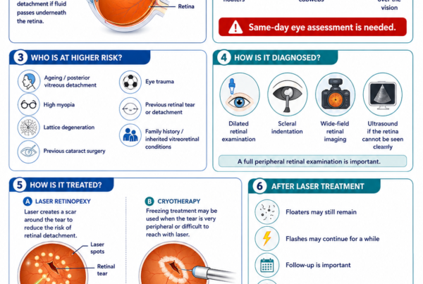
When a retinal tear is found without detachment, laser retinopexy may be performed.
Laser creates a surrounding adhesion between the retina and the underlying tissue.
The goal is to reduce the chance that fluid passes through the tear and causes retinal detachment.
Dense haemorrhage may make immediate laser impossible. Surgery may then be considered if the tear cannot be treated safely.
Panretinal Photocoagulation
Why Is Panretinal Laser Used?
Panretinal photocoagulation, or PRP, treats ischaemic peripheral retina.
This reduces the drive for abnormal new vessel growth.
It is used in conditions such as:
- Proliferative diabetic retinopathy
- Ischaemic retinal vein occlusion
- Ocular ischaemic syndrome
- Selected proliferative retinal vasculopathies
Can PRP Be Performed Through Vitreous Blood?
Only if sufficient retinal detail is visible.
When the view is poor:
- Treatment may be delayed while the blood clears
- Anti-VEGF treatment may be used
- Endolaser may be performed during vitrectomy
Anti-VEGF Injections
How Do They Work?
Anti-VEGF medicines reduce the activity of vascular endothelial growth factor.
They may cause abnormal vessels to:
- Regress
- Leak less
- Bleed less readily
Possible medicines include:
- Aflibercept
- Ranibizumab
- Bevacizumab
- Other locally approved anti-VEGF agents
Do Injections Remove the Blood?
Not directly.
The medicine does not dissolve or suction out blood already present.
Instead, it treats active abnormal vessels and may:
- Reduce recurrent bleeding
- Allow the haemorrhage to clear
- Permit later laser treatment
- Reduce neovascularisation
- Prepare the eye for surgery
Anti-VEGF for Diabetic Vitreous Haemorrhage
Anti-VEGF treatment may be a reasonable initial approach for selected eyes with proliferative diabetic retinopathy.
Its advantages may include:
- Avoiding immediate surgery
- Regression of active neovascularisation
- Reduced recurrent bleeding
- Opportunity to complete laser after clearing
Its limitations include:
- Slower restoration of clear vision than surgical removal
- Need for repeated injections
- Persistent obscuration of the retina
- Possible eventual need for vitrectomy
- Dependence on reliable follow-up
What Did Protocol AB Show?
The DRCR Retina Network compared initial aflibercept injections with initial vitrectomy and panretinal photocoagulation for vitreous haemorrhage from proliferative diabetic retinopathy.
Average visual acuity over the initial 24 weeks was not statistically different between the groups.
However, the clinical experience differed:
- Vitrectomy generally cleared the haemorrhage faster.
- Early visual recovery was often faster after surgery.
- Recurrent haemorrhage was more common in the injection-first group.
- Some injection-treated eyes later required vitrectomy.
- Some surgery-treated eyes later required anti-VEGF treatment.
Both approaches are therefore viable, but the choice should be individualised.
Can Anti-VEGF Cause Traction?
Rapid regression of neovascular tissue may sometimes increase contraction of associated fibrous tissue.
This is particularly relevant when there is:
- Extensive fibrovascular proliferation
- Pre-existing tractional retinal detachment
- Traction close to the macula
- A delay between injection and planned surgery
Preoperative injections are therefore timed carefully when used before diabetic vitrectomy.
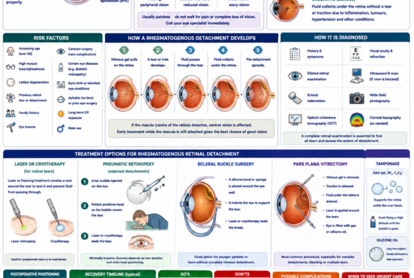
Pars Plana Vitrectomy
What Is Vitrectomy?
Vitrectomy is microsurgery performed through small openings in the white part of the eye.
The surgeon removes:
- Vitreous gel
- Suspended blood
- Blood clots
- Tractional tissue when necessary
The retina can then be inspected and treated directly.
What May Be Done During Surgery?
Depending on the cause, surgery may include:
- Removal of vitreous blood
- Induction or completion of posterior vitreous separation
- Identification of retinal tears
- Endolaser photocoagulation
- Removal of fibrovascular membranes
- Relief of retinal traction
- Repair of retinal detachment
- Drainage of subretinal fluid
- Treatment of bleeding vessels
- Air, gas or silicone-oil tamponade
- Cataract surgery in selected patients
When Is Vitrectomy Considered?
Possible indications include:
- Retinal detachment
- Retinal tear that cannot be treated adequately
- Tractional retinal detachment threatening the macula
- Combined tractional-rhegmatogenous detachment
- Dense non-clearing haemorrhage
- Recurrent haemorrhage
- Bilateral severe visual impairment
- Haemorrhage in a functionally one-eyed patient
- Inability to monitor or treat active retinal disease
- Suspicion of hidden retinal pathology
- Vitreous haemorrhage associated with intraocular foreign body or trauma
- Need for rapid visual rehabilitation
- Ghost-cell glaucoma related to persistent blood
- Premacular haemorrhage causing prolonged central visual obstruction
Early Versus Delayed Vitrectomy
Historically, dense haemorrhage was often observed for several months before surgery.
That approach was influenced by:
- Larger surgical instruments
- Longer operating times
- Higher historical complication rates
- More limited viewing systems
Modern vitrectomy uses:
- Smaller-gauge instruments
- Wide-angle viewing
- Faster cutting systems
- Improved endolaser
- Better fluidics
- More precise membrane dissection
As a result, earlier surgery is increasingly considered when the benefits outweigh the risks.
Reasons to Operate Earlier
Earlier surgery may:
- Restore the visual axis quickly
- Establish the diagnosis
- Detect hidden tears
- Treat retinal detachment before macular involvement
- Permit complete laser treatment
- Reduce prolonged functional disability
- Remove tractional tissue
- Reduce repeated clinic visits for obscured retinal views
Reasons to Observe Initially
Observation may avoid surgery when:
- The haemorrhage is expected to clear
- The retina is confidently attached
- The underlying disease has already been treated
- Vision remains adequate
- Surgical risk is relatively high
- The patient prefers a conservative approach
Systematic reviews suggest that early vitrectomy can be beneficial in selected vitreous haemorrhage, particularly when retinal pathology may remain hidden or when delayed recovery imposes substantial functional consequences.
Diabetic Vitreous Haemorrhage: Injection or Surgery?
The decision is not simply based on the amount of blood.
Important considerations include:
- Duration of haemorrhage
- First episode or recurrent bleeding
- Previous PRP
- Activity of neovascularisation
- Tractional retinal detachment
- Condition of the macula
- Vision in the other eye
- Patient occupation
- Ability to attend frequent reviews
- Preference for faster recovery
- Anaesthetic and surgical risk
- Access to injections and surgery
Injection-First May Be Considered When:
- There is no macula-threatening traction
- The retina appears attached on ultrasound
- The patient accepts slower clearing
- Follow-up is reliable
- Surgery can be arranged later if required
- Anti-VEGF treatment is otherwise indicated
Earlier Vitrectomy May Be Favoured When:
- The haemorrhage is dense
- Rapid visual rehabilitation is important
- Bleeding is recurrent
- Previous PRP is incomplete
- Traction is present or suspected
- The other eye has poor vision
- Follow-up is difficult
- The retina cannot be monitored
- The patient wishes to reduce prolonged uncertainty
The original Diabetic Retinopathy Vitrectomy Study demonstrated a visual advantage from early surgery particularly in eyes with severe proliferative disease associated with type 1 diabetes. Modern observational evidence and contemporary surgical practice also support considering earlier intervention in appropriate patients.
What Happens on the Day of Vitrectomy?
Before Surgery
The surgical team may:
- Confirm the eye and procedure
- Review medication
- Check blood glucose and blood pressure
- Discuss anaesthesia
- Dilate the pupil
- Perform ultrasound or retinal imaging
- Review the possibility of gas or silicone oil
- Discuss postoperative positioning
Anaesthesia
Vitrectomy may be performed under:
- Local anaesthesia with sedation
- Regional anaesthesia
- General anaesthesia
The choice depends on the patient and complexity of surgery.
During Surgery
Small instruments are inserted through the pars plana.
The surgeon may:
- Remove the blood-filled vitreous
- Control active bleeding
- Examine the peripheral retina
- Treat tears with laser
- Remove scar tissue
- Flatten a detached retina
- Apply endolaser
- Insert air, gas or silicone oil when required
After Surgery
The eye may feel:
- Gritty
- Watery
- Mildly sore
- Blurred
Vision will not always be clear immediately.
Recovery depends on:
- Retinal condition
- Macular involvement
- Presence of gas or oil
- Cataract
- Postoperative bleeding
- Previous optic-nerve or retinal damage
Risks of Vitrectomy
Possible risks include:
- Infection
- Recurrent vitreous haemorrhage
- Retinal tear
- Retinal detachment
- Cataract progression
- Raised eye pressure
- Low eye pressure
- Inflammation
- Corneal problems
- Macular oedema
- Need for further surgery
- Loss of vision
- Very rare loss of the eye
The risk must be balanced against the danger of untreated retinal disease and prolonged visual obstruction.
Gas Bubbles and Flying
A gas bubble may be inserted when retinal repair is required.
A patient with intraocular gas must not:
- Fly
- Travel to high altitude
- Receive nitrous-oxide anaesthesia
until the bubble has completely disappeared.
Reduced atmospheric pressure can cause the bubble to expand and dangerously increase eye pressure.
The restriction does not apply merely because a vitreous haemorrhage is present. It applies when gas has been placed inside the eye.
Cataract and Vitrectomy
Vitrectomy may accelerate cataract progression, particularly in older phakic patients.
Possible approaches include:
- Vitrectomy alone
- Cataract surgery at a later date
- Combined cataract surgery and vitrectomy
The decision depends on:
- Existing cataract
- Age
- Retinal pathology
- Expected surgical complexity
- Need for gas or silicone oil
- Ability to view the retina
Ghost-Cell Glaucoma
What Is It?
Older red blood cells may degenerate within the vitreous and move into the front of the eye.
These rigid cells can obstruct the drainage system.
Symptoms
Possible symptoms include:
- Eye pain
- Redness
- Headache
- Blurred vision
- Halos
- Nausea
Treatment
Management may involve:
- Pressure-lowering medication
- Anterior-chamber washout
- Vitrectomy to remove the source of blood
- Treatment of the underlying cause
Will Vision Return After Vitreous Haemorrhage?
Vision may recover well when:
- The retina remains attached
- The macula is healthy
- A retinal tear is treated promptly
- Bleeding stops
- The blood clears or is removed
- There is no significant optic-nerve disease
Vision may remain limited when there is:
- Macular ischaemia
- Diabetic macular damage
- Macular scarring
- Submacular haemorrhage
- Tractional retinal detachment
- Macula-off retinal detachment
- Proliferative vitreoretinopathy
- Advanced glaucoma
- Optic-nerve damage
- Severe trauma
- Age-related macular degeneration
Clearing the vitreous blood improves the optical pathway but cannot restore retinal tissue that has already been permanently damaged.
How Long Does Recovery Take?
With Observation
Clearing may take weeks or months.
Vision may fluctuate as blood moves and settles.
After Vitrectomy
The blood is removed during surgery, but visual recovery may still take time because of:
- Inflammation
- Gas
- Retinal swelling
- Macular disease
- Cataract
- Recurrent bleeding
- Healing of retinal laser
The surgeon will explain the likely recovery based on the underlying retinal condition.
Can Vitreous Haemorrhage Happen Again?
Yes.
Recurrence may occur if:
- Abnormal vessels remain active
- Retinal ischaemia is untreated
- Diabetic control remains poor
- Laser treatment is incomplete
- A new retinal tear develops
- Fibrovascular tissue contracts
- A macroaneurysm bleeds again
- Neovascular macular disease remains active
- Postoperative vessels bleed
Preventing recurrence requires treatment of the source rather than simply waiting for each episode to clear.
Can Vitreous Haemorrhage Be Prevented?
Not every episode is preventable.
Risk may be reduced by:
- Regular diabetic eye screening
- Timely treatment of proliferative diabetic retinopathy
- Completing retinal laser when advised
- Attending scheduled anti-VEGF injections
- Controlling blood pressure
- Managing diabetes
- Treating retinal tears promptly
- Wearing eye protection during hazardous activities
- Managing sickle-cell or inflammatory disease
- Seeking urgent care for new flashes and floaters
Diabetes and Systemic Health
For diabetic vitreous haemorrhage, long-term management should include attention to:
- HbA1c
- Blood pressure
- Cholesterol
- Kidney function
- Smoking
- Weight
- Medication adherence
- Regular retinal review
Rapid improvement of very poor glucose control should be medically supervised, as retinal disease may require closer monitoring during major systemic treatment changes.
Common Myths
“Vitreous Haemorrhage Is Just a Bruise Inside the Eye”
False.
It may be caused by a retinal tear, retinal detachment or active proliferative vascular disease.
“The Blood Must Clear Before the Retina Can Be Assessed”
False.
Ultrasound can assess major posterior-segment structures, and vitrectomy may be used to clear the blood and examine the retina directly.
“If There Is No Pain, It Is Not Urgent”
False.
Retinal tears, retinal detachment and diabetic vitreous haemorrhage are commonly painless.
“Eye Drops Can Dissolve the Blood”
False.
Standard eye drops do not remove blood from the vitreous cavity.
“Blood Thinners Are Always the Cause”
False.
They may increase bleeding from an existing lesion, but an underlying retinal or vascular source must still be sought.
“Blood Thinners Should Be Stopped Immediately”
False.
Stopping prescribed medication without medical advice may cause serious systemic harm.
“One Anti-VEGF Injection Will Clear Every Diabetic Haemorrhage”
False.
Some patients require repeated injections, retinal laser, vitrectomy or a combination of treatments.
“Waiting Is Always Safer Than Surgery”
False.
Observation may be appropriate, but delay can be harmful if a retinal tear, detachment or progressive traction is hidden behind the blood.
“Surgery Guarantees Normal Vision”
False.
Vitrectomy clears the media and treats structural problems, but the visual outcome depends on the health of the retina, macula and optic nerve.
“Once the Blood Clears, Follow-Up Is No Longer Necessary”
False.
The disease that caused the haemorrhage may remain active or recur.
Frequently Asked Questions
Is Vitreous Haemorrhage the Same as a Retinal Haemorrhage?
No.
A retinal haemorrhage is located within or beneath the retinal tissue.
A vitreous haemorrhage is blood within the vitreous cavity.
Both may occur together.
Is It the Same as a Subconjunctival Haemorrhage?
No.
A subconjunctival haemorrhage is a red patch on the white surface of the eye.
It usually does not affect vision.
A vitreous haemorrhage occurs inside the eye and may cause profound visual loss.
Can a Vitreous Haemorrhage Cause Blindness?
The blood alone may temporarily block vision.
Permanent visual loss is more commonly caused by the underlying disease, such as:
- Retinal detachment
- Macular ischaemia
- Diabetic retinal damage
- Glaucoma
- Severe trauma
- Macular scarring
Can It Affect Both Eyes?
Yes, although many episodes begin in one eye.
Bilateral disease may occur with:
- Proliferative diabetic retinopathy
- Terson syndrome
- Blood disorders
- Retinal vasculitis
- Sickle-cell retinopathy
- Severe systemic vascular disease
Can High Blood Pressure Cause It?
Hypertension may contribute to:
- Retinal vein occlusion
- Retinal arterial macroaneurysm
- Severe hypertensive retinal disease
- Increased vascular fragility
However, the exact ocular bleeding source must still be identified.
Can Coughing or Heavy Lifting Cause It?
A sudden rise in venous pressure may occasionally cause a Valsalva-related preretinal haemorrhage.
More commonly, activity reveals bleeding from an already fragile abnormal vessel.
Coughing or lifting should not be assumed to be the sole explanation until retinal disease has been excluded.
Can Exercise Make It Worse?
Temporary activity restriction may be advised when bleeding is fresh or when proliferative vessels are active.
Long-term regular exercise remains beneficial for cardiovascular and diabetic health once the ophthalmologist considers it safe.
Can I Drive?
Do not drive when vision is significantly impaired.
Driving safety depends on:
- Visual acuity
- Visual field
- Vision in the other eye
- Depth perception
- Glare
- Ability to detect hazards
- Local licensing requirements
Can I Fly?
An uncomplicated vitreous haemorrhage does not ordinarily prevent flying.
Flying is prohibited when an intraocular gas bubble is present following surgery.
Can I Continue Aspirin?
Do not change aspirin or other blood-thinning medication without discussing it with the doctor who prescribed it.
How Often Must Ultrasound Be Repeated?
The interval depends on:
- Cause
- Density
- Symptoms
- Retinal-tear risk
- Ultrasound findings
- Whether the haemorrhage is clearing
- Whether surgery is being considered
High-risk cases may require frequent early review.
What Happens if the Ultrasound Is Normal?
A normal ultrasound is reassuring regarding a major retinal detachment or mass.
It does not guarantee that there is no small retinal tear.
Clinical follow-up remains necessary until the retina can be examined adequately or surgery establishes the diagnosis.
How Long Should I Wait Before Vitrectomy?
There is no universal waiting period.
The decision depends on:
- Cause
- Retinal findings
- Visual needs
- Fellow-eye vision
- Duration
- Recurrence
- Traction
- Likelihood of spontaneous clearing
- Surgical risk
- Patient preference
Does Vitrectomy Remove All the Blood?
The surgeon removes the blood-filled vitreous and washes out the vitreous cavity.
Small residual red blood cells or mild postoperative bleeding may remain temporarily.
Can the Haemorrhage Return After Vitrectomy?
Yes.
Recurrence may arise from active neovascular tissue, incomplete laser, residual traction or postoperative vascular bleeding.
Will I Need Laser During Vitrectomy?
Laser is commonly performed when surgery reveals:
- Retinal tears
- Proliferative diabetic retinopathy
- Ischaemic retina
- Retinal neovascularisation
- Retinal detachment
Not every vitrectomy requires laser.
Will I Need Cataract Surgery?
Vitrectomy may accelerate cataract formation.
Some patients undergo combined cataract and retinal surgery, while others have cataract surgery later.
Is Vitrectomy Painful?
The procedure is performed with anaesthesia.
Mild soreness or grittiness may occur afterwards, but severe or increasing pain is not expected and requires review.
When to Seek Urgent Eye Care
Seek urgent same-day assessment for:
- A sudden shower of floaters
- A sudden red, brown or smoky haze
- Flashes of light
- A curtain or shadow
- Rapid visual deterioration
- New loss of part of the visual field
- Symptoms following trauma
- Vitreous haemorrhage in the only functional eye
- Eye pain
- Redness
- Headache, nausea or vomiting
- New symptoms after eye surgery or injection
After treatment or surgery, seek immediate advice for:
- Increasing pain
- Increasing redness
- Marked light sensitivity
- Sudden worsening of vision
- Thick discharge
- Increasing eyelid swelling
- Loss of previously improving vision
A Vitreous Haemorrhage Assessment Checklist
Symptoms to Report
- Sudden floaters
- Flashes
- Red or smoky vision
- Blurred vision
- Curtain or shadow
- Field loss
- Eye pain
- Recent trauma
- Exact time of onset
- Previous similar episodes
Medical Information to Report
- Diabetes
- HbA1c
- Hypertension
- High cholesterol
- Kidney disease
- Sickle-cell disease
- Blood disorders
- Autoimmune disease
- Recent severe headache or intracranial bleeding
- Antiplatelet or anticoagulant medication
Ocular Information to Report
- Previous retinal tear
- Retinal detachment
- Retinal laser
- Anti-VEGF injections
- Cataract surgery
- Vitrectomy
- Glaucoma
- Age-related macular degeneration
- Retinal vein occlusion
Tests That May Be Used
- Visual acuity
- Pupil examination
- Eye-pressure measurement
- Slit-lamp examination
- Dilated retinal examination
- B-scan ultrasound
- OCT
- Fundus photography
- Fluorescein angiography
- OCT angiography
- Systemic blood or vascular tests
Questions to Ask the Ophthalmologist
- What is the likely source of the bleeding?
- Is there a retinal tear?
- Is the retina attached?
- Is there diabetic neovascularisation?
- Is retinal traction present?
- Do I need laser?
- Would an anti-VEGF injection help?
- Is observation safe?
- How frequently must I return?
- How long is the haemorrhage likely to take to clear?
- Should I consider vitrectomy?
- What are the advantages of operating earlier?
- Will I need gas or silicone oil?
- Can I fly?
- Can I drive?
- What visual recovery is realistic?
The Bottom Line
A vitreous haemorrhage occurs when blood enters the normally clear vitreous cavity.
It may cause:
- New floaters
- Cobwebs
- Red or smoky vision
- Blurred vision
- Shadows
- Profound painless visual loss
Common causes include:
- Posterior vitreous detachment with a retinal tear
- Proliferative diabetic retinopathy
- Retinal vein occlusion
- Trauma
- Retinal neovascularisation
- Retinal arterial macroaneurysm
- Age-related macular degeneration
- Retinal vasculitis
- Terson syndrome
A vitreous haemorrhage is a clinical finding—not the final diagnosis.
The main priorities are to:
- Identify the source
- Exclude retinal tear and detachment
- Assess retinal ischaemia and traction
- Treat active abnormal vessels
- Preserve the macula
- Restore the visual axis
Diagnosis commonly involves:
- Dilated retinal examination
- B-scan ultrasound
- OCT
- Retinal imaging
- Systemic assessment when indicated
Treatment may include:
- Observation
- Retinal laser
- Anti-VEGF injection
- Pars plana vitrectomy
- Treatment of an underlying systemic condition
Observation can be appropriate when the retina is attached, the cause is understood and follow-up is reliable.
Vitrectomy may be recommended when the haemorrhage is dense, persistent or recurrent; when retinal pathology is hidden; when traction or retinal detachment is present; or when faster visual rehabilitation is important.
The most important message is:
A sudden shower of floaters, red haze or painless loss of vision should never be ignored. Prompt retinal assessment can identify a treatable retinal tear or proliferative vascular condition before further permanent vision is lost.
References
- Spraul CW, Grossniklaus HE. Vitreous hemorrhage. Surv Ophthalmol. 1997;42(1):3–39. doi:10.1016/S0039-6257(97)84041-6. PMID: 9265701.
- Shaikh N, Kumar V, Ramachandran A, et al. Vitreous hemorrhage: causes, diagnosis, and management. Indian J Ophthalmol. 2023. PMID: 36588205.
- Strand E, Meshkin RS, Hoyek S, Miller JB, Patel NA. Nondiabetic vitreous hemorrhage: a review of management strategy and outcomes. Ophthalmic Surg Lasers Imaging Retina. 2024;55(11):660–667. doi:10.3928/23258160-20240620-01. PMID: 39172227.
- Bond-Taylor M, Jakobsson G, Zetterberg M. Posterior vitreous detachment—prevalence of and risk factors for retinal tears. Clin Ophthalmol. 2017;11:1689–1695. doi:10.2147/OPTH.S143898. PMID: 29075095.
- Patel PR, et al. Analysis of posterior vitreous detachment and development of complications using a large database of retina specialists. Ophthalmol Retina. 2023;7(3). PMID: 36423892.
- Sarrafizadeh R, Hassan TS, Ruby AJ, et al. Incidence of retinal detachment and visual outcome in eyes presenting with posterior vitreous separation and dense fundus-obscuring vitreous hemorrhage. Ophthalmology. 2001;108(12):2273–2278. PMID: 11733270.
- Getahun H, Sharp CN, Kato K, et al. Clinical outcomes and predictors of retinal breaks in patients with vitreous hemorrhage secondary to a posterior vitreous detachment. J Vitreoretin Dis. 2026. doi:10.1177/24741264261455221. PMID: 42255511.
- Antoszyk AN, Glassman AR, Beaulieu WT, et al; DRCR Retina Network. Effect of intravitreous aflibercept versus vitrectomy with panretinal photocoagulation on visual acuity in patients with vitreous hemorrhage from proliferative diabetic retinopathy: a randomized clinical trial. JAMA. 2020;324(23):2383–2395. doi:10.1001/jama.2020.23027.
- Glassman AR, Beaulieu WT, Maguire MG, et al; DRCR Retina Network. Visual acuity, vitreous hemorrhage, and other ocular outcomes after vitrectomy versus aflibercept for vitreous hemorrhage due to diabetic retinopathy. JAMA Ophthalmol. 2021;139(7):725–733. doi:10.1001/jamaophthalmol.2021.1110. PMID: 33956075.
- Diabetic Retinopathy Vitrectomy Study Research Group. Early vitrectomy for severe vitreous hemorrhage in diabetic retinopathy: two-year results of a randomized trial. Arch Ophthalmol. 1985;103(11):1644–1652. PMID: 2865943.
- Diabetic Retinopathy Vitrectomy Study Research Group. Early vitrectomy for severe vitreous hemorrhage in diabetic retinopathy: four-year results of a randomized trial. Arch Ophthalmol. 1990;108(7):958–964. PMID: 2196036.
- Bhavsar AR, Torres K, Glassman AR, et al. Randomized clinical trial evaluating intravitreal ranibizumab or saline for vitreous hemorrhage from proliferative diabetic retinopathy. JAMA Ophthalmol. 2013;131(3):283–293. PMID: 23370902.
- Confalonieri F, Barone G, Ferraro V, et al. Early versus late pars plana vitrectomy in vitreous hemorrhage: a systematic review. J Clin Med. 2023;12(20):6652. doi:10.3390/jcm12206652. PMID: 37892789.
- Smith M, et al. Oral anticoagulation and the risk of vitreous hemorrhage and retinal tears in eyes with acute posterior vitreous detachment. Retina. 2013. PMID: 23108264.
- Iwase T, Baba T, Saito Y, et al. Surgical outcomes of vitrectomy for breakthrough vitreous hemorrhage in eyes with exudative age-related macular degeneration. Int Ophthalmol. 2021;41(5):1835–1844. doi:10.1007/s10792-021-01744-x. PMID: 33611763.
- Jung JH, et al. Prognostic factors and long-term surgical outcomes for exudative age-related macular degeneration with breakthrough vitreous hemorrhage. J Clin Med. 2020. PMID: 32783420.
- Stevanovic M, Eliott D. Terson syndrome: a review of the literature. Int Ophthalmol Clin. 2024;64(2):89–106. doi:10.1097/IIO.0000000000000502. PMID: 38525984.
- Czorlich P, et al. Terson’s syndrome, current concepts and management strategies: a review of the literature. Clin Neurol Neurosurg. 2021. PMID: 34775364.
- Biswas J, Sharma T, Gopal L, Madhavan HN, Sulochana KN, Ramakrishnan S. Eales disease—an update. Surv Ophthalmol. 2002;47(3):197–214. doi:10.1016/S0039-6257(02)00288-6. PMID: 12052408.
- Saxena S, Jalali S, Verma L, Pathengay A. Management of vitreous haemorrhage. Indian J Ophthalmol. 2003;51(2):189–196. PMID: 12831156.