Author: Dr Val Phua
Estimated reading time: 20 minutes
Cataract surgery removes the cloudy natural lens inside the eye and replaces it with a clear artificial intraocular lens.
It is one of the most frequently performed surgical procedures in medicine and usually provides substantial improvement in vision, visual function and quality of life. Modern cataract surgery is commonly performed through a small incision using ultrasound-assisted phacoemulsification. Most operations are completed under local anaesthesia without an overnight hospital stay.
Cataract surgery may improve:
- Blurred vision
- Glare
- Halos caused by the cataract
- Faded colours
- Difficulty reading
- Poor night vision
- Difficulty driving
- Loss of contrast
- Dependence on strong spectacle prescriptions
However, removing the cataract does not automatically correct every cause of poor vision.
The final visual result also depends on the health of the:
- Cornea
- Macula
- Retina
- Optic nerve
- Tear film
- Visual pathway
Cataract surgery has also become a form of refractive surgery. Before the operation, the patient and ophthalmologist must decide not only whether to remove the cataract, but also what type of vision to aim for afterwards.
Important decisions may include:
- Distance or near focus
- Monovision
- Astigmatism correction
- Monofocal or enhanced monofocal lenses
- Extended-depth-of-focus lenses
- Multifocal or trifocal lenses
- The acceptable balance between spectacle independence and optical side effects
A successful outcome therefore depends on careful examination, accurate measurements, appropriate intraocular lens selection, precise surgery and realistic expectations.
The Quick Answer
What Happens During Cataract Surgery?
The cloudy natural lens is broken into small pieces and removed through a small incision.
A clear artificial intraocular lens is then inserted into the thin natural lens capsule that remains inside the eye.
Is Cataract Surgery Painful?
Most patients feel little or no pain.
Anaesthetic eye drops are commonly used. Some patients also receive:
- Intracameral anaesthetic inside the eye
- Mild intravenous sedation
- A sub-Tenon’s or regional anaesthetic injection
- General anaesthesia in selected circumstances
Patients may notice bright lights, colours, movement or gentle pressure during the operation. Randomised studies have shown that topical anaesthesia provides acceptable comfort for routine cataract surgery, although additional intracameral or regional anaesthesia may improve comfort in selected patients.
How Long Does It Take?
The surgical portion commonly takes approximately 10 to 30 minutes in an uncomplicated case.
Additional time is required for:
- Registration
- Eye drops
- Pupil dilation
- Preparation
- Anaesthesia
- Postoperative observation
- Discharge instructions
Complex cataracts may take longer.
Will I Be Awake?
Most adults remain awake but comfortable.
The patient is usually asked to:
- Lie reasonably still
- Look towards the microscope light
- Avoid talking unless necessary
- Inform the surgical team if there is discomfort
The face is covered with a sterile drape, but the patient can breathe normally.
Will I Need Spectacles Afterwards?
Possibly.
The need for spectacles depends on:
- The selected intraocular lens
- The refractive target
- Corneal astigmatism
- Healing
- Measurement accuracy
- Retinal and optic-nerve health
- The visual demands of the patient
No lens can guarantee complete spectacle independence in every situation.
Is Cataract Surgery Permanent?
The removed natural lens does not grow back.
However, the thin capsule behind the artificial lens may become cloudy later. This is called posterior capsule opacification and can usually be treated with a brief YAG laser procedure.
What Is a Cataract?
A cataract is clouding of the natural crystalline lens inside the eye.
The lens is located behind the coloured iris and pupil.
A healthy lens helps focus light onto the retina. As it becomes cloudy, light may be:
- Scattered
- Absorbed
- Distorted
- Unevenly focused
This may reduce visual clarity even when a spectacle test appears reasonably good.
Why Is the Natural Lens Removed?
The cataract cannot be polished or cleaned while it remains inside the eye.
Once the lens proteins have become significantly cloudy, the only definitive treatment is to remove the cataract surgically.
Eye drops, supplements and exercises do not reliably reverse an established age-related cataract.
When Is Cataract Surgery Recommended?
Cataract surgery is generally considered when the cataract interferes meaningfully with the patient’s life or with the medical care of the eye.
Possible reasons include:
- Difficulty reading
- Difficulty driving
- Disabling glare
- Poor night vision
- Reduced work performance
- Difficulty recognising faces
- Loss of independence
- Increased risk of falls
- Difficulty examining or treating the retina
- Cataract-related inflammation or glaucoma
- A major difference in vision between the two eyes
The decision should be based on symptoms, functional impairment, examination findings and patient preference—not simply on a particular visual-acuity number.
Does the Cataract Need to Be “Ripe”?
No.
Modern cataract surgery does not require the cataract to become completely dense or mature.
Waiting until a cataract becomes extremely hard may make surgery more technically challenging and may increase the amount of ultrasound energy required.
A patient also does not need surgery merely because a cataract has been detected.
The appropriate time is usually when:
- The visual difficulty is significant
- The expected benefits outweigh the risks
- The patient feels ready
- Other causes of poor vision have been considered
Can Surgery Be Performed Before the Vision Becomes Very Poor?
Yes.
Some patients remain able to read a letter chart but experience major problems with:
- Night glare
- Headlight scatter
- Contrast
- Colour quality
- Changing prescriptions
- Monocular double vision
- Occupational visual demands
Visual acuity alone may underestimate the disability caused by a cataract.
What If the Cataract Is Not Bothering Me?
Observation may be reasonable when:
- Daily activities remain comfortable
- The cataract is not causing a medical complication
- The retina can still be examined
- The patient is satisfied with current vision
- Surgery is not currently desired
The cataract may be reviewed periodically.
What Tests Are Performed Before Cataract Surgery?
A complete preoperative evaluation is essential because cataract surgery removes the cloudy lens but cannot correct every eye condition.
Medical and Medication History
The ophthalmologist should know about:
- Diabetes
- High blood pressure
- Heart disease
- Stroke
- Lung disease
- Bleeding disorders
- Sleep apnoea
- Parkinson disease or tremor
- Dementia or severe anxiety
- Allergies
- Previous problems with anaesthesia
- Anticoagulant or antiplatelet medication
- Prostate medication
- Previous eye surgery
Medicines such as tamsulosin and some other alpha-blockers may affect the behaviour of the iris during surgery.
Do not stop prescribed blood-thinning medication unless instructed by the relevant treating doctor and surgical team.
Visual-Acuity and Refraction Testing
These tests assess:
- Unaided vision
- Spectacle-corrected vision
- Current refractive error
- Astigmatism
- Potential improvement with updated spectacles
If spectacles restore satisfactory vision, surgery may not yet be necessary.
Slit-Lamp Examination
The ophthalmologist assesses:
- Cataract type and density
- Corneal clarity
- Pupil size
- Iris abnormalities
- Depth of the anterior chamber
- Previous surgical wounds
- Lens stability
- Signs of inflammation
- Pseudoexfoliation
- Zonular weakness
Eye-Pressure Measurement
Eye pressure is measured because glaucoma may:
- Coexist with cataract
- Affect visual potential
- Influence lens selection
- Require simultaneous treatment
- Affect postoperative monitoring
Dilated Retinal Examination
The retina and optic nerve are examined for:
- Diabetic retinopathy
- Macular degeneration
- Epiretinal membrane
- Macular hole
- Retinal tears
- Retinal detachment
- Retinal vein occlusion
- Glaucoma damage
- Other causes of reduced vision
A very dense cataract may prevent a complete view.
Optical Coherence Tomography
OCT scans are often performed to detect subtle macular disease.
OCT may reveal:
- Epiretinal membrane
- Macular oedema
- Macular degeneration
- Vitreomacular traction
- Macular hole
- Photoreceptor damage
- Glaucoma-related nerve-fibre loss
These findings may affect:
- Expected visual recovery
- Timing of surgery
- Need for retinal treatment
- Suitability for a multifocal or trifocal lens
Ocular Biometry
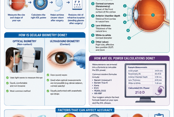
Biometry measures the eye so that an appropriate intraocular lens power can be calculated.
Measurements may include:
- Axial length
- Corneal curvature
- Anterior-chamber depth
- Lens thickness
- Corneal diameter
- Total corneal power
Modern formulas such as Barrett Universal II, Kane and other contemporary calculation methods have improved refractive prediction, although no formula eliminates all outliers.
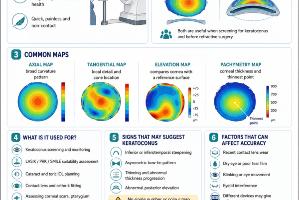
Corneal Topography or Tomography
Corneal mapping may be required when:
- Astigmatism is present
- A toric lens is being considered
- Keratoconus is suspected
- The patient previously underwent LASIK, PRK or SMILE
- Corneal shape is irregular
- A premium lens is being considered
- Previous contact-lens wear has altered the corneal measurements
The Ocular Surface
Dry eye disease can make corneal measurements unstable.
This may affect:
- Intraocular lens power calculations
- Astigmatism measurements
- Visual quality
- Postoperative comfort
- Satisfaction with premium lenses
Significant dry eye, blepharitis or meibomian gland dysfunction should therefore be treated before final measurements whenever possible. Cataract surgery itself may temporarily worsen dry-eye signs and symptoms.
Choosing the Refractive Target
Cataract surgery requires the patient and surgeon to decide what range of vision to prioritise.
Distance Vision in Both Eyes
Both eyes may be targeted for clear distance vision.
Advantages include:
- Good binocular distance vision
- Improved depth perception
- Easier adaptation
- Suitability for driving and outdoor activities
Reading spectacles will generally still be required with standard monofocal lenses.
Near Vision in One or Both Eyes
A patient who has been naturally short-sighted may prefer to retain some near vision.
Options include:
- Both eyes targeted for near
- One eye targeted for near
- Mild short-sightedness in both eyes
Distance spectacles may then be required.
Monovision
In monovision:
- One eye is targeted primarily for distance
- The other eye is left mildly short-sighted for intermediate or near vision
Advantages may include:
- Reduced dependence on reading spectacles
- Use of monofocal optical technology
- Fewer halos than with diffractive multifocal lenses
Possible disadvantages include:
- Reduced stereoacuity
- Reduced distance sharpness in the near eye
- Difficulty adapting
- Need for spectacles for demanding tasks
A contact-lens trial may be useful when the patient has never experienced monovision.
Mini-Monovision
Mini-monovision uses a smaller difference between the eyes.
It may provide:
- Good distance vision
- Improved intermediate vision
- Easier adaptation than stronger monovision
Reading spectacles may still be necessary for fine print.
Understanding Intraocular Lenses
The intraocular lens, or IOL, is the clear artificial lens inserted during surgery.
It usually remains inside the eye permanently.
Monofocal Intraocular Lenses
A monofocal lens has one principal focal range.
It may be targeted for:
- Distance
- Intermediate
- Near
Advantages include:
- High-quality distance optics
- Relatively low risk of halos
- Good contrast
- Suitability for many eyes with retinal or optic-nerve disease
Limitations include:
- Reading spectacles are usually needed when both eyes are targeted for distance
- Intermediate spectacles may be required for prolonged computer work
- Residual astigmatism may blur unaided vision
Enhanced Monofocal Lenses
Enhanced monofocal lenses are designed to extend functional vision towards the intermediate range while maintaining a mainly monofocal optical profile.
Compared with standard monofocal lenses, studies suggest that some enhanced monofocal designs provide better intermediate vision, with distance vision generally remaining comparable. The amount of near benefit varies, and reading spectacles are still commonly required.
Toric Intraocular Lenses
A toric IOL corrects regular corneal astigmatism.
It must be:
- Calculated accurately
- Inserted at the correct axis
- Rotationally stable after surgery
Randomised trials and meta-analyses show that toric lenses generally reduce residual astigmatism and improve unaided distance vision compared with non-toric lenses in appropriately selected patients.
A toric lens does not automatically provide near vision.
Toric correction can be incorporated into:
- Monofocal lenses
- Enhanced monofocal lenses
- Extended-depth-of-focus lenses
- Multifocal or trifocal lenses
Extended-Depth-of-Focus Lenses
Extended-depth-of-focus, or EDOF, lenses aim to produce a broader continuous range of focus.
They may improve:
- Distance vision
- Intermediate vision
- Some functional near vision
Fine reading may still require spectacles.
Possible side effects include:
- Halos
- Glare
- Starbursts
- Reduced contrast in dim lighting
- Incomplete spectacle independence
Different EDOF lenses use different optical principles and should not be assumed to perform identically.
Multifocal and Trifocal Lenses
Multifocal and trifocal lenses divide or distribute incoming light to provide more than one focal range.
They may provide:
- Distance vision
- Intermediate vision
- Near vision
- Greater spectacle independence
Compared with monofocal lenses, systematic reviews show that multifocal lenses generally provide better unaided near vision and greater spectacle independence. However, they also produce more halos, glare and other unwanted visual phenomena.
They require careful patient selection.
They may be less suitable when there is:
- Significant macular disease
- Advanced glaucoma
- Corneal irregularity
- Severe dry eye
- Unstable astigmatism
- Reduced contrast sensitivity
- Major optic-nerve disease
- Unrealistic expectations
- Frequent night-driving demands with low tolerance for halos
Is There a “Best” Intraocular Lens?
There is no single best lens for every patient.
The most appropriate lens depends on:
- Eye health
- Corneal astigmatism
- Pupil size
- Lifestyle
- Occupation
- Night-driving requirements
- Desire for spectacle independence
- Tolerance of halos
- Reading distance
- Computer use
- Previous refractive surgery
- Willingness to accept optical compromises
The lens with the widest range of focus is not automatically the lens with the best visual quality.
How Is Cataract Surgery Performed?
The most common modern technique is small-incision phacoemulsification with intraocular lens implantation.
Step 1: Anaesthesia and Preparation
The eye is numbed.
The surrounding skin and eyelids are cleaned with antiseptic.
A sterile drape is placed around the eye.
A small instrument gently keeps the eyelids open.
The patient does not need to worry about blinking.
Step 2: The Corneal Incision
The surgeon creates one or more small incisions at the edge of the cornea.
These incisions are commonly self-sealing and may not require stitches.
Step 3: Filling the Eye with Viscoelastic
A clear protective gel is placed inside the eye.
It helps:
- Maintain space
- Protect the corneal endothelium
- Stabilise the anterior chamber
- Facilitate lens insertion
Step 4: Opening the Lens Capsule
A circular opening is created in the front of the thin capsule surrounding the cataract.
This opening is called a capsulorhexis or capsulotomy.
The remaining capsule supports the artificial lens.
Step 5: Separating the Cataract
Fluid may be injected around the cataract to separate it gently from the capsule.
This is called hydrodissection.
Step 6: Phacoemulsification
A fine ultrasound probe breaks the cloudy lens into small pieces.
The fragments are aspirated from the eye.
Modern systems regulate:
- Fluid flow
- Vacuum
- Ultrasound energy
- Chamber stability
Step 7: Removing the Remaining Lens Material
Soft cortical lens material is removed while preserving the capsule.
The surgeon checks that the capsule is sufficiently clean and intact.
Step 8: Inserting the Intraocular Lens
The artificial lens is folded and inserted through the small incision.
It unfolds inside the capsular bag.
The supporting arms of the lens help keep it centred.
Step 9: Removing the Viscoelastic
The protective gel is removed to reduce the risk of postoperative pressure elevation.
Step 10: Sealing the Wound
The incision is checked for leakage.
The wound may seal naturally.
A stitch may be placed when necessary.
Antibiotic medication may be inserted into the eye at the end of surgery. A large European randomised study demonstrated that intracameral cefuroxime substantially reduced the risk of postoperative endophthalmitis, and intracameral antibiotic prophylaxis is now widely incorporated into cataract-surgery protocols.
What Is Femtosecond Laser-Assisted Cataract Surgery?
A femtosecond laser may be used to perform selected parts of the procedure, including:
- Corneal incisions
- The anterior capsule opening
- Lens fragmentation
- Selected astigmatic incisions
The surgeon must still:
- Enter the eye
- Remove the lens material
- Insert the intraocular lens
- Complete the operation
Is Laser Cataract Surgery Better?
Femtosecond assistance can produce a highly reproducible capsulotomy and may reduce ultrasound energy in selected cases.
However, large randomised trials have not demonstrated consistent superiority in final vision, refractive accuracy, patient-reported outcomes or overall safety compared with well-performed conventional phacoemulsification. The FEMCAT and FACT trials found broadly similar clinical outcomes, while laser-assisted surgery was more costly in the FEMCAT analysis.
In selected dense cataracts, femtosecond fragmentation may reduce ultrasound energy, although the overall benefit depends on the eye, laser platform and surgical strategy.
The skill of the surgeon, quality of the measurements, lens selection and management of complications remain more important than whether every preliminary step is performed by laser.
What Happens on the Day of Surgery?
Before Leaving Home
The patient should follow the specific instructions provided by the surgical facility.
These may include:
- When to stop eating or drinking
- Which regular medicines to take
- Whether to use preoperative eye drops
- Whether contact lenses must be stopped
- What time to arrive
Fasting requirements depend on the anaesthetic plan.
At the Surgical Facility
The team may:
- Confirm the patient’s identity
- Confirm the correct eye
- Review consent
- Check allergies and medication
- Measure vital signs
- Dilate the pupil
- Mark the cornea for a toric lens
- Administer sedative medication
- Repeat selected measurements when required
During the Operation
The patient may see:
- Bright light
- Colours
- Moving shapes
- Shadows
- Water-like patterns
The patient should not expect to watch the surgical instruments clearly.
There may be:
- Gentle pressure
- Cool fluid
- Awareness of movement
- Mild discomfort
Sharp or significant pain should be reported immediately.
Immediately Afterwards
A clear shield or dressing may be placed over the eye.
Vision may initially be:
- Blurred
- Misty
- Bright
- Distorted
- Coloured by the microscope light
The patient is usually discharged after a brief period of observation.
A responsible adult may be needed to accompany the patient home, particularly when sedation has been used.
How Soon Will Vision Improve?
Some patients notice improvement within hours.
Others improve over several days or weeks.
The speed of recovery depends on:
- Cataract density
- Corneal swelling
- Pupil dilation
- Dry eye
- Retinal health
- Optic-nerve health
- Surgical complexity
- The presence of astigmatism
- The refractive target
- The type of intraocular lens
The First 24 Hours
Possible temporary symptoms include:
- Blurred vision
- Mild grittiness
- Watering
- Light sensitivity
- A small red patch
- Mild aching
- Halos
- A foreign-body sensation
The patient should use prescribed medication and avoid rubbing the eye.
The First Week
Vision commonly becomes clearer as:
- The pupil returns to normal
- Corneal swelling settles
- Inflammation decreases
- The tear film stabilises
- The brain adapts to the new optical system
Fluctuation can occur.
The First Month
Most uncomplicated eyes become increasingly stable over several weeks.
Final spectacle testing is often delayed until the refraction is sufficiently stable.
Patients with:
- Diabetes
- Corneal disease
- Retinal disease
- Glaucoma
- Dense cataracts
- Complicated surgery
may require a longer recovery period.
Eye Drops After Surgery
Postoperative medication may include:
- Antibiotic drops
- Steroid drops
- Non-steroidal anti-inflammatory drops
- Lubricating drops
- Eye-pressure medication
The exact regimen varies.
Patients should:
- Wash their hands
- Avoid touching the bottle tip to the eye
- Space different drops apart
- Follow the prescribed taper
- Avoid stopping steroid medication abruptly unless advised
What Should Be Avoided?
The surgeon’s individual instructions take priority.
Temporary precautions may include avoiding:
- Eye rubbing
- Dirty water entering the eye
- Eye makeup
- Dusty environments
- Contact sports
- Heavy straining
- Swimming
- Hot tubs
- Saunas
- Direct trauma
- Driving before vision is safe
Ordinary walking, bending gently and routine household activity are usually possible, but recommendations vary according to the operation and patient.
Can I Shower?
Showering is generally possible, but water, soap and shampoo should be kept out of the operated eye during the early recovery period.
Do not press or rub the eye.
When Can I Drive?
Driving should resume only when:
- Vision meets the legal requirement
- The patient feels confident
- Depth perception is adequate
- Glare is manageable
- Sedative effects have resolved
- The surgeon has not advised otherwise
The patient should not drive home after surgery.
When Can I Return to Work?
This depends on:
- Occupation
- Visual demands
- Dust exposure
- Risk of eye trauma
- Need to drive
- Whether one or both eyes have been treated
- Speed of visual recovery
Office work may be possible within several days for some patients.
Heavy physical, outdoor or hazardous work may require a longer break.
Common Temporary Visual Effects
Blurred or Fluctuating Vision
Possible causes include:
- Corneal swelling
- Dry eye
- Dilating drops
- Residual refractive error
- Inflammation
- Macular swelling
- Adaptation to a new lens design
Glare and Halos
Glare may be present temporarily because of:
- A dilated pupil
- Corneal swelling
- dry eye
- Edge effects from the new lens
- Residual astigmatism
- Neuroadaptation
Multifocal and some EDOF lenses may produce more persistent halos than monofocal lenses.
A Dark Crescent at the Side of Vision
Some patients notice a peripheral shadow, often temporally.
This is called negative dysphotopsia.
It commonly improves with time, although persistent cases may require further assessment.
Flickering or Shimmering
The lens edge or reflections may be more noticeable shortly after surgery.
These sensations often reduce as the capsule contracts around the lens and the brain adapts.
Dryness and Grittiness
The tear film may be temporarily disturbed by:
- Eyelid preparation
- Microscopic incisions
- Postoperative drops
- Reduced corneal sensitivity
- Pre-existing meibomian gland dysfunction
Dry-eye symptoms may last for weeks or occasionally several months.
What Are the Risks of Cataract Surgery?
Cataract surgery is generally highly successful, but no intraocular operation is risk-free.
Possible complications range from temporary and treatable to rare and sight-threatening.
Posterior Capsule Rupture
The thin posterior lens capsule may tear during surgery.
This may require:
- Anterior vitrectomy
- A different lens position
- A different type of lens
- Additional medication
- Retinal monitoring
- Further surgery in selected cases
Large European registry data reported posterior capsule rupture in approximately 0.9% of cataract operations, although individual risk varies considerably with case complexity.
Risk may be higher with:
- Very dense cataracts
- Pseudoexfoliation
- Weak zonules
- Previous trauma
- Small pupils
- Previous vitrectomy
- Posterior polar cataracts
- Lens instability
Most affected eyes still improve visually, but the risk of other complications and refractive error is higher than after an uncomplicated operation.
Dropped Lens Material
Part of the cataract may move into the vitreous cavity.
Management may involve:
- Observation for a very small fragment
- Anti-inflammatory treatment
- Eye-pressure treatment
- Vitrectomy by a retinal surgeon
Endophthalmitis
Endophthalmitis is a serious infection inside the eye.
Possible symptoms include:
- Increasing pain
- Increasing redness
- Worsening vision
- Marked light sensitivity
- Eyelid swelling
- Discharge
Large registry studies generally report acute postoperative endophthalmitis in approximately 0.02% to 0.04% of routine adult cataract procedures, although rates vary by population and surgical circumstances.
It requires urgent treatment.
Corneal Oedema
The cornea may swell temporarily after surgery.
This is more likely with:
- A very dense cataract
- High ultrasound energy
- Fuchs endothelial dystrophy
- Low endothelial cell reserve
- Complicated surgery
- Pre-existing corneal disease
Mild swelling often clears with time.
Severe endothelial failure may require corneal transplantation.
Cystoid Macular Oedema
Inflammatory fluid may accumulate in the macula after surgery.
Symptoms may include:
- Blurred central vision
- Distortion
- Reduced contrast
- Initially good vision that later worsens
The reported frequency depends strongly on how macular oedema is defined and whether OCT is performed routinely. Risk is higher with diabetes, epiretinal membrane, uveitis, retinal vein occlusion and previous vitrectomy.
Treatment may include:
- Steroid drops
- Non-steroidal anti-inflammatory drops
- Periocular steroid
- Intravitreal treatment
- Treatment of the underlying retinal disease
Raised Eye Pressure
Eye pressure may rise because of:
- Retained viscoelastic
- Inflammation
- Steroid response
- Pre-existing glaucoma
- Lens particles
- Bleeding
- A narrow drainage angle
Medication may be required.
Retinal Tear or Retinal Detachment
Cataract surgery is associated with a small long-term risk of retinal detachment.
Risk is higher in:
- Younger patients
- Men
- Highly myopic eyes
- Long axial length
- Previous retinal detachment
- Lattice degeneration
- Eyes with posterior capsule rupture
- Eyes with trauma
A population-based study of more than 58,000 operations found an overall retinal-detachment rate of approximately 0.5% during the available follow-up, with markedly higher risk in younger highly myopic men.
Urgent examination is required for:
- New flashes
- A sudden shower of floaters
- A curtain or shadow
- Sudden visual-field loss
Residual Refractive Error
The final spectacle prescription may differ from the intended target.
Possible causes include:
- Biological healing variation
- Measurement limitations
- Dry eye
- Corneal irregularity
- Unusual eye dimensions
- Previous LASIK, PRK or SMILE
- Toric lens rotation
- Lens position
- Calculation-formula limitations
Management may include:
- Spectacles
- Contact lenses
- Corneal laser enhancement
- Piggyback intraocular lens
- Intraocular lens exchange
The appropriate option depends on the size and type of error.
Posterior Capsule Opacification
Cells may grow across the capsule behind the intraocular lens months or years after surgery.
Symptoms may resemble cataract:
- Blurred vision
- Glare
- Reduced contrast
- Faded colours
This is not a recurrence of the original cataract.
It is commonly treated with YAG laser capsulotomy.
Intraocular Lens Decentration or Dislocation
The lens may shift because of:
- Weak zonules
- Pseudoexfoliation
- Previous trauma
- High myopia
- Previous vitrectomy
- Capsular contraction
- Connective-tissue disease
Treatment may involve observation, lens repositioning or lens exchange.
Dysphotopsia
Dysphotopsia describes unwanted optical effects such as:
- Halos
- Glare
- Starbursts
- Arcs
- Edge shadows
- Streaks around lights
Symptoms may improve with:
- Time
- Dry-eye treatment
- Spectacle correction
- Neuroadaptation
Persistent severe symptoms require assessment for:
- Residual refractive error
- Lens decentration
- Capsule opacification
- Pupil-related effects
- Lens-design intolerance
Severe Loss of Vision
Profound permanent visual loss is rare but may result from:
- Severe infection
- Retinal detachment
- Suprachoroidal haemorrhage
- Corneal decompensation
- Major macular damage
- Uncontrolled glaucoma
- Anaesthetic or vascular complications
The risk must be balanced against the visual disability and medical risks of leaving the cataract untreated.
Cataract Surgery in Special Situations
Diabetes
Patients with diabetes may achieve substantial visual improvement.
However, diabetes increases the risk of:
- Macular oedema
- Retinal inflammation
- Progression or reactivation of diabetic eye disease
- Slower recovery
- Infection in selected circumstances
The retina should be assessed before surgery.
Active diabetic macular oedema or proliferative diabetic retinopathy may require treatment before, during or after cataract surgery.
Glaucoma
Cataract surgery may deepen the anterior chamber and lower eye pressure, particularly in angle-closure disease.
In open-angle glaucoma, the average pressure reduction is generally more modest and cataract surgery does not replace glaucoma monitoring.
Some patients may undergo combined cataract and glaucoma treatment, such as:
- Minimally invasive glaucoma surgery
- Goniosynechialysis
- Trabeculectomy
- Glaucoma drainage surgery
Advanced glaucoma may also affect the suitability of multifocal lenses because of reduced contrast sensitivity and visual-field loss.
High Myopia
Highly myopic eyes may have:
- Long axial length
- Peripheral retinal degeneration
- Macular degeneration
- Myopic macular changes
- Weaker zonules
- Greater refractive unpredictability
- Increased retinal-detachment risk
A careful retinal examination is important.
Previous LASIK, PRK or SMILE
Previous corneal refractive surgery alters the relationship between the front and back surfaces of the cornea.
This makes conventional intraocular lens calculations less reliable.
Modern approaches may use:
- Barrett True-K
- Total keratometry
- Corneal tomography
- Ray tracing
- Previous refractive records
- The ASCRS post-refractive calculator
- Intraoperative aberrometry
Accuracy has improved, but the risk of refractive surprise remains greater than in an untreated regular cornea.
Fuchs Endothelial Corneal Dystrophy
Fuchs dystrophy reduces the cornea’s ability to pump out fluid.
Cataract surgery may precipitate prolonged corneal swelling when endothelial reserve is poor.
The evaluation may include:
- Corneal thickness
- Endothelial cell assessment
- Corneal clarity
- Morning blur
- Tomography
- Degree of cataract
Options may include:
- Cataract surgery alone
- Cataract surgery followed by endothelial keratoplasty
- Combined cataract surgery and DMEK
- DMEK followed later by cataract surgery
The choice is individualised. Current evidence has not established universal superiority of combined over sequential surgery in every patient.
Retinal Disease
Cataract surgery may improve the clarity of the optical media but cannot reverse:
- Macular scarring
- Macular ischaemia
- Advanced macular degeneration
- Severe diabetic damage
- Optic atrophy
- Advanced glaucoma
- Photoreceptor loss
The patient should understand the realistic visual potential before surgery.
Small Pupils
A small pupil may require:
- Mechanical stretching
- Iris hooks
- A pupil-expansion ring
- Intracameral medication
Small-pupil surgery may have a higher risk of iris inflammation and complications.
Pseudoexfoliation
Pseudoexfoliation may cause:
- Poor dilation
- Weak zonules
- Lens instability
- Higher glaucoma risk
- Late intraocular lens dislocation
The surgeon may use:
- Capsular support devices
- Pupil-expansion devices
- Modified lens placement
- Additional postoperative monitoring
Previous Vitrectomy
Eyes that have undergone retinal surgery may have:
- Deeper or less stable anterior chambers
- Weak zonules
- Dense nuclear cataracts
- Silicone oil
- Retinal pathology
- Less predictable lens position
Surgery may therefore be more complex.
Cataract Surgery in the Only Functional Eye
The decision may understandably cause anxiety.
Planning should consider:
- Degree of visual disability
- Visual potential
- Surgical complexity
- Ability to function during recovery
- Availability of assistance
- Need for an experienced surgical team
- Risk of delaying surgery
Only-eye cataract surgery can provide major functional benefit, but careful counselling is important.
One Eye at a Time or Both Eyes Together?
Cataract surgery is commonly performed on separate dates.
Advantages include:
- Allowing the first eye to heal
- Assessing the first-eye refractive result
- Modifying the second-eye lens calculation
- Avoiding simultaneous bilateral recovery
Immediate sequential bilateral cataract surgery may be offered to selected patients under strict protocols.
Each eye must be treated as a separate sterile operation with:
- Separate instruments
- Separate medication
- Separate preparation
- Independent safety checks
Large registry studies have reported low infection rates when these principles are followed, but the decision remains individualised.
Why Might the Second Eye Feel Different?
The two eyes may differ in:
- Cataract density
- Anatomy
- Pupil behaviour
- Sensitivity
- Surgical difficulty
- Corneal healing
- Refractive target
A different experience does not necessarily indicate that something went wrong.
Common Myths
“The Cataract Must Be Ripe Before Surgery”
False.
Surgery is generally considered when the cataract causes meaningful visual or medical problems.
“Cataract Surgery Is Performed with a Laser”
Usually incomplete.
Standard cataract surgery uses phacoemulsification ultrasound. A femtosecond laser may assist with selected preliminary steps, but the surgeon must still complete the intraocular operation.
“Laser-Assisted Surgery Always Gives Better Vision”
False.
Large randomised trials have shown similar final visual and refractive outcomes between femtosecond-assisted and conventional phacoemulsification in routine cases.
“The Artificial Lens Can Never Develop a Problem”
False.
The lens itself usually remains clear, but the capsule may become cloudy, the lens may shift or optical side effects may occur.
“Premium Lenses Guarantee Freedom from Spectacles”
False.
Premium lenses may reduce spectacle dependence but cannot guarantee complete independence for every distance, lighting condition or visual task.
“Multifocal Means Better Vision Than Monofocal”
Not necessarily.
Multifocal lenses provide a wider range of focus but may cause more halos and reduced contrast. Monofocal lenses may provide cleaner optical quality.
“Cataract Surgery Corrects Retinal Disease”
False.
It clears the optical pathway but does not repair the macula, retina or optic nerve.
“I Cannot Bend Over After Surgery”
Ordinary gentle bending is not automatically prohibited after uncomplicated surgery, although the surgeon may recommend activity restrictions based on the individual case.
“The Cataract Can Grow Back”
False.
The natural lens does not return. Posterior capsule opacification may develop and can often be treated with YAG laser.
“Vision Should Be Perfect the Next Morning”
False.
Some patients recover very quickly, while others need days or weeks for corneal swelling, inflammation, dry eye and neuroadaptation to settle.
Frequently Asked Questions
Can Cataract Surgery Be Repeated?
The original cataract surgery is performed once because the natural lens has been removed.
Further procedures may still be required for:
- Posterior capsule opacification
- Residual refractive error
- Lens rotation
- Lens dislocation
- Lens exchange
- Retinal or glaucoma disease
Can Both Cataracts Be Removed?
Yes.
Each eye is assessed separately.
The second eye may be treated after the first has recovered or, in selected cases, on the same day under an immediate sequential bilateral protocol.
Will Colours Look Different?
Many patients notice that colours appear:
- Brighter
- Whiter
- Cooler
- More vivid
A cataract often adds a yellow or brown tint to vision.
The untreated eye may temporarily perceive colours differently.
Why Does Everything Look Too Bright?
The new lens allows more light to enter the eye than the cloudy cataract did.
Brightness sensitivity usually improves as the brain adapts.
Sunglasses may help outdoors.
Will My Reading Vision Become Worse?
A patient who was previously short-sighted may have been able to read without spectacles.
When both eyes are targeted for distance with monofocal lenses, unaided near vision will usually be reduced.
This should be discussed before surgery.
Can the Lens Power Be Changed Afterwards?
The power cannot be adjusted routinely after a standard lens has been implanted.
Residual error may be managed with:
- Spectacles
- Contact lenses
- Laser enhancement
- An additional lens
- Lens exchange
Some specialised adjustable-lens technologies exist, but they are not used in every surgical setting.
Can I Choose Any Lens I Want?
The final recommendation depends on whether the eye is suitable.
A lens may be inappropriate because of:
- Retinal disease
- Corneal irregularity
- Glaucoma
- Severe dry eye
- Zonular weakness
- Inaccurate measurements
- Occupational requirements
- Unrealistic expectations
Can Cataract Surgery Remove Astigmatism?
It may reduce regular corneal astigmatism through:
- A toric intraocular lens
- Corneal relaxing incisions
- Incision placement
- Additional corneal laser treatment
Astigmatism from an irregular cornea may not be fully correctable with a toric lens.
Why Do I Still Need Spectacles?
Possible reasons include:
- The lens was intentionally targeted for one distance
- Residual astigmatism
- Refractive prediction error
- The other eye has a different prescription
- The patient requires fine near vision
- Retinal or optic-nerve disease
- Dry eye
- The selected lens does not cover every distance
Can a Multifocal Lens Be Removed?
Yes, but lens exchange is another intraocular operation.
It may be technically more difficult after the capsule has contracted or after YAG capsulotomy.
A careful assessment should identify whether symptoms are actually caused by:
- Dry eye
- Residual refractive error
- Lens decentration
- Posterior capsule opacification
- Retinal disease
- Neuroadaptation
- The lens design
Does Cataract Surgery Lower Eye Pressure?
It often lowers eye pressure modestly and may produce a larger reduction in angle-closure disease.
However, glaucoma may still require drops, laser or surgery.
Can I Fly After Cataract Surgery?
Routine uncomplicated cataract surgery without an intraocular gas bubble does not usually prohibit flying.
The patient should still consider:
- Timing of postoperative reviews
- Access to emergency eye care
- Dry aircraft cabin conditions
- Carrying medication
- The surgeon’s advice
Flying is prohibited when an intraocular gas bubble has been placed during associated retinal surgery.
When Can I Swim?
The patient should avoid swimming until the surgical wound has healed sufficiently and the surgeon considers it safe.
This is often several weeks, depending on the individual case and type of water exposure.
When Can I Exercise?
Gentle walking is usually possible early.
Strenuous exercise, heavy lifting, contact sports and activities with a risk of eye trauma may need to be avoided temporarily.
Can I Wash My Hair?
Yes, but avoid allowing water, soap or shampoo to enter the operated eye during the early postoperative period.
What Happens if I Cough or Sneeze?
An ordinary cough or sneeze is unlikely to damage an uncomplicated self-sealing wound.
Do not deliberately hold back a forceful sneeze.
Seek advice if coughing is severe or prolonged.
Can Cataract Surgery Be Performed During Pregnancy?
Elective surgery is usually postponed when possible.
Urgent treatment requires coordination between the ophthalmologist, obstetrician, anaesthetist and patient.
Can Cataract Surgery Fail?
The cataract can usually be removed successfully.
The final vision may nevertheless remain limited by:
- Retinal disease
- Optic-nerve disease
- Corneal disease
- Complications
- Refractive error
- Amblyopia
- Neurological disease
When to Seek Urgent Eye Care After Surgery
Contact the surgical team urgently for:
- Increasing pain
- Rapidly worsening vision
- Increasing redness
- Thick discharge
- Marked light sensitivity
- New flashes or floaters
- A curtain or shadow
- Severe headache
- Nausea or vomiting
- A sudden increase in halos
- Trauma to the eye
- Sudden loss of previously improving vision
Do not wait for the next routine appointment when symptoms are severe or worsening.
A Cataract Surgery Planning Checklist
Symptoms to Discuss
- Blurred vision
- Glare
- Night-driving difficulty
- Halos
- Poor contrast
- Monocular double vision
- Difficulty reading
- Frequent spectacle changes
- Loss of independence
- Occupational visual needs
Medical Information to Report
- Diabetes
- High blood pressure
- Heart or lung disease
- Previous stroke
- Sleep apnoea
- Bleeding disorders
- Anticoagulants
- Antiplatelet medication
- Prostate medication
- Allergies
- Previous anaesthetic problems
Eye History to Report
- LASIK, PRK or SMILE
- Retinal tear or detachment
- Retinal laser
- Intravitreal injections
- Glaucoma
- Uveitis
- Corneal disease
- Eye trauma
- Previous vitrectomy
- Contact-lens wear
- Lazy eye
- Previous cataract surgery in the other eye
Questions About the Refractive Target
- Do I want distance or near vision?
- Do I currently remove my spectacles to read?
- Would I accept monovision?
- How important is spectacle independence?
- How much night driving do I do?
- Am I willing to accept halos for a wider range of focus?
- Do I have astigmatism?
- Is a toric lens appropriate?
- Is my retina suitable for a premium lens?
Questions About Surgery
- Which surgical technique is recommended?
- Will topical anaesthesia be used?
- Is the cataract complex?
- Will a pupil-expansion device be needed?
- Is femtosecond assistance likely to provide a specific benefit?
- Will glaucoma or retinal treatment be combined?
- What are the individual risk factors?
- What follow-up is required?
The Bottom Line
Cataract surgery removes the cloudy natural lens and replaces it with a clear artificial intraocular lens.
Modern surgery is commonly performed:
- Through a small corneal incision
- With ultrasound-assisted phacoemulsification
- Under local anaesthesia
- Without an overnight hospital stay
The operation may improve:
- Clarity
- Contrast
- Colour perception
- Glare
- Night vision
- Reading ability
- Driving confidence
- Visual independence
However, cataract surgery is not simply a mechanical lens-removal procedure.
A successful outcome depends on:
- Identifying other eye disease
- Treating the ocular surface
- Accurate biometry
- Selecting an appropriate refractive target
- Choosing a suitable intraocular lens
- Precise surgery
- Appropriate postoperative care
- Realistic expectations
Monofocal lenses provide reliable optical quality but usually require reading spectacles when targeted for distance.
Toric lenses correct regular corneal astigmatism.
Enhanced monofocal and EDOF lenses may extend intermediate vision.
Multifocal and trifocal lenses provide greater spectacle independence but may cause more halos, glare and contrast-related symptoms.
Femtosecond laser assistance can perform selected steps of surgery, but large randomised trials have not shown consistent superiority over well-performed conventional phacoemulsification for routine cases.
Most patients recover well, but possible complications include:
- Posterior capsule rupture
- Infection
- Corneal swelling
- Macular oedema
- Raised eye pressure
- Retinal detachment
- Residual refractive error
- Posterior capsule opacification
- Intraocular lens problems
The most important message is:
Cataract surgery should be planned around the individual eye and the individual patient. The best result is not simply a clear lens—it is useful, comfortable vision that matches the patient’s eye health, lifestyle and expectations.
References
- Miller KM, Oetting TA, Tweeten JP, et al. Cataract in the Adult Eye Preferred Practice Pattern. Ophthalmology. 2022;129(1)–P126. doi:10.1016/j.ophtha.2021.10.006. PMID: 34780842.
- Day AC, Burr JM, Bennett K, et al. Femtosecond laser-assisted cataract surgery versus phacoemulsification cataract surgery: the FACT randomised non-inferiority trial. Ophthalmology. 2020;127(8):1012–1019. doi:10.1016/j.ophtha.2020.02.028. PMID: 32386810.
- Schweitzer C, Brezin A, Cochener B, et al. Femtosecond laser-assisted versus phacoemulsification cataract surgery: the FEMCAT randomised superiority and cost-effectiveness trial. Lancet. 2020;395(10219):212–224. doi:10.1016/S0140-6736(19)32481-X. PMID: 31954466.
- Chee SP, Yang Y, Wong MH. Randomized controlled trial comparing femtosecond laser-assisted with conventional phacoemulsification for dense cataracts. Am J Ophthalmol. 2021;229:1–7. doi:10.1016/j.ajo.2020.12.024. PMID: 33662301.
- ESCRS Endophthalmitis Study Group. Prophylaxis of postoperative endophthalmitis following cataract surgery: results of the ESCRS multicentre study and identification of risk factors. J Cataract Refract Surg. 2007;33(6):978–988. doi:10.1016/j.jcrs.2007.02.032. PMID: 17531690.
- Melles RB, Holladay JT, Chang WJ. Accuracy of intraocular lens calculation formulas. Ophthalmology. 2018;125(2):169–178. doi:10.1016/j.ophtha.2017.08.027. PMID: 28951074.
- Cooke DL, Cooke TL. Comparison of nine intraocular lens power calculation formulas. J Cataract Refract Surg. 2016;42(8):1157–1164. doi:10.1016/j.jcrs.2016.06.029. PMID: 27531292.
- Kessel L, Andresen J, Tendal B, Erngaard D, Flesner P, Hjortdal J. Toric intraocular lenses in the correction of astigmatism during cataract surgery: a systematic review and meta-analysis. Ophthalmology. 2016;123(2):275–286. doi:10.1016/j.ophtha.2015.10.002. PMID: 26601819.
- Khandelwal SS, Jun JJ, Mak S, Booth MS, Shekelle PG. Effectiveness of multifocal and monofocal intraocular lenses for cataract surgery and lens replacement: a systematic review and meta-analysis. Graefes Arch Clin Exp Ophthalmol. 2019;257(5):863–875. doi:10.1007/s00417-018-04218-6. PMID: 30627791.
- de Silva SR, Evans JR, Kirthi V, Ziaei M, Leyland M. Multifocal versus monofocal intraocular lenses after cataract extraction. Cochrane Database Syst Rev. 2016;12. doi:10.1002/14651858.CD003169.pub4. PMID: 27943250.
- Ta H, McCann P, Xiao M, et al. Dry eye post-cataract surgery: a systematic review and meta-analysis. BMC Ophthalmol. 2025;25(1):18. doi:10.1186/s12886-024-03841-8. PMID: 39806338.
- Smarlamaki R, Koutsoumpi EA, Psaltopoulou T, et al. Risk factors and cumulative incidence of cystoid macular edema after simple cataract surgery: a systematic review and meta-analysis. Cureus. 2025;17(5). doi:10.7759/cureus.84212. PMID: 40525008.
- Chu CJ, Johnston RL, Buscombe C, Sallam AB, Mohamed Q, Yang YC. Risk factors and incidence of macular edema after cataract surgery: a database study of 81,984 eyes. Ophthalmology. 2016;123(2):316–323. doi:10.1016/j.ophtha.2015.10.001. PMID: 26681390.
- Thylefors J, Jakobsson G, Zetterberg M, Sheikh R. Retinal detachment after cataract surgery: a population-based study. Acta Ophthalmol. 2022;100(8)–e1599. doi:10.1111/aos.15142. PMID: 35338568.
- Lundström M, Barry P, Henry Y, Rosen P, Stenevi U. Outcomes of cataract surgery complicated by posterior capsule rupture in the European Registry of Quality Outcomes for Cataract and Refractive Surgery. J Cataract Refract Surg. 2022;48(8):921–926. doi:10.1097/j.jcrs.0000000000000901. PMID: 35179858.
- Pershing S, Lum F, Hsu S, et al. Endophthalmitis after cataract surgery in the United States: a report from the Intelligent Research in Sight Registry, 2013–2017. Ophthalmology. 2020;127(2):151–158. doi:10.1016/j.ophtha.2019.08.026. PMID: 31611015.
- Ferguson TJ, Downes RA, Randleman JB. Intraocular lens power calculations after LASIK or PRK: Barrett True-K biometer-only calculation strategy yields equivalent outcomes to a multiple-formula approach. J Cataract Refract Surg. 2022;48(7):784–789. doi:10.1097/j.jcrs.0000000000000883. PMID: 35067661.
- Azuara-Blanco A, Burr J, Ramsay C, et al. Effectiveness of early lens extraction for the treatment of primary angle-closure glaucoma: the EAGLE randomised controlled trial. Lancet. 2016;388(10052):1389–1397. doi:10.1016/S0140-6736(16)30956-4. PMID: 27707497.
- Romano V, Passaro ML, Bachmann B, et al. Combined or sequential DMEK in cases of cataract and Fuchs endothelial corneal dystrophy: a systematic review and meta-analysis. Acta Ophthalmol. 2024;102(1)–e30. doi:10.1111/aos.15691. PMID: 37155336.
- Kessel L, Erngaard D, Flesner P, Andresen J, Tendal B, Hjortdal J. Cataract surgery and age-related macular degeneration: an evidence-based update. Acta Ophthalmol. 2015;93(7):593–600. doi:10.1111/aos.12665. PMID: 25773930.