Author: Dr Val Phua
Estimated reading time: 20 minutes
A retinal tear is a full-thickness break in the retina—the thin layer of light-sensitive nerve tissue lining the inside of the back of the eye.
Retinal tears commonly develop when the vitreous gel pulls strongly on a small area of the peripheral retina as it separates with age. They may also occur because of:
- Eye trauma
- High myopia
- Lattice degeneration
- Previous eye surgery
- Abnormal vitreoretinal attachment
- Inherited vitreoretinal conditions
A retinal tear is important because liquefied vitreous can pass through the opening and collect beneath the retina. This may cause a rhegmatogenous retinal detachment, in which the retina separates from the tissue supporting it.
The principal warning symptoms are:
- A sudden increase in floaters
- A shower of black dots
- Cobwebs or strands
- Flashes of light
- A curtain or shadow
- Missing peripheral vision
- Sudden blurred vision
Retinal tears are usually painless.
A prospective study of patients presenting with flashes or floaters found a retinal tear or detachment in approximately one in ten eyes with an acute posterior vitreous detachment. A complete peripheral retinal examination—including indirect ophthalmoscopy and scleral indentation—is therefore essential when symptoms begin.
Most acute symptomatic horseshoe tears should be treated promptly with laser retinopexy or cryotherapy. Treatment creates a scar around the tear to reduce the risk of fluid passing beneath the retina.
Laser greatly reduces the risk of retinal detachment, but it does not eliminate it. New retinal tears may develop elsewhere, and an incompletely surrounded tear may require additional treatment. Long-term follow-up and continued awareness of warning symptoms remain important after successful laser.
The Quick Answer
What Is a Retinal Tear?
A retinal tear is an opening through the full thickness of the retina.
It most commonly forms when the vitreous gel pulls on the peripheral retina during a posterior vitreous detachment.
A tear may allow fluid to pass underneath the retina and produce a retinal detachment.
Is a Retinal Tear an Emergency?
A newly symptomatic retinal tear requires urgent treatment.
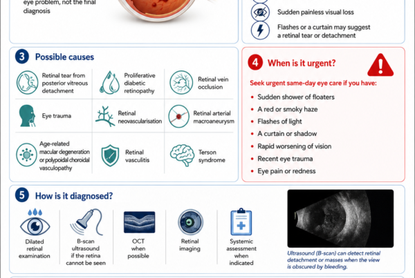
Seek same-day ophthalmic assessment for:
- New flashes
- A sudden increase in floaters
- A shower of dark spots
- Cobwebs
- A curtain or shadow
- Sudden loss of side vision
- Sudden blurred vision
The treatment may not always need to be performed within minutes, but the eye should be examined promptly because a tear can progress to retinal detachment without pain.
Does Every Retinal Tear Need Laser?
No.
Treatment depends on:
- Type of break
- Presence of symptoms
- Whether vitreous traction remains attached
- Presence of fluid beneath the retina
- Location and size
- Lattice degeneration
- Previous retinal detachment
- Condition of the other eye
- Ability to attend follow-up
Acute symptomatic horseshoe tears are usually treated promptly.
Many asymptomatic atrophic holes or operculated holes without traction may be safely observed. Current guidance does not recommend automatically treating every retinal hole or area of lattice degeneration.
Can Eyedrops Heal a Retinal Tear?
No.
Eyedrops cannot:
- Seal the retinal opening
- Release vitreous traction
- Create a protective retinal scar
- Prevent fluid passing through an untreated high-risk tear
Treatment, when required, usually involves:
- Laser retinopexy
- Cryotherapy
- Retinal-detachment surgery if fluid has already spread significantly beneath the retina
Can Laser Guarantee That the Retina Will Not Detach?
No.
Laser substantially reduces the risk but does not provide a complete guarantee.
In a 2026 study of 600 eyes treated for symptomatic retinal breaks:
- 7.2% later developed a rhegmatogenous retinal detachment.
- 11% required additional laser treatment.
- Risk was higher with multiple tears, vitreous haemorrhage and pseudophakia.
These findings reinforce the importance of follow-up and returning immediately if symptoms worsen.
Understanding the Retina
What Is the Retina?
The retina is the thin nerve layer lining the inside of the back of the eye.
It contains:
- Rod photoreceptors
- Cone photoreceptors
- Retinal nerve cells
- Supporting cells
- Retinal blood vessels
The retina converts light into electrical signals that travel through the optic nerve to the brain.
The Peripheral Retina
The peripheral retina provides much of the side visual field.
Most retinal tears develop in the peripheral retina rather than at the centre.
A peripheral tear may therefore be present while:
- Central visual acuity remains 6/6
- Reading vision remains clear
- There is no pain
- The patient notices only floaters or flashes
The Macula
The macula is the central part of the retina responsible for:
- Reading
- Recognising faces
- Driving
- Colour vision
- Fine visual detail
A peripheral retinal tear does not initially affect the macula.
If the resulting detachment spreads to the macula, central vision may deteriorate permanently.
Understanding the Vitreous
What Is the Vitreous?
The vitreous is the transparent gel filling most of the eye.
In youth, it is relatively firm and attached to the retinal surface.
With age, it gradually:
- Becomes more liquid
- Shrinks
- Separates from the retina
This process is called a posterior vitreous detachment, or PVD.
Is a PVD the Same as a Retinal Detachment?
No.
A PVD is separation of the vitreous gel from the retina.
A retinal detachment is separation of the retina from its supporting layer.
Most PVDs occur without retinal detachment. However, the vitreous may pull strongly enough to create a tear during the separation process.
Why Does the Vitreous Pull on Certain Areas?
The vitreous is not attached to the retina with equal strength everywhere.
Stronger attachment may occur:
- Around retinal blood vessels
- At the vitreous base
- Around areas of lattice degeneration
- Around previous scars or inflammation
- At sites of congenital vitreoretinal adhesion
When the surrounding vitreous separates but remains attached at one focal point, traction may lift and tear the retina.
How Does a Retinal Tear Develop?
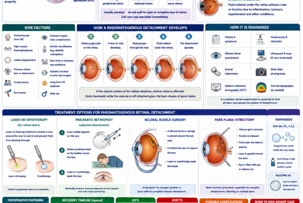
Step 1: The Vitreous Becomes More Liquid
Ageing and myopia accelerate vitreous liquefaction.
Step 2: The Vitreous Begins to Separate
The back surface of the vitreous detaches from the retina.
Step 3: Focal Traction Persists
A small area remains abnormally adherent.
Step 4: The Retina Tears
Continued pulling creates a full-thickness break.
Step 5: Fluid May Pass Through the Tear
Liquefied vitreous enters the space beneath the retina.
Step 6: A Retinal Detachment May Develop
The fluid spreads and lifts the retina away from its supporting tissue.
Without treatment, acute tractional tears have a substantial risk of progressing to retinal detachment. Retinopexy reduces this risk markedly, although published rates vary with tear characteristics and patient selection.
Symptoms of a Retinal Tear
A Sudden Increase in Floaters
Floaters may appear as:
- Small dots
- Threads
- Rings
- Cobwebs
- Insects
- Wispy strands
- A cloud of spots
A few stable longstanding floaters are common.
The concerning pattern is:
- A sudden onset
- A clear increase in number
- A shower of many small dots
- Floaters accompanied by flashes
- Floaters accompanied by blurred vision
A Shower of Black Dots
Numerous small dark spots may represent:
- Pigment cells released through a retinal tear
- Small amounts of blood within the vitreous
- Debris associated with an acute PVD
Pigment cells seen during slit-lamp examination are sometimes called Shafer’s sign or “tobacco dust” and increase suspicion of a retinal break.
Flashes of Light
Flashes occur when vitreous traction mechanically stimulates the retina.
They may appear as:
- Lightning streaks
- Camera flashes
- Sparks
- Brief arcs
- Flickering at the edge of vision
They are often most noticeable:
- In a dark room
- When moving the eye
- At the temporal side of the visual field
- During the early phase of a PVD
Cobwebs or a Moving Ring
A large ring-shaped floater may represent separation of vitreous tissue from around the optic nerve.
This is called a Weiss ring.
A Weiss ring supports the diagnosis of PVD but does not prove that the peripheral retina is intact.
Blurred or Hazy Vision
Blur may occur because of:
- Vitreous haemorrhage
- Numerous floaters
- A developing retinal detachment
- Inflammation
- Another associated retinal problem
A Curtain or Shadow
A curtain, veil or missing area of side vision suggests that fluid has already spread beneath the retina.
This indicates retinal detachment rather than an isolated tear and requires emergency retinal assessment.
Retinal Tears Are Usually Painless
Do not wait for:
- Eye pain
- Redness
- Discharge
- Headache
Most retinal tears and detachments cause none of these symptoms.
Floaters and Flashes: When Are They Concerning?
Lower-Risk Features
Symptoms may be less concerning when:
- Floaters have been unchanged for years.
- The pattern is identical to previously assessed symptoms.
- No flashes, blur or field defect is present.
- A recent complete retinal examination was reassuring.
Even then, a definite change warrants reassessment.
Higher-Risk Features
Risk is greater when symptoms include:
- Sudden numerous floaters
- A shower of dots
- Vitreous haemorrhage
- Retinal haemorrhage
- New flashes
- Reduced vision
- A field defect
- High myopia
- Lattice degeneration
- Previous tear or detachment
- Recent cataract surgery
- Recent trauma
Vitreous or retinal haemorrhage is particularly important. Studies have found a strong association between haemorrhage and the presence of one or multiple retinal tears.
Types of Retinal Break
Horseshoe Tear
A horseshoe tear is also called:
- Flap tear
- U-shaped tear
- Tractional retinal tear
The torn retinal flap remains attached to the vitreous.
This indicates persistent traction and is the classic high-risk retinal tear associated with acute PVD.
Acute symptomatic horseshoe tears are generally treated promptly with laser or cryotherapy.
Operculated Retinal Hole
An operculated hole forms when vitreous traction pulls a small circular piece of retina completely free.
The detached fragment is called the operculum.
Once the operculum has separated, active traction at the hole may be reduced.
Many asymptomatic operculated holes without surrounding fluid can be monitored, although treatment may be considered according to:
- Symptoms
- Residual traction
- Fluid beneath the retina
- Fellow-eye history
- Other risk factors
Atrophic Round Hole
An atrophic hole forms through gradual thinning rather than an acute pulling event.
It is frequently associated with lattice degeneration.
Many atrophic holes:
- Cause no symptoms
- Remain stable
- Do not require treatment
Selected holes may cause a slowly progressive retinal detachment, particularly in younger myopic patients.
Retinal Dialysis
A retinal dialysis is a tear at the front edge of the retina, near the ora serrata.
It is commonly associated with blunt trauma.
The resulting detachment may:
- Develop slowly
- Remain unnoticed initially
- Present months or years after injury
- Occur in a child or young adult
Retinal dialysis often requires scleral-buckle or other retinal-detachment surgery rather than simple office laser when significant detachment is present.
Giant Retinal Tear
A giant retinal tear extends through at least 90 degrees of the retinal circumference.
It is a complex condition associated with:
- Trauma
- High myopia
- Stickler syndrome
- Certain inherited vitreoretinal disorders
- Previous eye surgery
- Spontaneous vitreous change
It requires urgent specialist retinal surgery.
Macular Hole
A macular hole is an opening at the centre of the retina.
It causes:
- Central distortion
- Blurred central vision
- Missing letters
- A central grey spot
It is different from a peripheral retinal tear and is treated using vitrectomy and macular membrane surgery rather than ordinary peripheral laser retinopexy.
Lattice Degeneration
What Is Lattice Degeneration?
Lattice degeneration is an area of thinned peripheral retina.
It may appear:
- Linear
- Oval
- Pigmented
- Associated with white retinal vessels
- Associated with atrophic holes
- Associated with abnormal vitreous attachment
Is Lattice Degeneration a Retinal Tear?
No.
Lattice is a peripheral retinal degeneration that may increase the risk of a retinal tear or detachment.
Most people with lattice degeneration never develop retinal detachment.
Does All Lattice Need Laser?
No.
Routine laser is generally not recommended for every asymptomatic area of lattice degeneration.
Observation is often appropriate when there is:
- No symptomatic tractional tear
- No progressive subretinal fluid
- No relevant high-risk fellow-eye history
- No specific inherited vitreoretinal condition
- Reliable patient awareness and follow-up
The decision becomes more individualised when the patient has:
- Previous detachment in the other eye
- High myopia
- Aphakia
- Multiple retinal breaks
- Strong inherited risk
- Poor access to urgent care
- Planned intraocular surgery
- High-risk lattice features
Evidence for prophylactic treatment of asymptomatic lattice remains more limited than the evidence supporting treatment of acute symptomatic horseshoe tears.
Risk Factors
Increasing Age
Ageing causes vitreous liquefaction and posterior vitreous separation.
Retinal tears associated with acute PVD become more common in middle age and later life.
Myopia
Myopic eyes—particularly highly myopic eyes—may have:
- Longer axial length
- A thinner peripheral retina
- Earlier vitreous liquefaction
- Earlier PVD
- More lattice degeneration
- Greater risk of retinal tears and detachment
In prospective studies of symptomatic PVD, myopia has been associated with a higher likelihood of retinal breaks or delayed tears.
Cataract Surgery
Cataract surgery alters the vitreous environment and may be followed by PVD, retinal tears or detachment.
Risk is higher in selected patients, including those who are:
- Younger
- Male
- Highly myopic
- Long in axial length
- Affected by lattice degeneration
- Complicated by posterior capsule rupture or vitreous loss
Large registry data estimated that rhegmatogenous retinal detachment occurred in approximately one in 500 cataract operations among patients older than 40 during the first postoperative year, although individual risk varies substantially.
Previous Retinal Tear
A tear in one part of the retina indicates a vitreoretinal environment capable of producing additional breaks.
New tears can occur:
- Before laser scars fully mature
- During the continuing PVD process
- Months later
- In another quadrant
- In the fellow eye
Previous Retinal Detachment
A previous detachment increases the risk of a tear or detachment in:
- The repaired eye
- The fellow eye
Lattice Degeneration
Lattice is an important risk factor for delayed retinal tears after symptomatic PVD.
Vitreous Haemorrhage
Blood in the vitreous substantially increases the likelihood of a retinal tear.
A dense haemorrhage may prevent the tear from being seen and require:
- Repeated examination
- Ultrasound
- Early vitrectomy in selected cases
Eye Trauma
Blunt or penetrating injury may cause:
- Horseshoe tears
- Retinal dialysis
- Giant retinal tears
- Vitreous haemorrhage
- Retinal detachment
Symptoms may occur immediately or after a delay.
Family History
A family history may reflect inherited tendencies towards:
- High myopia
- Lattice degeneration
- Abnormal vitreous
- Stickler syndrome
- Other vitreoretinal disorders
Inherited Conditions
Risk may be particularly high in conditions such as:
- Stickler syndrome
- Marfan syndrome
- Familial exudative vitreoretinopathy
- Certain collagen disorders
- Previous retinopathy of prematurity
Previous Intraocular Surgery
Risk may be altered after:
- Cataract surgery
- Vitrectomy
- Glaucoma surgery
- Lens surgery
- Retinal procedures
- Eye trauma repair
Can a Retinal Tear Occur Without Symptoms?
Yes.
An asymptomatic break may be discovered during:
- Routine dilated examination
- Myopia screening
- Cataract assessment
- Examination of the fellow eye
- Follow-up after retinal detachment
- Diabetic retinal screening
The absence of symptoms does not by itself determine whether treatment is needed.
The tear type, traction and surrounding fluid are more important.
How Is a Retinal Tear Diagnosed?
Symptom History
The ophthalmologist will ask about:
- Time of onset
- Number of floaters
- Flashes
- Blurred vision
- A curtain or shadow
- Previous similar symptoms
- Myopia
- Trauma
- Previous surgery
- Previous tear or detachment
- Family history
Visual-Acuity Testing
Central vision may remain normal with an isolated peripheral tear.
Reduced acuity may suggest:
- Vitreous haemorrhage
- Macular involvement
- Retinal detachment
- Another ocular condition
Pupil Examination
A large retinal detachment may produce an abnormal pupil response.
An isolated peripheral tear usually does not.
Slit-Lamp Examination
The anterior vitreous is examined for:
- Pigment cells
- Blood
- Inflammatory cells
- Evidence of an acute PVD
Shafer’s sign strongly increases suspicion of a retinal break.
Dilated Retinal Examination
The pupil must generally be dilated to examine the peripheral retina adequately.
The ophthalmologist may use:
- Slit-lamp retinal lenses
- Indirect ophthalmoscopy
- Scleral indentation
- Wide-field photography as an adjunct
Scleral Indentation
Scleral indentation involves gently pressing the outside of the eye while examining the peripheral retina.
This brings the far peripheral retina into view and can reveal:
- Small horseshoe tears
- Retinal dialysis
- Multiple breaks
- Anterior tears hidden near the vitreous base
A prospective PVD study emphasised that some tears were detected only through indented indirect examination.
Wide-Field Photography
Wide-field imaging may document:
- Tear location
- Laser treatment
- Lattice degeneration
- Associated haemorrhage
It does not reliably replace a complete examination with scleral indentation.
Very anterior tears may fall outside the photographed area or be obscured by eyelids, eyelashes or poor image quality.
Ultrasound B-Scan
Ultrasound is useful when the retina cannot be seen because of:
- Dense vitreous haemorrhage
- Cataract
- Corneal opacity
- Small pupil
- Severe inflammation
Ultrasound may identify:
- Retinal detachment
- Vitreous haemorrhage
- Posterior vitreous separation
- Intraocular mass
A negative ultrasound does not always exclude a small retinal tear, so repeat examination remains necessary when suspicion is high.
Why Can Tears Be Missed During the First Examination?
The Tear Is Very Peripheral
Small anterior tears may be difficult to visualise without indentation.
Vitreous Haemorrhage Obscures the Retina
Blood may block the view.
The PVD Is Still Progressing
A new tear may develop after an initially normal examination.
The Pupil Does Not Dilate Well
A small pupil limits peripheral examination.
Cataract or Corneal Disease Reduces the View
Media opacity may obscure a retinal break.
More Than One Tear Is Present
Finding one tear should not end the examination.
The entire peripheral retina must be checked because multiple breaks may coexist.
Delayed Retinal Tears After PVD
A Normal First Examination Does Not End All Risk
In one long-term study, 7.39% of eyes with an acute symptomatic PVD but no tear at the initial visit later developed a retinal tear.
Half occurred within approximately 4.6 months, but some occurred substantially later. Lattice degeneration was the strongest identified risk factor.
Who May Need Closer Follow-Up?
Closer or extended follow-up may be appropriate for patients with:
- Vitreous haemorrhage
- Retinal haemorrhage
- Numerous floaters
- Persistent flashes
- Lattice degeneration
- Myopia
- Pseudophakia
- Fellow-eye tear or detachment
- Incomplete peripheral visualisation
Large database analyses similarly show that a clinically relevant proportion of PVD complications are diagnosed after the initial presentation.
Return Earlier if Symptoms Change
Do not wait for the scheduled review if there is:
- A new shower of floaters
- Increasing flashes
- Sudden haze
- A curtain
- Missing peripheral vision
- Reduced central vision
Which Retinal Tears Are Usually Treated?
Acute Symptomatic Horseshoe Tears
These are generally treated promptly because persistent vitreous traction increases the risk of retinal detachment.
Tears With Subretinal Fluid
Fluid extending beneath the retina suggests that the tear is already allowing separation.
Treatment urgency depends on:
- Amount of fluid
- Progression
- Location
- Proximity to the macula
Tears Associated With Vitreous Haemorrhage
These are higher-risk and require careful examination for:
- Multiple tears
- Retinal detachment
- Inadequate visibility
Tears With Persistent Traction
Active pulling at the tear increases risk.
Selected High-Risk Asymptomatic Tears
Treatment may be considered according to:
- Tear configuration
- Fellow-eye retinal detachment
- High myopia
- Aphakia
- Inherited vitreoretinal disease
- Inability to recognise symptoms
- Access to urgent care
- Planned ocular surgery
Which Retinal Breaks May Be Observed?
Observation may be appropriate for selected:
- Asymptomatic atrophic holes
- Operculated holes without traction
- Stable holes within lattice degeneration
- Longstanding pigmented breaks
- Lesions with no surrounding subretinal fluid
- Incidental breaks judged to have low detachment risk
Observation still requires:
- Patient education
- Appropriate follow-up
- Immediate review if symptoms develop
Laser Retinopexy
What Is Laser Retinopexy?
Laser retinopexy uses focused light energy to create small controlled burns around the retinal tear.
These burns produce a chorioretinal scar that adheres:
- The retina
- The retinal pigment epithelium
- The tissue beneath
The barrier reduces the risk of fluid passing through the tear and spreading beneath the retina.
Does the Laser Close the Opening?
The retinal opening remains anatomically present.
The treatment surrounds it with adhesive scar tissue.
How Is the Procedure Performed?
The eye is dilated.
Anaesthetic drops are applied.
Laser may be delivered using:
- A slit-lamp contact lens
- An indirect ophthalmoscope
- An operating-room laser system
The ophthalmologist applies several rows of laser spots around the tear.
The exact pattern depends on:
- Tear size
- Location
- Visibility
- Presence of fluid
- Nearby blood vessels
- Ability to surround the anterior edge
What Will I Experience?
Patients may notice:
- Bright flashes
- Coloured lights
- After-images
- Mild stinging
- Pressure from the contact lens
- Brief sharp sensations
More peripheral or extensive laser may be less comfortable.
How Long Does It Take?
The laser application often takes several minutes.
The full visit is longer because of:
- Pupil dilation
- Examination
- Consent
- Anaesthesia
- Post-treatment pressure or retinal checks
When Does the Laser Scar Become Strong?
The adhesion begins developing over the following days and continues strengthening.
This is why follow-up is required to assess whether:
- The tear is fully surrounded
- The laser uptake was adequate
- Additional treatment is required
- Subretinal fluid has progressed
Cryotherapy
What Is Cryotherapy?
Cryotherapy uses a freezing probe placed against the outside of the eye.
The freezing effect creates a controlled scar around the retinal break.
When Is Cryotherapy Used?
Cryotherapy may be chosen when:
- The tear is extremely peripheral
- Laser cannot reach the anterior edge
- The view is partially obscured
- The pupil is small
- A contact lens cannot be used
- Retinal-detachment surgery is being performed
What Is the Procedure Like?
Anaesthesia is required.
The patient may feel:
- Pressure
- Cold
- Temporary discomfort
- Eyelid swelling afterwards
Laser Versus Cryotherapy
Both aim to create retinal adhesion.
Laser is commonly preferred for accessible tears because it generally produces less external inflammation.
Cryotherapy remains valuable when the tear cannot be completely surrounded with laser.
Some retrospective evidence suggests that epiretinal-membrane formation may differ between cryotherapy and laser-treated eyes, although the choice is usually determined primarily by anatomy and access to the break.
What Happens After Laser Retinopexy?
Vision May Be Temporarily Blurred
Temporary blur may result from:
- Pupil dilation
- The contact lens
- Corneal surface disturbance
- Bright after-images
- Existing floaters or haemorrhage
Mild Discomfort May Occur
The eye may feel:
- Achy
- Gritty
- Light-sensitive
- Tired
Severe or increasing pain is not typical.
Floaters May Persist
Laser does not remove vitreous floaters.
The original floater may:
- Remain visible
- Move differently
- Become less noticeable with time
A sudden increase in floaters remains a warning sign even after treatment.
Flashes May Continue Temporarily
The PVD process may continue after the tear has been treated.
Flashes may gradually reduce as the vitreous completes separation.
Increasing or changing flashes require reassessment.
Activity After Laser Treatment
Follow the Treating Doctor’s Instructions
Recommendations vary according to:
- Tear size
- Amount of traction
- Presence of subretinal fluid
- Vitreous haemorrhage
- Whether a detachment is beginning
- Type of work or sport
Ordinary Visual Activity
Reading, watching television and using a computer do not usually pull a treated tear open.
Physical Activity
The surgeon may recommend temporarily avoiding:
- Heavy lifting
- Vigorous straining
- High-impact exercise
- Contact sports
- Activities involving rapid acceleration
- Eye trauma
These restrictions are usually precautionary and should be individualised.
Driving
Do not drive immediately after dilation if vision is blurred or light-sensitive.
Ongoing driving depends on:
- Visual acuity
- Floaters
- Field of vision
- Whether a detachment is present
- Vision in the other eye
Flying
An uncomplicated external laser retinopexy does not place gas inside the eye, so flying restrictions related to gas do not ordinarily apply.
Flying may still be inappropriate when:
- A retinal detachment is developing
- Surgery is planned urgently
- The treating specialist advises against travel
- Follow-up access would be limited
Follow-Up After Laser
Why Is Follow-Up Needed?
Follow-up determines whether:
- Laser scars are adequate
- The anterior edge is fully treated
- The original tear has extended
- A new tear has developed
- Fluid has progressed
- Retinal detachment has occurred
Additional Laser Is Not Unusual
In studies of treated retinal breaks, additional laser has been required because of:
- Incomplete initial treatment
- Extension of the original tear
- A new tear elsewhere
- Continued vitreous traction
A large series found that approximately 19% of treated eyes received additional laser and approximately 6% later required retinal-detachment surgery.
More recent data similarly found that follow-up laser and retinal detachment remain uncommon but clinically important after initial treatment.
Treatment Does Not End Symptom Awareness
Return immediately for:
- New floaters
- More flashes
- Sudden haze
- A curtain
- Missing side vision
- Reduced central vision
Why Can Retinal Detachment Occur Despite Laser?
A New Tear Develops Elsewhere
The vitreous may continue separating and pulling on another retinal area.
The Original Tear Extends
Persistent traction can enlarge the break beyond the treated border.
The Anterior Edge Was Difficult to Reach
A tear close to the ora serrata may be difficult to surround completely.
The Laser Scar Was Inadequate
Possible reasons include:
- Poor retinal visibility
- Vitreous haemorrhage
- Subretinal fluid
- Lightly pigmented retina
- Patient movement
- Limited laser uptake
Fluid Was Already Progressing
A detachment may have begun before the laser was applied.
Multiple Tears Were Present
An additional tear may have been hidden or developed later.
Proliferative Vitreoretinopathy Develops
This is uncommon after simple isolated tears but can complicate established retinal detachment.
Possible Complications of Laser Retinopexy
Laser retinopexy is generally safe, but possible complications include:
- Temporary blur
- Discomfort
- Inflammation
- Small retinal or vitreous haemorrhage
- Accidental laser to an unintended retinal area
- Epiretinal membrane
- Macular oedema
- Additional retinal tears
- Retinal detachment despite treatment
- Rare visual-field effects with extensive treatment
The risk of treatment must be balanced against the risk of leaving a high-risk symptomatic tear untreated.
Retinal Tear After Cataract Surgery
Why Can Cataract Surgery Be Followed by a Tear?
Cataract surgery may accelerate vitreous separation.
The risk is influenced by:
- Age
- Myopia
- Axial length
- Lattice degeneration
- Surgical complications
- Previous retinal tear or detachment
How Long Does the Risk Last?
A tear or detachment may occur:
- Soon after surgery
- Months later
- Several years later
New flashes, floaters or a curtain require assessment regardless of when the cataract surgery was performed.
Should Every Patient Receive Preventive Laser Before Cataract Surgery?
No.
Routine prophylactic laser for every asymptomatic peripheral lesion is not supported.
Treatment may be appropriate when examination identifies:
- An acute symptomatic horseshoe tear
- A high-risk untreated break
- Progressive subretinal fluid
- Another lesion with a specific significant risk profile
Retinal Tear After YAG Laser Capsulotomy
YAG laser capsulotomy treats clouding behind an intraocular lens.
It is performed at the front of the vitreous cavity rather than directly treating the retina.
Patients should seek urgent assessment after YAG if they develop:
- New floaters
- Flashes
- A curtain
- Sudden blur
- Missing peripheral vision
The presence of a retinal risk factor does not automatically mean that necessary YAG treatment should never be performed. The risks and benefits should be individualised.
Retinal Tears After Eye Trauma
Blunt Trauma
Possible examples include:
- Ball injury
- Racquet or shuttlecock injury
- Fist or elbow impact
- Airbag injury
- Fall
- Workplace accident
The eye may rapidly compress and expand, transmitting force to the retina.
Symptoms May Be Delayed
A traumatic tear or dialysis may not cause an immediate curtain.
Retinal detachment can develop after:
- Days
- Weeks
- Months
- Occasionally years
Protective Eyewear
Appropriate polycarbonate protective eyewear should be used during:
- Racquet sports
- High-velocity ball sports
- Industrial work
- Construction
- Activities with projectile risk
Retinal Tears in Children and Young Adults
Retinal tears are less commonly related to ordinary age-related PVD in children.
Possible causes include:
- Trauma
- High myopia
- Stickler syndrome
- Previous cataract surgery
- Retinopathy of prematurity
- Familial exudative vitreoretinopathy
- Other inherited vitreoretinal disorders
Children may not clearly report:
- Floaters
- Flashes
- A visual-field defect
- Reduced vision in one eye
A child with unexplained reduced vision, trauma or a relevant genetic condition requires specialist assessment.
The Fellow Eye
Is the Other Eye at Risk?
Yes.
Many risk factors affect both eyes, including:
- Myopia
- Lattice degeneration
- Abnormal vitreous
- Family history
- Inherited disease
- Previous cataract surgery
Should the Fellow Eye Receive Laser?
Not automatically.
A complete dilated examination should identify:
- Existing tears
- Lattice degeneration
- Atrophic holes
- PVD status
- High-risk abnormalities
Prophylactic treatment is considered individually.
Laser applied to visible lattice cannot prevent every future tear because a new tear may arise in previously normal retina.
Can Retinal Tears Be Prevented?
Not Every Tear Is Preventable
Age-related PVD may occur without warning.
Recognise Symptoms Early
Prompt assessment of flashes and floaters allows many tears to be treated before retinal detachment develops.
Use Eye Protection
Protective eyewear reduces trauma-related risk.
Control Myopia Progression in Children
Myopia-control treatment may reduce future high-myopia-related retinal risk, although it cannot completely remove inherited or structural susceptibility.
Attend Follow-Up
Patients with:
- Recent PVD
- Previous retinal tear
- Previous detachment
- High myopia
- Lattice degeneration
- Inherited vitreoretinal disease
should follow the recommended examination schedule.
Can Exercise Cause a Retinal Tear?
Ordinary Exercise
Routine walking, jogging and general exercise do not cause most retinal tears.
The principal mechanism is usually vitreoretinal traction during PVD.
Contact or High-Impact Activities
Eye trauma may cause retinal tears.
Patients with a newly diagnosed PVD, untreated tear, evolving detachment or recent retinopexy should discuss:
- Contact sports
- Heavy lifting
- High-impact exercise
- Roller coasters or rapid acceleration activities
- Combat sports
with their retinal specialist.
Case reports suggest that extreme acceleration forces may trigger retinal events in structurally predisposed eyes, although this is uncommon and does not mean that all ordinary movement is dangerous.
Can Coughing, Sneezing or Bending Cause a Tear?
Routine coughing, sneezing or bending does not usually create a retinal tear in a healthy eye.
Symptoms may coincidentally become noticeable during an activity when a PVD and tear are already developing.
Patients should not blame themselves for ordinary movement.
Retinal Tear Versus Migraine Aura
Vitreoretinal Flashes
These are often:
- Brief
- Lightning-like
- At the side of one eye
- Triggered by eye movement
- Associated with floaters
Migraine Visual Aura
Migraine aura commonly:
- Appears in both eyes or the same half of vision in both eyes
- Has a shimmering or zigzag pattern
- Expands gradually
- Lasts several minutes
- May be followed by headache
Patients may find it difficult to distinguish them.
A new one-sided flash pattern—particularly with floaters—requires retinal examination.
Retinal Tear Versus Retinal Detachment
Retinal Tear
A retinal tear is an opening in an otherwise largely attached retina.
Possible symptoms include:
- Flashes
- Floaters
- Mild haze
Retinal Detachment
Fluid has passed through the tear and lifted the retina.
Possible symptoms include:
- A curtain
- Missing peripheral vision
- Increasing blur
- Central visual loss when the macula detaches
A tear may often be treated with office laser.
An established spreading detachment usually requires:
- Pneumatic retinopexy
- Scleral-buckle surgery
- Vitrectomy
- A combination of procedures
Common Myths
“A Retinal Tear Always Causes Pain”
False.
Retinal tears are usually painless.
“All Floaters Are Harmless”
False.
Longstanding stable floaters are common, but sudden new floaters may indicate a tear or haemorrhage.
“Laser Removes the Floater”
False.
Laser retinopexy treats the retinal break. It does not remove the vitreous opacity causing the floater.
“Once Laser Is Done, No Follow-Up Is Needed”
False.
The tear may require additional treatment, and new tears may develop.
“Every Retinal Hole Must Be Lasered”
False.
Many asymptomatic atrophic or operculated holes can be observed.
“Every Area of Lattice Degeneration Must Be Treated”
False.
Management is risk-based.
“Reading or Using a Phone Will Make the Tear Open”
Ordinary visual use does not generally pull a treated retinal tear open.
“A Normal First Examination Means No Tear Can Develop Later”
False.
Delayed tears may develop as the PVD progresses.
“Laser Guarantees That Retinal Detachment Cannot Occur”
False.
Laser reduces risk but does not eliminate it.
“Good Central Vision Means the Retina Is Safe”
False.
A peripheral tear or detachment may be present while central acuity remains excellent.
Frequently Asked Questions
How Quickly Can a Retinal Tear Become a Detachment?
Progression is unpredictable.
It may occur:
- Within hours
- Over several days
- More gradually
- Not at all
This uncertainty is why a symptomatic high-risk tear is treated promptly.
How Soon Should Laser Be Performed?
An acute symptomatic horseshoe tear is generally treated as soon as practical after diagnosis.
Timing may depend on:
- Whether retinal detachment has begun
- Tear location
- Visibility
- Availability of the necessary equipment
- Medical considerations
Is Laser Retinopexy Painful?
Most patients tolerate it with anaesthetic drops.
Peripheral treatment may cause brief stinging, pressure or aching.
Is One Laser Session Enough?
Often, but not always.
Additional treatment may be needed because:
- Laser uptake was incomplete.
- The tear extended.
- An untreated gap remains.
- A new tear developed.
Can the Laser Scar Be Seen?
The patient does not usually perceive the peripheral laser scars.
They are visible during retinal examination and may appear pale or pigmented as they mature.
Will My Flashes Stop Immediately?
Not necessarily.
Flashes may continue while the vitreous completes separation.
They should gradually improve.
A sudden increase or change requires review.
Will My Floaters Disappear?
Some become less noticeable as:
- They move away from the visual axis.
- The blood clears.
- The brain adapts.
Laser itself does not remove them.
Can I Shower After Laser?
Ordinary showering does not usually affect an internal retinal laser scar.
Follow any specific instructions if a contact lens caused corneal irritation or another procedure was performed.
Can I Swim After Laser?
Swimming restrictions vary according to:
- Whether only laser was performed
- Corneal surface condition
- Additional eye procedures
- The clinician’s protocol
Retinal laser alone does not create an external wound, but strenuous swimming and delayed access to care may be discouraged during the initial observation period.
Can I Fly After Laser?
Usually yes, because no gas bubble is inserted during ordinary laser retinopexy.
Do not travel when:
- A detachment is suspected
- Urgent follow-up is required
- Surgery may be needed
- The retinal specialist advises against it
Can I Exercise After Laser?
Light ordinary activity is commonly permitted.
Temporary restriction of strenuous or high-impact exercise may be advised while the laser scar develops.
Can a Retinal Tear Heal Naturally?
The retinal opening usually remains.
An operculated or low-risk hole may remain stable without treatment, but a symptomatic tractional horseshoe tear should not be expected to seal safely by itself.
Can Laser Damage Vision?
Serious visual damage is uncommon when peripheral laser is correctly applied.
Possible complications exist, but the risk is usually much lower than the risk posed by an untreated high-risk tear.
Why Do I Need Repeat Dilation?
A repeat peripheral examination checks for:
- New tears
- Tear extension
- Incomplete laser
- Subretinal fluid
- Retinal detachment
Can a Retinal Tear Recur?
The original tear remains anatomically present but is surrounded by scar.
A new tear may develop elsewhere.
Can Both Eyes Develop Tears?
Yes.
Symptoms in the other eye require a new urgent examination.
Should My Family Members Be Screened?
Routine screening of every relative is unnecessary.
Examination may be appropriate when there is:
- Strong family history
- High myopia
- Stickler syndrome
- Previous detachment at a young age
- Another inherited vitreoretinal disorder
Can a Tear Cause Complete Blindness?
An isolated treated tear usually does not.
An untreated tear may progress to retinal detachment and severe permanent visual loss.
When to Seek Emergency Eye Care
Seek immediate same-day assessment for:
- Sudden new floaters
- A marked increase in floaters
- A shower of black dots
- Cobwebs
- New flashes
- Sudden haze
- A curtain or shadow
- Missing peripheral vision
- Sudden blurred vision
- Visual symptoms after trauma
- New symptoms after cataract or YAG laser treatment
After retinal laser, return urgently for:
- New or increasing floaters
- Increasing flashes
- A curtain
- Missing side vision
- Sudden visual deterioration
- Significant pain or redness
- Trauma to the eye
Do not drive yourself when vision is substantially impaired.
A Retinal-Tear Emergency Checklist
Report
Tell the ophthalmologist about:
- When symptoms began
- Whether floaters increased suddenly
- Flashes
- A curtain or field defect
- Eye trauma
- High myopia
- Previous cataract surgery
- Previous tear or detachment
- Family history
- Blood-thinning medication
Expect
Assessment may include:
- Visual acuity
- Pupil examination
- Slit-lamp examination
- Dilating drops
- Indirect ophthalmoscopy
- Scleral indentation
- Wide-field photography
- Ultrasound when the retina cannot be seen
Ask
- Is there a retinal tear or detachment?
- What type of tear is present?
- Is vitreous traction still attached?
- Is there fluid beneath the retina?
- Is laser or cryotherapy required?
- Is the tear fully surrounded?
- When is the follow-up examination?
- What activity restrictions apply?
- What symptoms require immediate return?
- Does the other eye have high-risk changes?
The Bottom Line
A retinal tear is a full-thickness break in the peripheral retina.
It commonly develops when the vitreous gel separates and pulls strongly on a focal area of retina.
The principal warning symptoms are:
- A sudden increase in floaters
- A shower of black dots
- Cobwebs
- Flashes of light
- A curtain or shadow
- Sudden blurred or missing vision
Retinal tears are generally painless.
The most important high-risk tear is an acute symptomatic horseshoe tear with persistent vitreous traction.
Treatment usually involves:
- Laser retinopexy
- Cryotherapy when laser cannot adequately reach the break
The treatment creates a scar around the tear to reduce the risk of retinal detachment.
Not every retinal hole or area of lattice degeneration requires treatment. Management depends on:
- Symptoms
- Tear type
- Traction
- Subretinal fluid
- Myopia
- Previous retinal detachment
- Fellow-eye findings
- Other individual risk factors
Laser greatly reduces the likelihood of retinal detachment but does not eliminate it.
After treatment:
- The PVD may continue evolving.
- A new retinal tear may form.
- Additional laser may be required.
- Retinal detachment remains possible.
The most important message is:
Sudden new floaters, flashes or a curtain over the vision require an urgent dilated retinal examination—even when there is no pain and central vision remains clear.
References
- Kim SJ, Bailey ST, Kovach JL, et al. Posterior vitreous detachment, retinal breaks, and lattice degeneration Preferred Practice Pattern®. Ophthalmology. 2025;132(4)–P196. doi:10.1016/j.ophtha.2024.12.023.
- Nixon TRW, Davie RL, Snead MP. Posterior vitreous detachment and retinal tear: a prospective study of community referrals. Eye. 2024;38:2865–2871. doi:10.1038/s41433-023-02779-3. PMID: 37798362.
- Jindachomthong W, Zheng A, Akintilo L, et al. Incidence and risk factors for delayed retinal tears after an acute, symptomatic posterior vitreous detachment. Ophthalmol Retina. 2023;7(2):143–150. doi:10.1016/j.oret.2022.10.013. PMID: 36307014.
- Uhr JH, Obeid A, Wibbelsman TD, et al. Delayed retinal breaks and detachments after acute posterior vitreous detachment. Ophthalmology. 2020;127(4):516–522. doi:10.1016/j.ophtha.2019.10.020. PMID: 31767432.
- Choi S, Waheed NK, et al. Clinical outcomes of symptomatic horseshoe tears after laser retinopexy. J Vitreoretin Dis. 2023;7(6):471–476. doi:10.1177/24741264231196337. PMID: 37927320.
- Zhao MY, Yuan Z, Rohowetz LJ, Yannuzzi NA, Smiddy WE, Flynn HW Jr. Outcomes of symptomatic retinal breaks treated with laser retinopexy. J Vitreoretin Dis. 2026. doi:10.1177/24741264261453044. PMID: 42221732.
- Moussa G, Bassilious K, Mathews N. Primary retinopexy in preventing retinal detachment in a tertiary eye hospital: a study of 1,157 eyes. Eye. 2022;36:976–983. doi:10.1038/s41433-021-01548-8. PMID: 34035492.
- Garoon RB, Smiddy WE, Flynn HW Jr. Treated retinal breaks: clinical course and outcomes. Graefes Arch Clin Exp Ophthalmol. 2018;256(6):1053–1057. doi:10.1007/s00417-018-3958-2. PMID: 29523992.
- Goh YW, Ehrlich R, Stewart J, Polkinghorne P. The incidence of retinal breaks in the presenting and fellow eyes in patients with acute symptomatic posterior vitreous detachment and their associated risk factors. Asia Pac J Ophthalmol. 2015;4(1):5–8. doi:10.1097/APO.0000000000000072. PMID: 26068606.
- Karahan E, et al. Risk factors for multiple retinal tears in patients with acute posterior vitreous detachment. Int Ophthalmol. 2018;38(1):257–263. PMID: 28160191.
- Blindbæk S, Grauslund J. Prophylactic treatment of retinal breaks: a systematic review. Acta Ophthalmol. 2015;93(1):3–8. doi:10.1111/aos.12447.
- Wilkinson CP. Evidence-based analysis of prophylactic treatment of asymptomatic retinal breaks and lattice degeneration. Ophthalmology. 2000;107(1):12–15. PMID: 10647712.
- Morano MJ, Khan MA, Zhang Q, et al. Incidence and risk factors for retinal detachment after cataract surgery in the IRIS Registry. Ophthalmol Sci. 2023;3(4):100314. PMID: 37274012.
- Moussa G, Kottegoda Y, Andreatta W, et al. Epiretinal membrane formation after treatment of retinal breaks: cryoretinopexy versus laser retinopexy. Ophthalmol Retina. 2019;3(12):1087–1092. PMID: 31446030.
- Chronopoulos A, Schutz JS, Finger RP. Prevention of rhegmatogenous retinal detachment. Surv Ophthalmol. 2025;70(6):1061–1066. doi:10.1016/j.survophthal.2025.04.006. PMID: 40339663.