Author: Dr Val Phua
Estimated reading time: 20 minutes
A retinal detachment occurs when the light-sensitive retina separates from the layer beneath it that normally provides support, nourishment and oxygen.
The retina lines the inside of the back of the eye and converts light into electrical signals that travel through the optic nerve to the brain.
When the retina becomes detached, its function begins to deteriorate. Without prompt treatment, the condition may cause:
- Permanent loss of peripheral vision
- Severe reduction in central vision
- Retinal scarring
- Recurrent detachment
- Low eye pressure
- Shrinkage and loss of function of the eye
- Blindness in the affected eye
The most common form is rhegmatogenous retinal detachment, in which fluid passes through a tear or hole in the retina and accumulates underneath it.
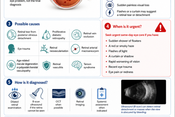
Retinal detachment is generally painless. The principal warning symptoms are:
- A sudden increase in floaters
- Flashes of light
- A dark curtain or shadow
- Missing peripheral vision
- Sudden blurred or reduced vision
These symptoms require urgent same-day ophthalmic assessment.
Early treatment is particularly important while the macula—the central part of the retina responsible for detailed vision—remains attached. Evidence suggests that macula-on detachments repaired within approximately 24 hours may achieve slightly better final vision than those repaired later, while macula-off detachments may have better outcomes when treated within the first few days after central vision is affected. The precise timing depends on the retinal configuration, duration, surgical resources and individual patient factors.
The Quick Answer
What Is a Retinal Detachment?
A retinal detachment is a separation of the neurosensory retina from the retinal pigment epithelium beneath it.
The detached retina cannot function normally because:
- Its photoreceptors are displaced from their supporting tissue.
- Oxygen and nutrient delivery become impaired.
- Fluid accumulates beneath the retina.
- Retinal cells may suffer permanent damage if separation persists.
Is a Retinal Detachment an Emergency?
Yes.
A suspected retinal detachment requires urgent assessment by an ophthalmologist or retinal specialist.
Do not wait for:
- Pain
- Redness
- Complete loss of vision
- The shadow to reach the centre
- A routine appointment several weeks later
A detachment may enlarge without causing discomfort.
What Are the Main Warning Symptoms?
Seek urgent care for:
- A sudden shower of new floaters
- Numerous black dots
- Cobwebs or strands
- Flashes of light
- A curtain or veil
- A dark shadow moving across the vision
- Missing side vision
- Sudden blur
- A sudden reduction in central vision
The symptoms are usually present in one eye, although patients may initially find it difficult to identify which eye is affected.
Can Eyedrops Treat a Retinal Detachment?
No.
Eyedrops cannot:
- Seal a retinal tear
- Remove fluid beneath the retina
- Reattach the retina
- Prevent an established detachment from spreading
Treatment usually requires:
- Laser or cryotherapy for selected retinal tears
- Pneumatic retinopexy
- Scleral-buckle surgery
- Pars plana vitrectomy
- A combination of procedures
Can Vision Return After Surgery?
Many patients regain useful vision after successful retinal reattachment.
The result depends on:
- Whether the macula was detached
- How long it was detached
- The extent and location of the detachment
- The cause
- The presence of retinal scarring
- The health of the optic nerve and macula
- Whether the retina remains attached after surgery
- Other conditions such as cataract, glaucoma or diabetic retinopathy
Successful anatomical reattachment does not guarantee a complete return to the vision present before the detachment.
Understanding the Retina
What Is the Retina?
The retina is a thin layer of nerve tissue lining the inside of the back of the eye.
It contains:
- Rod photoreceptors
- Cone photoreceptors
- Retinal nerve cells
- Supporting cells
- Blood vessels
The retina detects light and begins the process of vision.
What Is the Retinal Pigment Epithelium?
The retinal pigment epithelium, or RPE, lies immediately beneath the neurosensory retina.
It helps:
- Support photoreceptor function
- Remove metabolic waste
- Transport nutrients
- Maintain the outer blood-retinal barrier
- Pump fluid out from beneath the retina
When the retina separates from the RPE, this close support system is disrupted.
What Is the Macula?
The macula is the central area of the retina responsible for:
- Reading
- Recognising faces
- Driving
- Colour vision
- Fine visual detail
- Seeing directly in front of you
The centre of the macula is called the fovea.
Whether the macula is still attached is one of the most important factors influencing urgency and visual prognosis.
The Three Main Types of Retinal Detachment
Rhegmatogenous Retinal Detachment
A rhegmatogenous retinal detachment develops when fluid passes through a full-thickness retinal break.
“Rhegma” means a tear or rupture.
The sequence commonly involves:
- The vitreous gel pulls on the retina.
- A tear or hole develops.
- Liquefied vitreous passes through the break.
- Fluid accumulates beneath the retina.
- The detachment enlarges.
Rhegmatogenous retinal detachment is the most common type treated surgically.
Tractional Retinal Detachment
A tractional detachment occurs when scar tissue attached to the retinal surface contracts and mechanically pulls the retina away from the underlying tissue.
There may initially be no retinal tear.
Common causes include:
- Proliferative diabetic retinopathy
- Retinopathy of prematurity
- Sickle-cell retinopathy
- Previous trauma
- Proliferative vitreoretinopathy
- Other retinal vascular or inflammatory disorders
If traction creates a retinal tear, the condition becomes a combined tractional and rhegmatogenous detachment.
Exudative Retinal Detachment
An exudative or serous detachment occurs when fluid accumulates beneath the retina without a retinal tear or direct vitreoretinal traction.
Possible causes include:
- Inflammation
- Posterior scleritis
- Uveal effusion
- Severe hypertension
- Pre-eclampsia
- Retinal or choroidal tumours
- Central serous chorioretinopathy
- Certain vascular disorders
Treatment is directed at the underlying disease rather than automatically performing retinal-tear surgery.
Retinal Tear Versus Retinal Detachment
What Is a Retinal Tear?
A retinal tear is a full-thickness break in the retina through which fluid may pass.
A tear may occur before a detachment develops.
Does Every Tear Cause a Detachment?
No.
Some tears are detected and treated before fluid accumulates beneath the retina.
Others may remain stable, particularly selected small atrophic holes without vitreous traction.
Why Are Some Tears Treated?
Laser or cryotherapy may be recommended when a tear has a significant risk of progressing to retinal detachment.
Risk is greater when the tear is:
- Symptomatic
- Associated with an acute posterior vitreous detachment
- Horseshoe-shaped
- Associated with subretinal fluid
- Located in a high-risk area
- Present in a patient with relevant fellow-eye or surgical history
The American Academy of Ophthalmology recommends prompt treatment of acute symptomatic horseshoe tears, while many asymptomatic atrophic holes within lattice degeneration can be observed.
Posterior Vitreous Detachment
What Is the Vitreous?
The vitreous is the transparent gel filling most of the inside of the eye.
In youth, it is relatively solid and attached to the retinal surface.
With age, it gradually:
- Becomes more liquid
- Shrinks
- Pulls away from the retina
This process is called posterior vitreous detachment, or PVD.
Is PVD the Same as Retinal Detachment?
No.
A PVD is separation of the vitreous gel from the retina.
A retinal detachment is separation of the retina from the RPE beneath it.
A PVD is common with ageing and often causes no lasting damage. However, vitreous traction during separation may tear the retina.
Symptoms of PVD
Possible symptoms include:
- A new large floater
- Cobwebs
- Small black dots
- Flashes in the peripheral vision
- A moving ring-shaped floater
Because a symptomatic PVD may be accompanied by a retinal tear, new flashes or floaters require a dilated retinal examination.
Can a Tear Develop After the First Examination?
Yes.
A retinal tear may develop after an initial examination that shows only a PVD.
One long-term study found that delayed tears occurred in a meaningful minority of patients with acute symptomatic PVD, including tears developing beyond the traditional six-week review period. Risk was greater in patients with lattice degeneration, younger age and myopia.
Patients should return urgently if symptoms increase, even if the first examination was reassuring.
Common Symptoms of Retinal Detachment
New Floaters
Floaters may appear as:
- Dots
- Threads
- Rings
- Cobwebs
- Insects
- Wispy clouds
- A shower of black specks
A few longstanding stable floaters are common.
A sudden increase is more concerning.
Flashes of Light
Flashes may result from vitreous traction stimulating the retina mechanically.
Patients may describe:
- Lightning streaks
- Camera flashes
- Sparks
- Flickering at the side of the vision
- Brief flashes in a dark room
Flashes from vitreoretinal traction usually occur in the peripheral visual field.
A Curtain or Shadow
As the retina detaches, the corresponding visual field may become:
- Dark
- Grey
- Blurred
- Missing
- Covered by a curtain or veil
The perceived shadow appears opposite the anatomical retinal location.
A superior retinal detachment may therefore produce an inferior field defect.
Blurred Vision
Blur may occur because of:
- Vitreous haemorrhage
- Macular involvement
- Retinal folds
- Loss of part of the visual field
- Associated inflammation
- Cataract or corneal changes
Sudden Central Visual Loss
Central vision falls when:
- The detachment reaches the macula
- The fovea becomes detached
- Vitreous haemorrhage obscures the view
- Another central retinal complication occurs
Distorted Vision
After or during macular detachment, straight lines may appear:
- Wavy
- Bent
- Compressed
- Stretched
- Misaligned
Distortion may persist after successful repair.
Retinal Detachment Is Usually Painless
The retina does not contain pain fibres that produce ordinary eye pain.
A painless emergency can still be extremely serious.
Pain or redness may indicate:
- Acute glaucoma
- Inflammation
- Infection
- Trauma
- A postoperative complication
- Another ocular condition
Macula-On and Macula-Off Detachment
Macula-On Retinal Detachment
In a macula-on detachment, the central fovea remains attached.
Central visual acuity may still be relatively good, although peripheral vision may be missing.
The objective is to repair the retina before the detachment reaches the fovea.
This commonly requires urgent surgery, frequently on the same day or within approximately 24 hours, depending on the anatomy and surgical circumstances.
Macula-Off Retinal Detachment
In a macula-off detachment, the fovea has separated.
The patient commonly has:
- Reduced central vision
- Distortion
- A dense central shadow
- Difficulty reading or recognising faces
Surgery remains urgent.
A systematic review found that repair within approximately three days of macular involvement was associated with better average final visual acuity than repair during days four to seven, although the evidence was observational and individual outcomes vary.
How Does the Doctor Know Whether the Macula Is Attached?
Assessment may involve:
- Dilated retinal examination
- Macular OCT
- Symptom history
- Retinal photography
- Ultrasound when the retina cannot be seen
OCT may detect shallow foveal fluid that is difficult to identify clinically.
Risk Factors for Rhegmatogenous Retinal Detachment
Increasing Age
Age-related vitreous liquefaction and PVD increase the likelihood of retinal tears.
Myopia
Myopic eyes, particularly highly myopic eyes, are at increased risk because they may have:
- Greater axial length
- A thinner peripheral retina
- Earlier PVD
- Lattice degeneration
- Peripheral retinal holes
- Greater susceptibility to traction
High-myopia-related retinal complications include retinal tears and detachment.
Lattice Degeneration
Lattice degeneration is an area of thinned peripheral retina.
It may contain:
- Atrophic holes
- Abnormal vitreous attachment
- Pigmentation
- White retinal vessels
Most people with lattice degeneration never develop a retinal detachment.
Routine preventive laser is not required for every asymptomatic lattice lesion. Management depends on symptoms, retinal breaks and individual risk.
Previous Retinal Detachment
A previous detachment increases the risk in the same or fellow eye.
Family History
A close family history may increase risk, partly through inherited tendencies towards:
- High myopia
- Lattice degeneration
- Abnormal vitreous
- Connective-tissue disorders
Cataract Surgery
Retinal detachment is an uncommon but recognised complication after cataract surgery.
Risk is greater in selected patients, including:
- Younger patients
- Men
- Highly myopic eyes
- Eyes with long axial length
- Eyes with surgical complications
- Eyes with posterior capsular rupture or vitreous loss
- Patients with previous retinal detachment
A 2024 systematic review involving more than five million patients estimated a pooled retinal-detachment incidence of approximately 0.66% after cataract surgery, although individual risk varied considerably.
Previous Eye Trauma
Blunt or penetrating trauma may cause:
- Retinal dialysis
- Horseshoe tears
- Giant retinal tears
- Vitreous haemorrhage
- Direct retinal injury
- Detachment developing weeks, months or years later
Traumatic retinal detachment may be complex and can affect children or younger adults.
Previous Retinal or Intraocular Surgery
Vitrectomy, glaucoma surgery and other intraocular procedures may alter vitreoretinal relationships and risk.
Genetic and Connective-Tissue Disorders
Higher risk may occur with conditions such as:
- Stickler syndrome
- Marfan syndrome
- Certain hereditary vitreoretinopathies
- Retinopathy of prematurity
- Familial exudative vitreoretinopathy
Aphakia
An aphakic eye has no natural or artificial lens.
Aphakia is associated with greater retinal-detachment risk than an uncomplicated eye with an intraocular lens.
Diabetic Retinopathy
Proliferative diabetic retinopathy can produce tractional or combined retinal detachment.
Inflammation and Infection
Conditions such as:
- Uveitis
- Acute retinal necrosis
- Posterior scleritis
- Severe choroidal inflammation
may cause rhegmatogenous, tractional or exudative detachment.
How Common Is Retinal Detachment?
Rhegmatogenous retinal detachment is uncommon but occurs often enough to represent an important cause of emergency retinal surgery.
A 2024 international systematic review and meta-analysis found that incidence varies between countries and has increased over time in several populations, partly reflecting ageing, myopia and cataract-surgery patterns.
The condition can occur at any age, but the underlying causes differ:
- PVD-related tears are common in older adults.
- Myopia-related detachments may occur earlier.
- Trauma and hereditary disorders are particularly important in children and young adults.
How Is Retinal Detachment Diagnosed?
Symptom History
The ophthalmologist will ask about:
- Onset of floaters
- Flashes
- A shadow or curtain
- Central vision loss
- Eye trauma
- Myopia
- Previous surgery
- Family history
- Previous retinal tear or detachment
- Symptoms in the fellow eye
Visual-Acuity Testing
Central visual acuity helps assess:
- Macular involvement
- Baseline function
- Visual prognosis
- Postoperative recovery
Good central acuity does not exclude a peripheral retinal detachment.
Pupil Examination
A relative afferent pupillary defect may be present in a large or severe detachment.
Dilated Retinal Examination
The pupil is dilated so that the ophthalmologist can examine the retina using:
- Indirect ophthalmoscopy
- Slit-lamp lenses
- Scleral indentation
- Wide-field imaging when available
The examination aims to identify:
- Every retinal tear or hole
- Extent of detachment
- Macular status
- Lattice degeneration
- Vitreous haemorrhage
- Proliferative vitreoretinopathy
- Other retinal disease
Scleral Indentation
Gentle pressure is applied externally to bring the peripheral retina into view.
This helps detect small anterior tears that may otherwise be missed.
Optical Coherence Tomography
OCT may help assess:
- Whether the fovea is attached
- Shallow macular fluid
- Outer-retinal damage
- Postoperative subretinal fluid
- Epiretinal membrane
- Macular folds
Ultrasound B-Scan
Ultrasound is particularly useful when the retina cannot be seen because of:
- Dense vitreous haemorrhage
- Cataract
- Corneal opacity
- Severe inflammation
- Small pupil
It may demonstrate:
- Retinal detachment
- Vitreous haemorrhage
- Choroidal detachment
- Intraocular mass
- Posterior vitreous separation
Retinal Photography
Wide-field photographs may document the detachment but do not replace a complete peripheral retinal examination.
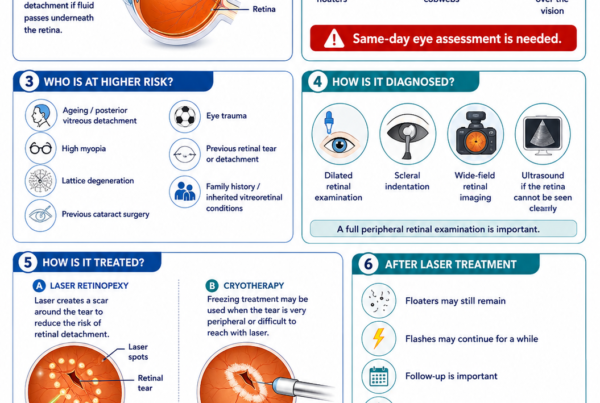
Laser Treatment for Retinal Tears
What Is Laser Retinopexy?
Laser retinopexy places burns around a retinal tear.
The laser produces a controlled scar between:
- The retina
- The retinal pigment epithelium
- The underlying tissue
This scar forms a barrier intended to prevent fluid from spreading through the tear.
When Is Laser Used?
Laser may be used for:
- Symptomatic horseshoe tears
- Selected retinal holes with subretinal fluid
- Tears found before cataract or other surgery when clinically indicated
- Additional retinal breaks identified during follow-up
- Retinal breaks surrounding a localised detachment in selected situations
Does Laser Immediately Seal the Tear?
No.
The scar strengthens over days.
Patients must continue monitoring for:
- New floaters
- Increasing flashes
- A curtain
- Visual-field loss
- Sudden blur
Laser substantially reduces risk but does not guarantee that detachment will never occur.
What Happens During Laser?
The eye is dilated and numbed.
A contact lens may be placed on the eye.
The patient may see:
- Bright flashes
- Coloured lights
- After-images
Some patients feel:
- Mild stinging
- Pressure
- Brief sharp sensations
Cryotherapy
Cryotherapy uses a freezing probe on the outside of the eye to create a scar around the retinal break.
It may be used when:
- The tear is very peripheral
- The view is limited
- Laser access is difficult
- Scleral-buckle surgery is planned
Treatment of Established Rhegmatogenous Retinal Detachment
The Principles of Repair
Successful repair usually requires:
- Identifying the retinal break.
- Closing or supporting the break.
- Relieving relevant vitreous traction.
- Removing or displacing subretinal fluid.
- Keeping the retina in position while scarring develops.
There Is No Single Best Operation for Every Detachment
The procedure is selected according to:
- Location and number of tears
- Lens status
- Age
- Presence of PVD
- Extent of detachment
- Macular status
- Vitreous haemorrhage
- Proliferative vitreoretinopathy
- High myopia
- Previous surgery
- Ability to maintain postoperative positioning
The principal options are pneumatic retinopexy, scleral buckling and pars plana vitrectomy.
Pneumatic Retinopexy
What Is Pneumatic Retinopexy?
Pneumatic retinopexy is a minimally invasive procedure in which:
- A gas bubble is injected into the eye.
- The patient positions the head so the bubble covers the tear.
- Laser or cryotherapy seals the retinal break.
It may be performed in a procedure room or operating theatre, depending on local practice.
Who May Be Suitable?
It is generally considered for selected uncomplicated detachments with:
- Superior retinal break or breaks
- Breaks located close together
- A clear view of the retina
- Limited proliferative vitreoretinopathy
- Ability to maintain positioning
- Reliable access to close follow-up
It is not suitable for every retinal configuration.
Advantages
Possible advantages include:
- No large incision
- Faster initial procedure
- Less induced short-sightedness than a buckle
- Less cataract progression than vitrectomy in some phakic eyes
- Less retinal displacement
- Potentially better visual quality in appropriately selected patients
The PIVOT randomised trial found that pneumatic retinopexy produced better average visual acuity, less vertical distortion and lower morbidity than primary vitrectomy among patients meeting its strict eligibility criteria.
Limitations
Pneumatic retinopexy has a lower single-procedure success rate in many studies than vitrectomy.
Failure may occur because of:
- Missed retinal tear
- New tear
- Inadequate positioning
- Gas not covering the break
- Extension of the detachment
- Proliferative vitreoretinopathy
Long-term studies nevertheless support pneumatic retinopexy as a durable first-line option for appropriately selected detachments.
Scleral-Buckle Surgery
What Is a Scleral Buckle?
A scleral buckle is a silicone band or segment secured to the outside of the eye.
It indents the wall of the eye towards the retinal tear.
This:
- Supports the break
- Reduces vitreous traction
- Brings the retinal layers closer together
- Allows laser or cryotherapy scars to secure the retina
The buckle normally remains permanently unless it causes a specific problem.
Who May Benefit?
A buckle may be particularly useful for:
- Younger phakic patients
- Retinal dialysis after trauma
- Detachments without a complete PVD
- Selected inferior or anterior tears
- Multiple breaks suitable for buckle support
- Eyes in which preserving the natural lens is desirable
Advantages
Possible advantages include:
- Avoiding removal of the vitreous
- Lower cataract risk than vitrectomy
- Good results in selected phakic eyes
- Effective support for anterior retinal pathology
The multicentre SPR randomised study found better improvement in best-corrected visual acuity with scleral buckling than primary vitrectomy in phakic eyes, while outcomes differed in pseudophakic eyes.
Possible Effects
A buckle may cause:
- Increased short-sightedness
- Astigmatism
- Temporary double vision
- Eyelid swelling
- Eye discomfort
- Change in eye shape
- Rare buckle infection or exposure
Pars Plana Vitrectomy
What Is Vitrectomy?
Vitrectomy removes the vitreous gel from inside the eye.
The surgeon can then:
- Release vitreoretinal traction
- Identify retinal tears
- Remove blood or membranes
- Drain subretinal fluid
- Apply internal laser
- Insert gas or silicone oil
When Is Vitrectomy Commonly Used?
Vitrectomy is frequently selected for:
- Pseudophakic retinal detachment
- Vitreous haemorrhage
- Large or posterior retinal tears
- Multiple retinal breaks
- Giant retinal tears
- Proliferative vitreoretinopathy
- Complex detachments
- Recurrent detachment
- Tractional or combined detachment
Modern small-gauge vitrectomy has become a principal treatment for rhegmatogenous retinal detachment.
What Happens During Surgery?
A typical operation may involve:
- Creating small openings in the white part of the eye.
- Removing the vitreous.
- Releasing traction around retinal tears.
- Draining fluid beneath the retina.
- Flattening the retina.
- Applying laser around the breaks.
- Filling the eye with gas, air or silicone oil.
- Closing the entry sites when required.
Advantages
Vitrectomy allows direct treatment of:
- Vitreous traction
- Multiple tears
- Posterior breaks
- Haemorrhage
- Retinal membranes
- Proliferative vitreoretinopathy
Possible Limitations
Potential disadvantages include:
- Cataract progression
- Raised or reduced eye pressure
- Gas-related restrictions
- Retinal displacement
- New retinal tears
- Infection
- Recurrent detachment
Combined Scleral Buckle and Vitrectomy
Some detachments are treated using both methods.
A combined operation may be considered for:
- Complex tears
- Inferior pathology
- Proliferative vitreoretinopathy
- Trauma
- Recurrent detachment
- Selected high-risk configurations
The buckle supports the peripheral retina while vitrectomy relieves internal traction.
Gas Tamponade
Why Is Gas Used?
A gas bubble floats upwards and presses the retina against the eye wall.
The bubble supports the retinal break while the laser or cryotherapy scar develops.
Common Gases
The surgeon may use:
- Air
- Sulphur hexafluoride, or SF6
- Perfluoropropane, or C3F8
The duration varies according to:
- Gas type
- Concentration
- Volume
- Individual eye
What Will the Patient See?
While gas is present, vision is usually very blurred.
As the bubble shrinks, patients may see:
- A moving horizontal line
- Clearer vision above the line
- A dark or shimmering bubble below
- Several small bubbles near the end
Positioning
The head may need to be positioned so the bubble supports a specific retinal break.
Instructions may include:
- Face-down positioning
- Left-side positioning
- Right-side positioning
- Upright positioning
- Avoiding lying on the back
Follow the surgeon’s exact instructions.
Flying and Altitude With Gas
Do Not Fly
Air travel is prohibited while any intraocular gas remains.
Reduced cabin pressure can cause the bubble to expand rapidly, leading to:
- Severe eye pain
- Extreme pressure elevation
- Retinal-artery blockage
- Optic-nerve damage
- Permanent blindness
Avoid High Altitude
Mountain travel or rapid altitude increase may also expand the gas.
Discuss all travel plans with the retinal surgeon.
Nitrous Oxide Anaesthesia
Nitrous oxide can rapidly expand intraocular gas.
Tell every:
- Doctor
- Dentist
- Anaesthetist
- Paramedic
- Emergency department
that a gas bubble is present.
Do not receive nitrous oxide until the bubble is completely gone.
Silicone Oil
What Is Silicone Oil?
Silicone oil is a clear liquid used to support the retina internally.
Unlike gas, it does not disappear on its own.
When May It Be Used?
Silicone oil may be selected for:
- Proliferative vitreoretinopathy
- Giant retinal tear
- Complex trauma
- Recurrent detachment
- Severe diabetic tractional detachment
- Inability to position with gas
- Need for more prolonged tamponade
- Selected patients who must travel by air
Flying restrictions differ with silicone oil, but travel should still be discussed with the surgeon.
Does Silicone Oil Need Removal?
It is commonly removed in a later operation after the retina is stable.
In selected eyes it may remain longer.
Possible Complications
Silicone oil may cause:
- Cataract
- Raised eye pressure
- Corneal damage
- Emulsification into small droplets
- Inflammation
- Visual-quality reduction
- Recurrent detachment after removal
Gas tamponade may provide better functional results than silicone oil in less complex detachments, but oil remains essential in selected difficult cases.
Proliferative Vitreoretinopathy
What Is PVR?
Proliferative vitreoretinopathy, or PVR, is a scarring response that may occur before or after retinal-detachment surgery.
Cells grow and form membranes on:
- The retinal surface
- Beneath the retina
- The vitreous base
These membranes contract and may:
- Wrinkle the retina
- Hold it stiffly detached
- Reopen retinal breaks
- Create new tears
- Cause recurrent detachment
Why Is PVR Important?
PVR is the most common cause of surgical failure after rhegmatogenous retinal-detachment repair and complicates a minority of cases.
Treatment
Treatment may require:
- Repeat vitrectomy
- Membrane peeling
- Retinectomy
- Scleral buckle
- Extensive laser
- Silicone oil
- Multiple operations
No medication has yet replaced surgery as the established treatment for clinically significant PVR.
Recovery After Retinal-Detachment Surgery
The First Day
Vision may be extremely blurred because of:
- Gas
- Air
- Silicone oil
- Dilating drops
- Ointment
- Corneal swelling
- Inflammation
- The retinal condition itself
The eye may feel:
- Gritty
- Watery
- Mildly sore
- Sensitive to light
- Swollen
The First Week
Patients may notice:
- Redness
- Eyelid swelling
- Foreign-body sensation
- Fluctuating vision
- Awareness of the gas bubble
- Mild aching
- Temporary double vision
The First Several Weeks
The retina may be attached, but vision continues recovering gradually.
Visual quality may be affected by:
- Persistent subretinal fluid
- Macular swelling
- Retinal folds
- Cataract
- Gas
- Changing prescription
- Photoreceptor recovery
Several Months
Macular recovery may continue for months.
Some patients experience improvement for up to a year or longer.
Vision After Macula-Off Detachment
Why May Vision Remain Blurred?
Photoreceptors may be damaged while detached.
Even after successful reattachment, the patient may have:
- Reduced fine detail
- Distortion
- Poor contrast
- Unequal image size
- Difficulty reading
- Reduced colour quality
Persistent Distortion
Postoperative metamorphopsia is common after macula-off retinal detachment.
A systematic review found that distortion can remain despite successful anatomical repair and may relate to macular displacement, folds, subretinal fluid and photoreceptor changes.
Retinal Displacement
The reattached retina may not return to precisely its original microscopic position.
This may contribute to:
- Bent lines
- Tilting
- Double outlines
- Unequal images
Pneumatic retinopexy has been associated with less retinal displacement than vitrectomy in selected detachments.
Possible Complications of Surgery
Recurrent Retinal Detachment
The retina may detach again because of:
- Missed tear
- New tear
- PVR
- Inadequate scar formation
- Persistent traction
- Reopening of the original break
Further surgery may be required.
Cataract
Vitrectomy commonly accelerates cataract development in eyes that retain the natural lens.
Symptoms may include:
- Haze
- Glare
- Faded colour
- Poor night vision
- Increasing short-sightedness
Raised Eye Pressure
Eye pressure may rise because of:
- Gas expansion
- Steroid response
- Silicone oil
- Inflammation
- Pupil block
- Pre-existing glaucoma
Low Eye Pressure
Low pressure may result from:
- Wound leakage
- Reduced aqueous production
- Ciliary-body dysfunction
- Severe inflammation
- Complex retinal disease
Infection
Endophthalmitis is rare but sight-threatening.
Warning signs include:
- Increasing pain
- Increasing redness
- Worsening vision
- Marked light sensitivity
- Discharge
- Eyelid swelling
Bleeding
Bleeding may occur:
- Into the vitreous
- Beneath the retina
- In the anterior chamber
- Around the surgical wounds
Macular Oedema
Postoperative macular swelling may reduce vision.
Epiretinal Membrane
Scar-like tissue may form over the macula and cause:
- Distortion
- Retinal wrinkling
- Reduced vision
Double Vision
Temporary or persistent double vision may occur after a buckle because of:
- Swelling
- Changed eye shape
- Muscle imbalance
- Scar tissue
- Mechanical restriction
Buckle Exposure or Infection
A scleral buckle may rarely become exposed or infected and require removal.
Postoperative Activity
Do Not Rub the Eye
Rubbing may:
- Injure the surface
- Disturb wounds
- Increase infection risk
- Cause bleeding
Eye Shield
A protective shield may be recommended during sleep.
Washing
Avoid getting soap, shampoo or dirty water directly into the eye during early healing.
Swimming
Do not swim until the surgeon confirms that the eye has healed sufficiently.
Exercise
Gentle walking may be allowed early.
Avoid:
- Heavy lifting
- Straining
- Contact sports
- High-impact exercise
- Activities risking eye trauma
until cleared by the surgeon.
Driving
Do not drive while:
- Gas remains
- Vision is blurred
- Depth perception is impaired
- A field defect remains
- The surgeon has not confirmed that driving is safe
Work
Return to work depends on:
- Vision in the fellow eye
- Gas or oil
- Positioning
- Physical demands
- Dust or infection exposure
- Driving requirements
- Type of surgery
Warning Signs After Surgery
Seek urgent review for:
- Increasing eye pain
- Increasing redness
- Sudden visual deterioration
- Nausea or vomiting
- Severe headache
- Thick discharge
- A new curtain
- New flashes
- A sudden shower of floaters
- Trauma to the eye
- Loss of the previously visible gas-fluid line unexpectedly
- Inability to follow the required position
These symptoms may indicate:
- Raised IOP
- Infection
- Recurrent detachment
- Bleeding
- Gas-related complication
- Wound problem
Tractional Retinal Detachment From Diabetes
How Does It Develop?
Proliferative diabetic retinopathy produces fragile abnormal vessels and fibrous tissue.
The tissue may contract and pull the retina away from the eye wall.
Symptoms
Possible symptoms include:
- Progressive blur
- Distortion
- Missing visual field
- Floaters
- Sudden deterioration after bleeding
- A central shadow
When Is Surgery Required?
Vitrectomy may be considered when traction:
- Involves the macula
- Threatens the macula
- Is progressively worsening
- Causes significant vitreous haemorrhage
- Creates a retinal tear
- Produces combined tractional and rhegmatogenous detachment
Role of Injections and Laser
Anti-VEGF injections and panretinal photocoagulation may treat abnormal blood vessels, but they do not physically remove established retinal scar tissue.
Preoperative anti-VEGF may reduce bleeding in selected diabetic vitrectomy cases but must be timed and used carefully because rapid membrane contraction may occasionally worsen traction.
Exudative Retinal Detachment
Symptoms
Exudative detachments may cause:
- Blurred vision
- A field defect
- Distortion
- Shifting subretinal fluid
- Symptoms from the underlying inflammatory or vascular disease
Treatment
Management may involve:
- Corticosteroids
- Immunosuppressive therapy
- Blood-pressure control
- Treatment of a tumour
- Treatment of infection
- Management of uveal effusion
- Another cause-specific therapy
Laser or vitrectomy is not automatically appropriate unless another surgical indication is present.
Retinal Detachment in Children
Causes
Important causes include:
- Trauma
- High myopia
- Stickler syndrome
- Retinopathy of prematurity
- Familial exudative vitreoretinopathy
- Congenital cataract surgery
- Developmental eye abnormalities
- Inflammation
- Previous surgery
Why Is Childhood Detachment Different?
Children may:
- Report symptoms poorly
- Present late
- Have chronic detachment
- Develop significant PVR
- Have underlying genetic disease
- Require amblyopia treatment after repair
A family or genetic assessment may be appropriate when hereditary vitreoretinal disease is suspected.
Retinal Detachment After Cataract Surgery
When Can It Occur?
Detachment may occur:
- Soon after surgery
- Months later
- Several years later
The ongoing risk reflects vitreous changes and individual retinal susceptibility rather than only the immediate postoperative period.
Does Cataract Surgery Cause Every Later Detachment?
No.
Many patients already have risk factors such as:
- Myopia
- Lattice degeneration
- Age-related PVD
- Previous trauma
- Fellow-eye detachment
Should Every Patient Have Preventive Retinal Laser Before Cataract Surgery?
No.
Preventive laser is not routinely required for every asymptomatic lattice lesion or atrophic retinal hole.
A retinal specialist may recommend treatment when a specific tear or high-risk lesion is present.
Symptoms After Cataract Surgery
New flashes, floaters or a curtain require urgent retinal examination regardless of how long ago the cataract operation was performed.
Retinal Detachment and YAG Laser
YAG laser capsulotomy treats posterior capsule opacification behind an intraocular lens.
Patients should report new:
- Flashes
- Floaters
- Field loss
- Sudden blur
after the procedure.
The need for YAG treatment should be based on visual symptoms and capsule opacity rather than avoided solely because retinal detachment is possible.
High-risk patients may benefit from a careful retinal history and examination.
Can Retinal Detachment Be Prevented?
Not Every Detachment Is Preventable
A retinal tear may develop without warning.
Treat Symptomatic High-Risk Tears
Prompt laser or cryotherapy can prevent many tears from progressing.
Recognise Warning Symptoms
The most important preventive strategy is urgent assessment of:
- New floaters
- Flashes
- A curtain
- Sudden field loss
Protect the Eyes From Trauma
Use suitable protective eyewear during:
- Racquet sports
- Ball sports
- Industrial work
- Construction
- High-risk recreational activities
Attend Follow-Up
People with:
- Previous retinal tear
- Previous detachment
- High myopia
- Lattice degeneration
- Hereditary vitreoretinal disease
- Recent symptomatic PVD
should follow the recommended review schedule.
The Fellow Eye
Is the Other Eye at Risk?
Yes.
Risk depends on:
- Myopia
- Lattice degeneration
- Retinal tears
- Family history
- Cataract surgery
- Hereditary disease
- Whether a PVD has occurred
- Cause of the first detachment
Should the Other Eye Receive Preventive Laser?
Not automatically.
The fellow eye should be examined carefully.
Treatment may be considered for specific high-risk tears or lesions, but routine prophylaxis of all lattice degeneration is not supported by strong evidence.
Retinal Detachment and Driving
Before Surgery
Do not drive with:
- A new curtain
- Missing peripheral vision
- Sudden blur
- Marked floaters obscuring vision
- Impaired depth perception
After Surgery
Driving depends on:
- Visual acuity
- Visual field
- Vision in the other eye
- Gas bubble
- Distortion
- Depth perception
- Local licensing requirements
A patient may meet a central acuity standard but remain unsafe because of peripheral field loss.
Common Myths
“A Retinal Detachment Is Painful”
Usually false.
Most rhegmatogenous detachments are painless.
“I Can Wait Until Tomorrow if I Can Still See”
False.
The macula may still be attached and could become detached during the delay.
“Floaters Are Always Harmless”
False.
Longstanding stable floaters are common, but a sudden shower may indicate a retinal tear or bleeding.
“Flashes Mean a Migraine”
Not always.
Migraine visual aura and vitreoretinal flashes have different patterns, but new one-sided peripheral flashes require retinal assessment.
“A Retina Can Be Reattached With Eyedrops”
False.
An established rhegmatogenous detachment requires a procedure or operation.
“Laser Can Repair Every Retinal Detachment”
False.
Laser treats retinal tears before or alongside limited detachments. A mobile established detachment generally requires pneumatic retinopexy or surgery.
“Once the Retina Is Reattached, Vision Returns Immediately”
False.
Retinal and photoreceptor recovery may take months, and permanent damage may remain.
“Gas Means the Eye Is Filled With Gas Forever”
False.
The gas is gradually absorbed and replaced by the eye’s natural fluid.
“A Tiny Gas Bubble Is Safe for Flying”
False.
Flying is prohibited until the bubble is completely gone.
“Successful Surgery Means the Retina Can Never Detach Again”
False.
New tears, PVR or recurrent traction can cause redetachment.
Frequently Asked Questions
How Quickly Can a Retinal Detachment Spread?
Some detachments progress within hours.
Others enlarge over days or remain relatively localised for longer.
It is not possible to predict safely from symptoms alone.
Should I Lie Down While Waiting for Assessment?
Do not delay treatment to experiment with positioning.
The clinical team may recommend a position according to the suspected tear location, but urgent examination is the priority.
Can Exercise Cause a Retinal Detachment?
Ordinary activity does not cause most detachments.
Trauma and significant vitreoretinal traction are more important.
Patients with an acute retinal tear or recent surgery should follow specific activity restrictions.
Can Sneezing or Coughing Detach the Retina?
Routine coughing or sneezing does not usually cause retinal detachment.
The timing may occasionally coincide with symptoms from an already evolving vitreous or retinal event.
Does High Blood Pressure Cause Rhegmatogenous Detachment?
High blood pressure is not a typical direct cause of a tear-related detachment.
Severe hypertension may contribute to exudative retinal detachment.
Can Retinal Detachment Occur With Good Vision?
Yes.
Central vision may remain 6/6 while a peripheral detachment approaches the macula.
Can the Retina Reattach Naturally?
A true rhegmatogenous retinal detachment rarely resolves safely without treatment.
Small amounts of subretinal fluid surrounding selected holes may occasionally remain stable, but this requires specialist assessment.
What Is the Success Rate of Surgery?
Modern surgery reattaches most primary rhegmatogenous detachments, often after one operation.
Success varies according to:
- Complexity
- PVR
- Number and location of breaks
- Trauma
- High myopia
- Previous surgery
- Surgical method
Review articles report high primary anatomical success rates with contemporary techniques, but some eyes require additional surgery.
Which Operation Is Best?
There is no universal best operation.
Pneumatic retinopexy may offer excellent functional outcomes in selected superior uncomplicated detachments.
Scleral buckling may be advantageous in selected younger phakic eyes.
Vitrectomy is often favoured for pseudophakic, haemorrhagic or complex detachments.
Will I Need Cataract Surgery After Vitrectomy?
Many older phakic patients develop faster cataract progression after vitrectomy.
The timing varies.
Will a Scleral Buckle Be Visible?
The buckle is placed beneath the conjunctiva and eye muscles and is not normally visible externally.
It may produce a subtle change in eye shape or prescription.
Can a Scleral Buckle Be Removed?
Usually it remains permanently.
Removal may be considered for:
- Infection
- Exposure
- Persistent pain
- Significant double vision
- Other specific complications
How Long Does the Gas Bubble Last?
The duration depends on the gas used.
Ask the surgeon to provide:
- The gas name
- Expected duration
- Positioning instructions
- Flight restrictions
Can I Read With a Gas Bubble?
Reading does not generally harm the repair, but vision may be too blurred and positioning may limit activity.
When Can I Return to Exercise?
The timing depends on:
- Surgical technique
- Gas or oil
- Wound healing
- Retinal stability
- Type of exercise
Obtain clearance from the surgeon.
Why Is My Vision Wavy After Successful Surgery?
Possible reasons include:
- Macular detachment
- Retinal displacement
- Persistent subretinal fluid
- Epiretinal membrane
- Macular folds
- Photoreceptor damage
- Macular oedema
Can the Retina Detach Again?
Yes.
Report any recurrence of:
- Floaters
- Flashes
- A curtain
- Sudden blur
Can Retinal Detachment Cause Complete Blindness?
Yes.
A severe untreated or repeatedly detached retina may lose most or all useful vision.
Should My Children Be Checked?
Routine retinal screening of all children is unnecessary.
Examination may be appropriate when there is:
- Stickler syndrome
- High myopia
- A strong hereditary history
- Previous trauma
- Prematurity-related retinal disease
- Another congenital risk factor
When to Seek Emergency Eye Care
Seek immediate same-day ophthalmic assessment for:
- A sudden increase in floaters
- A shower of black spots
- New flashes of light
- A curtain or shadow
- Missing peripheral vision
- Sudden blurred vision
- Sudden central visual loss
- Visual symptoms after eye trauma
- New symptoms after cataract or retinal surgery
After retinal surgery, seek urgent care for:
- Increasing pain
- Increasing redness
- Severe headache
- Nausea or vomiting
- Thick discharge
- Sudden worsening of vision
- A new curtain
- Trauma
- Accidental air travel with a gas bubble
Do not drive yourself when vision is significantly affected.
A Retinal-Detachment Emergency Checklist
Report Immediately
- New floaters
- Flashes
- Curtain or shadow
- Sudden blur
- Recent trauma
- Previous retinal detachment
- High myopia
- Recent eye surgery
Expect Possible Tests
- Visual acuity
- Pupil examination
- Dilated retinal examination
- Scleral indentation
- OCT
- Wide-field photography
- Ultrasound
Questions to Ask
- Is there a retinal tear or a detachment?
- Is the macula still attached?
- Which retinal break caused it?
- How urgently is surgery required?
- Which procedure is recommended?
- Will gas or silicone oil be used?
- What position must I maintain?
- How long must I avoid flying?
- What visual recovery is realistic?
- What is the risk of redetachment?
- Does the fellow eye require treatment or monitoring?
- When can I drive, work and exercise?
The Bottom Line
A retinal detachment occurs when the retina separates from the supporting tissue beneath it.
The principal types are:
- Rhegmatogenous retinal detachment
- Tractional retinal detachment
- Exudative retinal detachment
The most important warning symptoms are:
- A sudden increase in floaters
- Flashes of light
- A curtain or shadow
- Missing side vision
- Sudden blurred or reduced vision
Retinal detachment is usually painless.
Do not wait for pain before seeking help.
Diagnosis requires a prompt dilated retinal examination, often supported by:
- OCT
- Retinal photography
- Ultrasound
- Scleral indentation
Treatment may include:
- Laser retinopexy
- Cryotherapy
- Pneumatic retinopexy
- Scleral-buckle surgery
- Pars plana vitrectomy
- Gas or silicone-oil tamponade
- Combined retinal surgery
The choice of procedure depends on the location and number of tears, lens status, patient age, retinal scarring, macular involvement and overall complexity.
Modern surgery successfully reattaches most primary retinal detachments, but final vision depends heavily on whether the macula was detached and how long it remained separated.
The most important message is:
New floaters, flashes or a curtain over the vision are not symptoms to watch at home. Arrange an urgent same-day retinal examination before potentially preventable central vision is lost.
References
- American Academy of Ophthalmology Retina/Vitreous Preferred Practice Pattern Panel. Posterior vitreous detachment, retinal breaks, and lattice degeneration Preferred Practice Pattern®. Ophthalmology. 2025;132. doi:10.1016/j.ophtha.2024.12.021.
- Warren A, et al. Rhegmatogenous retinal detachment surgery: a review. Clin Exp Ophthalmol. 2023;51(3):271–284. PMID: 36640144.
- Davidović S, et al. Updates on treatment modalities for primary rhegmatogenous retinal detachment repair. Int J Retina Vitreous. 2024;10. PMID: 39061630.
- Ge JY, et al. International incidence of rhegmatogenous retinal detachment: a systematic review and meta-analysis. Br J Ophthalmol. 2024. PMID: 38000699.
- Sothivannan A, et al. Impact of time to surgery on visual outcomes for rhegmatogenous retinal detachment repair: a meta-analysis. Am J Ophthalmol. 2022;244:41–50. PMID: 35932819.
- Hillier RJ, Felfeli T, Berger AR, et al. The Pneumatic Retinopexy versus Vitrectomy for the Management of Primary Rhegmatogenous Retinal Detachment Outcomes Randomized Trial. Ophthalmology. 2019;126(4):531–539. PMID: 30468761.
- Chen TS, et al. Long-term redetachment rates of pneumatic retinopexy versus pars plana vitrectomy in the PIVOT trial. Ophthalmol Retina. 2025. PMID: 39182651.
- Heimann H, Bartz-Schmidt KU, Bornfeld N, et al. Scleral buckling versus primary vitrectomy in rhegmatogenous retinal detachment: a prospective randomized multicentre clinical study. Ophthalmology. 2007;114(12):2142–2154. PMID: 18054633.
- Pecaku A, et al. Reattachment rate with pneumatic retinopexy versus pars plana vitrectomy for rhegmatogenous retinal detachment meeting PIVOT criteria. Br J Ophthalmol. 2025. PMID: 39159999.
- Muni RH, et al. Postoperative photoreceptor integrity following pneumatic retinopexy versus pars plana vitrectomy for retinal detachment repair. JAMA Ophthalmol. 2021;139(6):620–627. PMID: 33885738.
- Chen Z, et al. Metamorphopsia after successful rhegmatogenous retinal-detachment surgery: a systematic review and meta-analysis. Ophthalmic Res. 2024. PMID: 37940621.
- Alshammari AJ, et al. Retinal detachment following cataract surgery: a systematic review and meta-analysis of incidence, preoperative risk factors, and postoperative complications. Ophthalmic Surg Lasers Imaging Retina. 2024;55(11):668–674. PMID: 39172224.
- Desideri LF, et al. Proliferative vitreoretinopathy: an update on current and emerging treatment options. Prog Retin Eye Res. 2024. PMID: 37843566.
- Visioli G, et al. Proliferative vitreoretinopathy in retinal detachment. Prog Retin Eye Res. 2024. PMID: 39456855.
- Chauhan K, et al. Traumatic retinal detachment: a contemporary update. Surv Ophthalmol. 2025. PMID: 39222801.