Author: Dr Val Phua
Estimated reading time: 22 minutes
Corneal cross-linking, commonly abbreviated as CXL, is a treatment designed to strengthen a structurally weakened cornea and reduce the risk of further distortion.
It is used primarily for:
- Progressive keratoconus
- Progressive corneal ectasia after LASIK, PRK or another corneal procedure
- Selected cases of pellucid marginal degeneration
- Other uncommon ectatic corneal disorders
During corneal cross-linking, riboflavin eye drops and controlled ultraviolet A light are applied to the cornea.
The treatment triggers a photochemical reaction that increases molecular bonding within the corneal stroma. This makes the cornea more resistant to progressive deformation and enzymatic breakdown. Oxygen-dependent reactions play an important role in the cross-linking process.
The principal goal of CXL is to stabilise the cornea.
It is not primarily intended to:
- Eliminate spectacles
- Eliminate contact lenses
- Restore a perfectly regular corneal shape
- Correct all myopia or astigmatism
- Reverse established corneal scarring
Some patients experience modest flattening or improvement in corrected vision after treatment, but this should be regarded as a possible secondary benefit rather than the main purpose.
High-quality randomised trials and an American Academy of Ophthalmology evidence review support epithelium-off CXL as an effective treatment for progressive keratoconus and post-refractive-surgery ectasia. Long-term studies show that stabilisation can persist for more than a decade in many treated eyes.
However, CXL is not risk-free.
Possible complications include:
- Pain during early healing
- Delayed epithelial closure
- Infection
- Sterile corneal infiltrates
- Corneal haze
- Scarring
- Dry eye
- Reduced corneal clarity
- Endothelial damage in an inadequately protected thin cornea
- Rare continuing progression despite treatment
- Rare loss of corrected vision
The decision to perform CXL should therefore be based on evidence of progression, the patient’s age, corneal thickness, corneal shape, visual function and risk of future deterioration.
The Quick Answer
What Is Corneal Cross-Linking?
Corneal cross-linking is a procedure that uses riboflavin and ultraviolet A light to increase the biomechanical strength of the corneal stroma.
Riboflavin acts as:
- A photosensitiser
- A substance that absorbs ultraviolet energy
- A protective shield that helps limit ultraviolet penetration into deeper eye structures
When activated by ultraviolet A light in the presence of oxygen, riboflavin generates reactive molecules that promote additional chemical bonding within the corneal extracellular matrix.
What Is the Main Purpose?
The main purpose is to slow or stop progressive corneal ectasia.
CXL aims to reduce the risk that the cornea will become:
- Thinner
- Steeper
- More irregular
- More astigmatic
- More difficult to correct with spectacles or contact lenses
Does CXL Cure Keratoconus?
No.
CXL does not remove the underlying diagnosis.
After successful treatment, the patient still has keratoconus and may continue to require:
- Spectacles
- Soft contact lenses
- Rigid gas-permeable contact lenses
- Hybrid lenses
- Scleral lenses
- Further visual rehabilitation procedures
The intended outcome is a stable keratoconic cornea rather than a normal cornea.
Does CXL Improve Vision?
It may.
Some patients experience:
- Mild corneal flattening
- Reduced irregularity
- Improved corrected vision
- Improved contact-lens tolerance
- A small reduction in myopia or astigmatism
Others experience little measurable improvement in vision despite successful stabilisation.
A cornea can be successfully cross-linked even if the spectacle prescription or visual acuity does not improve.
Is CXL Painful?
The procedure itself is usually not painful because anaesthetic eye drops are used.
Epithelium-off CXL commonly causes pain or significant discomfort after the anaesthetic wears off.
Possible symptoms during the first few days include:
- Burning
- Stinging
- Watering
- Light sensitivity
- Grittiness
- Difficulty opening the eye
- A foreign-body sensation
- Fluctuating pain
Discomfort generally improves as the epithelium heals.
Epithelium-on protocols usually cause less early discomfort because the surface layer is not removed, although efficacy depends on the specific riboflavin formulation, ultraviolet system and protocol.
How Long Does CXL Take?
The complete appointment may take one to two hours.
The ultraviolet-treatment portion varies according to protocol.
Examples include:
- Standard CXL: approximately 30 minutes of ultraviolet exposure
- Accelerated CXL: approximately 5 to 10 minutes in commonly used protocols
- Device-specific epithelium-on treatment: duration varies
- Individualised thin-cornea protocols: duration is adjusted according to intraoperative thickness
How Long Does Recovery Take?
After epithelium-off CXL:
- The epithelium commonly heals within approximately three to five days.
- Pain is usually greatest during the first one to three days.
- Functional vision may remain blurred for one to two weeks.
- Vision can fluctuate for several weeks or months.
- Tomographic and refractive stabilisation may take six to twelve months or longer.
Understanding Keratoconus
What Is Keratoconus?
Keratoconus is a corneal ectatic disorder in which the cornea becomes progressively thinner, weaker and more irregular.
Instead of maintaining a smooth dome shape, the cornea becomes increasingly steep or cone-shaped.
This may cause:
- Myopia
- Increasing astigmatism
- Irregular astigmatism
- Ghosting
- Multiple images
- Glare
- Halos
- Reduced night vision
- Poor spectacle-corrected vision
- Contact-lens intolerance
Does Keratoconus Affect Both Eyes?
Keratoconus is usually bilateral, but the severity may be very different between the eyes.
One eye may have obvious disease while the other has:
- Mild changes
- Suspicious tomography
- Subclinical keratoconus
- No measurable progression yet
Each eye should be assessed and treated independently.
Why Does Keratoconus Progress?
The precise cause is multifactorial.
Factors associated with keratoconus or progression include:
- Genetic susceptibility
- Family history
- Eye rubbing
- Atopic disease
- Ocular allergy
- Younger age
- Abnormal corneal biomechanics
- Oxidative stress
- Certain systemic or connective-tissue conditions
Eye rubbing should be actively discouraged, particularly in patients with allergy or itchy eyes.
When Does Keratoconus Progress?
Progression is often more active during:
- Childhood
- Adolescence
- Early adulthood
Progression can continue later in life, particularly in patients with:
- Advanced disease
- Persistent eye rubbing
- Atopy
- Pregnancy-related changes
- A previously unstable cornea
- Post-refractive-surgery ectasia
Age reduces average risk but does not guarantee stability.
What Is Corneal Ectasia?
Corneal ectasia is progressive thinning, steepening and distortion of the cornea.
It may occur because of:
- Keratoconus
- Pellucid marginal degeneration
- Previous LASIK
- Previous PRK
- Previous radial keratotomy
- Another procedure that has reduced corneal biomechanical stability
Post-LASIK ectasia may cause:
- Increasing myopia
- Increasing astigmatism
- Reduced unaided vision
- Reduced corrected vision
- Progressive inferior or central steepening
- Corneal thinning
- Irregular astigmatism
A multicentre randomised controlled trial found that epithelium-off CXL reduced progression in post-refractive-surgery ectasia compared with a control group.
What Does Cross-Linking Do to the Cornea?
Increasing Corneal Resistance
The treatment creates additional chemical links within the stromal collagen and surrounding matrix.
This increases the cornea’s resistance to deformation.
Increasing Resistance to Enzymatic Breakdown
Cross-linked corneal tissue becomes more resistant to certain enzymes that degrade collagen.
This may contribute to the stabilising effect in ectatic disease.
Treating Mainly the Anterior Stroma
Conventional CXL primarily affects the anterior and middle stromal layers.
The treatment effect is not uniform throughout the full corneal thickness.
This is useful because the anterior cornea contributes disproportionately to overall biomechanical strength.
The Demarcation Line
A stromal demarcation line may be visible on anterior-segment OCT several weeks after treatment.
It represents a transition between treated anterior tissue and deeper untreated tissue.
The depth of this line may give information about treatment penetration.
However:
- A visible demarcation line does not guarantee successful stabilisation.
- An absent or indistinct line does not prove treatment failure.
- Clinical success must be assessed with serial tomography and examination.
Who Should Consider CXL?
CXL is usually considered when there is:
- Documented keratoconus progression
- Documented post-refractive ectasia progression
- A young patient with a high likelihood of progression
- Increasing corneal steepness
- Increasing irregular astigmatism
- Progressive corneal thinning
- Worsening corrected vision attributable to ectasia
- Increasing contact-lens difficulty related to changing shape
How Is Progression Diagnosed?
There is no single measurement that defines progression in every patient.
The ophthalmologist considers serial changes in:
- Maximum keratometry
- Anterior corneal curvature
- Posterior corneal elevation
- Thinnest corneal thickness
- Pachymetric progression
- Corneal asymmetry
- Manifest refraction
- Corrected visual acuity
- Contact-lens fit
- Ectasia indices
- Overall tomographic pattern
A reproducible increase in maximum keratometry of approximately 1.0 dioptre over a year is commonly used in studies, but progression should not be judged from Kmax alone. Measurement quality, contact-lens warpage and changes in other tomographic parameters must also be considered.
Can CXL Be Performed Without Proving Progression?
This depends on the patient.
In a young child or teenager with clear keratoconus, waiting for substantial documented deterioration may allow avoidable structural damage.
Factors that may support earlier treatment include:
- Young age
- Clearly abnormal tomography
- Advanced disease at presentation
- Significant asymmetry
- Strong family history
- Frequent eye rubbing
- Rapidly changing refraction
- Reduced spectacle-corrected vision
- An unreliable likelihood of returning for close monitoring
In adults with mild or uncertain disease, repeated high-quality measurements may be appropriate before proceeding.
CXL in Children and Teenagers
Keratoconus may progress more rapidly in younger patients.
Children may present with:
- More advanced disease
- Rapid steepening
- Marked asymmetry
- Frequent eye rubbing
- Allergy
- Reduced awareness of monocular visual loss
Systematic reviews support standard and accelerated CXL as effective treatments for paediatric keratoconus, but progression after treatment appears more frequent than in typical adult populations. Long-term studies therefore support continued surveillance rather than assuming that one treatment guarantees lifelong stability.
A 2026 meta-analysis found broadly similar short-to-medium-term visual and topographic outcomes between commonly used standard and accelerated epithelium-off protocols in paediatric patients. Longer-term durability remains an important consideration.
CXL in Older Adults
Keratoconus often becomes less active with age, but progression can still occur.
CXL may remain appropriate when serial examinations show:
- Increasing steepness
- Progressive thinning
- Worsening irregular astigmatism
- Reduced corrected vision
- Progressive post-LASIK ectasia
The decision should be based on measured progression rather than age alone.
Who May Not Be Suitable?
CXL may be inappropriate or require modification in patients with:
- Active corneal infection
- Active herpetic keratitis
- Severe ocular-surface disease
- Severe dry eye
- Significant corneal scarring that already limits vision
- Very thin corneas without an appropriate modified protocol
- Poor epithelial healing
- Uncontrolled autoimmune disease
- Unrealistic expectations
- Pregnancy or breastfeeding in an elective setting
- An eye that is already more appropriately treated with corneal transplantation
Pregnancy and Breastfeeding
Elective CXL is generally postponed during pregnancy and breastfeeding when clinically reasonable.
Pregnancy may be associated with:
- Corneal biomechanical changes
- Refractive fluctuation
- Possible ectatic progression
- Medication considerations
A patient with known keratoconus who is pregnant should remain under ophthalmic surveillance if visual symptoms or refraction change.
Herpes Simplex Keratitis
Previous herpetic eye disease requires particular caution.
Ultraviolet exposure, epithelial removal and postoperative steroid use may potentially reactivate herpetic disease.
The ophthalmologist should assess:
- Previous episodes
- Corneal sensation
- Scar location
- Time since the last recurrence
- Need for antiviral prophylaxis
- Whether the expected benefit justifies the risk
Severe Dry Eye and Blepharitis
Dry eye and eyelid disease should be treated before epithelium-off CXL whenever possible.
Poor ocular-surface health may increase the risk of:
- Pain
- Delayed epithelial healing
- Infection
- Persistent epithelial defects
- Irregular vision
The Pre-CXL Assessment
The preoperative assessment determines:
- Whether ectasia is present
- Whether it is progressing
- Whether the cornea is thick enough for the intended protocol
- Whether another disease explains the visual loss
- Which CXL technique is most appropriate
Visual-Acuity Testing
The assessment includes:
- Unaided visual acuity
- Spectacle-corrected visual acuity
- Comparison with previous visits
- Assessment of visual potential
Manifest Refraction
Refraction measures:
- Myopia
- Astigmatism
- Axis
- Corrected visual acuity
Changes in refraction can support progression but should not be interpreted alone.
Cycloplegic Refraction
Cycloplegic refraction may be useful in:
- Children
- Teenagers
- Patients with variable responses
- Suspected accommodative spasm
- Significant discrepancies between measurements
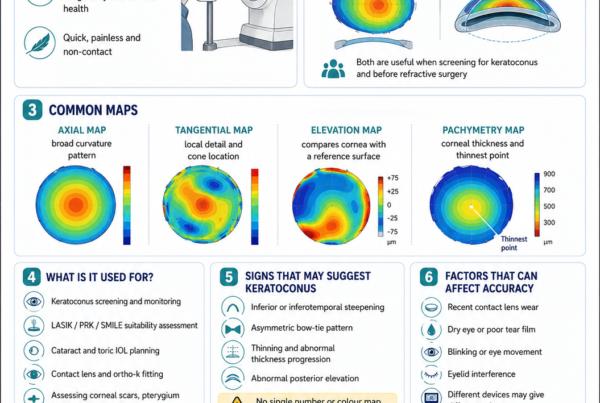
Corneal Topography
Topography maps the anterior corneal curvature.
It may show:
- Inferior steepening
- Central steepening
- Asymmetric bow-tie patterns
- Skewed radial axes
- Irregular astigmatism
- Contact-lens warpage
Corneal Tomography
Tomography evaluates the three-dimensional cornea.
It may assess:
- Anterior elevation
- Posterior elevation
- Corneal curvature
- Thinnest-point location
- Pachymetric progression
- Corneal volume
- Ectasia indices
Serial tomography is central to monitoring progression.
Pachymetry
Pachymetry measures corneal thickness.
The ophthalmologist considers:
- Preoperative thinnest thickness
- Thickness after epithelial removal
- Thickness during riboflavin saturation
- Thickness immediately before ultraviolet treatment
- The intended protocol
- Endothelial safety
Epithelial Thickness Mapping
Epithelial mapping may help identify:
- Epithelial compensation
- Early keratoconus
- Contact-lens warpage
- The relationship between epithelium and stromal shape
- Previous treatment patterns
Corneal Biomechanical Assessment
Selected devices assess the cornea’s response to an air pulse.
These measurements may provide additional information about biomechanical weakness.
They complement rather than replace:
- Tomography
- Topography
- Pachymetry
- Clinical history
- Serial measurements
Corneal Endothelial Cell Count
An endothelial cell count may be performed when:
- The cornea is thin
- Previous intraocular surgery has occurred
- Endothelial disease is suspected
- A modified protocol is planned
- The patient is older
- There are additional risk factors
Ocular-Surface Examination
The examination assesses:
- Tear-film quality
- Corneal staining
- Blepharitis
- Meibomian gland dysfunction
- Allergy
- Active infection
- Epithelial integrity
Contact Lenses Before Measurements
Contact lenses can temporarily distort corneal shape.
They should be discontinued before definitive progression assessment and treatment planning.
The required interval depends on:
- Soft or rigid lens type
- Duration of wear
- Scleral lens use
- Degree of warpage
- Stability of repeated scans
Rigid and scleral lenses may require a longer discontinuation period than ordinary soft lenses.
Dilated Retinal Examination
A retinal examination may be appropriate when:
- Myopia is significant
- Visual loss is not fully explained by the cornea
- Flashes or floaters are present
- Retinal disease is suspected
- A combined refractive procedure is being considered
Standard Epithelium-Off CXL
The traditional epithelium-off procedure is often called the Dresden protocol.
Its classic ultraviolet settings are:
- 3 mW/cm²
- 30 minutes
- Total fluence of 5.4 J/cm²
The original protocol required a minimum stromal thickness of approximately 400 micrometres during treatment to reduce ultraviolet exposure to the corneal endothelium. Long-term clinical studies support durable stabilisation with this protocol.
How Is Epithelium-Off CXL Performed?
Step 1: Confirming the Treatment
The clinical team confirms:
- Patient identity
- Correct eye
- Diagnosis
- Progression
- Intended protocol
- Corneal measurements
- Allergies
- Consent
Step 2: Anaesthetic Drops
Anaesthetic drops numb the corneal surface.
The eyelids and surrounding skin are cleaned.
An eyelid holder keeps the eye open.
Step 3: Removing the Epithelium
The central corneal epithelium is removed.
Possible techniques include:
- Mechanical removal
- Brush-assisted removal
- Alcohol-assisted loosening
- Laser epithelial removal in selected systems
Removing the epithelium allows riboflavin to penetrate the corneal stroma more reliably.
Step 4: Applying Riboflavin
Riboflavin drops are applied repeatedly.
The formulation may differ according to:
- Corneal thickness
- Ultraviolet system
- Intended protocol
- Need to swell or avoid thinning the cornea
Step 5: Confirming Stromal Saturation
The surgeon checks for adequate riboflavin penetration.
Yellow riboflavin may become visible within the cornea and anterior chamber.
Step 6: Measuring Corneal Thickness
The cornea may be measured again after:
- Epithelial removal
- Riboflavin loading
- Any required swelling procedure
Treatment should not proceed under a standard protocol if the cornea is below the safe thickness for that protocol.
Step 7: Ultraviolet A Treatment
Controlled ultraviolet A light is directed onto the cornea.
Riboflavin drops may continue during exposure.
The patient looks towards the treatment light while:
- Centration is maintained
- Corneal hydration is monitored
- Exposure duration is controlled
- Riboflavin coverage is maintained
Step 8: Irrigation
The eye may be rinsed with sterile solution after ultraviolet exposure.
Step 9: Applying Medication
Antibiotic and other prescribed drops are applied.
Step 10: Placing a Bandage Contact Lens
A bandage contact lens protects the exposed surface while the epithelium heals.
It reduces:
- Eyelid friction
- Exposure of corneal nerves
- Early discomfort
The lens remains until epithelial closure is adequate.
What Is Accelerated CXL?
Accelerated CXL uses greater ultraviolet intensity for a shorter duration.
Common examples include:
- 9 mW/cm² for 10 minutes
- 18 mW/cm² for 5 minutes
- Other device-specific protocols
The total intended ultraviolet energy may be similar to the standard protocol.
However, biological CXL is not determined by energy alone.
Other important factors include:
- Oxygen availability
- Riboflavin concentration
- Ultraviolet delivery pattern
- Continuous versus pulsed exposure
- Corneal hydration
- Epithelial status
- Treatment depth
Meta-analyses and registry studies suggest that commonly used standard and accelerated epithelium-off protocols can provide broadly comparable stabilisation in many patients. Some studies have found differences in flattening, treatment depth or other secondary outcomes, and not every accelerated protocol should be regarded as equivalent.
Advantages of Accelerated CXL
Possible advantages include:
- Shorter ultraviolet-treatment time
- Reduced total appointment time
- Easier cooperation in children
- Less exposure-related dehydration in selected protocols
- Improved patient comfort during treatment
Limitations of Accelerated CXL
Possible limitations include:
- Shallower treatment effect with some high-intensity protocols
- Reduced oxygen availability during continuous high-intensity exposure
- Less long-term evidence for certain protocols
- Differences between devices and riboflavin formulations
- Uncertainty about whether extremely rapid protocols provide equal durability
What Is Pulsed CXL?
Pulsed CXL alternates ultraviolet exposure with periods when the light is switched off.
The theoretical purpose is to allow oxygen to diffuse back into the cornea between pulses.
Whether pulsed delivery offers meaningful superiority depends on:
- Irradiance
- Pulse cycle
- Riboflavin formulation
- Epithelial status
- Treatment device
What Is Epithelium-On CXL?
Epithelium-on CXL is also called:
- Epi-on CXL
- Transepithelial CXL
The corneal epithelium is left intact.
Potential advantages include:
- Less pain
- Faster recovery
- Lower infection risk
- No large epithelial defect
- Less early haze
- Reduced need for a bandage contact lens in some protocols
The main challenge is that the intact epithelium limits penetration of:
- Riboflavin
- Oxygen
- Ultraviolet energy
Earlier transepithelial techniques often produced a shallower or less consistent treatment effect than conventional epithelium-off CXL.
A 2025 systematic review of randomised trials found that epi-on treatment had safety and recovery advantages, while efficacy depended heavily on the specific protocol and was not uniformly equivalent to epithelium-off CXL.
In October 2025, the United States FDA approved a specific epithelium-on riboflavin and oxygen-assisted system for keratoconus in patients aged 13 years and older. This approval applies to that particular drug-device protocol and does not establish that all forms of epi-on CXL are interchangeable or equally effective. Availability and regulatory status differ between countries.
How Do Modern Epi-On Protocols Improve Penetration?
Approaches may include:
- Special riboflavin formulations
- Chemical penetration enhancers
- Iontophoresis
- Supplemental oxygen
- Longer loading times
- Altered ultraviolet delivery
- Device-specific epithelial preparation
The outcomes of one epi-on system should not automatically be applied to another.
Is Epi-On Better Than Epi-Off?
Neither description alone is enough to determine the best procedure.
Epithelium-off CXL has:
- The most established long-term evidence
- Reliable stromal riboflavin penetration
- A deeper treatment effect
- Greater early discomfort and surface risk
Epithelium-on CXL may offer:
- Less pain
- Faster functional recovery
- Lower epithelial-defect-related risk
- A potentially shallower or protocol-dependent effect
The decision depends on:
- Age
- Disease severity
- Progression rate
- Corneal thickness
- Previous surgery
- Ocular-surface health
- Available validated protocols
- Surgeon experience
CXL for Thin Corneas
The traditional 400-micrometre threshold was introduced to protect the endothelium during standard epithelium-off treatment.
Some progressive keratoconic corneas are thinner than this after epithelial removal.
Modified options include:
- Hypo-osmolar riboflavin swelling
- Contact-lens-assisted CXL
- Individualised sub-400 protocols
- Reduced ultraviolet exposure time
- Epithelium-on treatment
- Specialised targeted protocols
Hypo-Osmolar Riboflavin
Hypo-osmolar riboflavin may temporarily swell a thin cornea.
Limitations include:
- Variable swelling
- Corneal thinning during ultraviolet exposure
- Uncertain effective stromal concentration
- A potentially less predictable biomechanical effect
Contact-Lens-Assisted CXL
A riboflavin-soaked ultraviolet-transparent soft contact lens is placed on the cornea to increase the functional treatment thickness.
The technique permits epithelium-off CXL in selected thin corneas.
Published reviews report encouraging outcomes, but ultraviolet absorption by the lens and altered oxygen availability must be considered.
The Sub-400 Protocol
The sub-400 approach individualises ultraviolet exposure according to the intraoperative stromal thickness.
Thinner corneas receive shorter exposure to limit ultraviolet dose at the endothelium.
Early clinical studies report promising safety and stabilisation in ultrathin corneas, but the evidence base is smaller and less mature than for the standard Dresden protocol.
Is a Thin Cornea Untreatable?
Not necessarily.
A thin cornea requires:
- Precise intraoperative pachymetry
- An appropriate modified protocol
- Careful endothelial protection
- Realistic counselling
- Experienced surgical planning
A thin cornea should not simply receive the standard protocol without adjustment.
What Is Customised CXL?
Customised or topography-guided CXL applies a non-uniform ultraviolet pattern.
Greater energy may be directed towards the ectatic cone while less energy is applied to more normal tissue.
Potential goals include:
- Targeted biomechanical strengthening
- Greater cone flattening
- Reduced unnecessary treatment of normal tissue
- Improved corneal regularity
Early clinical research is promising, but conventional broad-zone CXL has a larger long-term evidence base.
Can CXL Be Combined with Topography-Guided PRK?
Yes, in carefully selected patients.
CXL stabilises the cornea.
Topography-guided PRK removes a limited amount of tissue to improve corneal regularity and visual function.
The combined strategy may be performed:
- Simultaneously
- Sequentially after stabilisation
This approach is sometimes associated with the term Athens Protocol.
It is not ordinary refractive laser surgery.
The objective is usually to:
- Regularise the cornea
- Improve corrected vision
- Reduce irregular astigmatism
- Improve contact-lens or spectacle function
It does not necessarily produce spectacle independence.
Ten-year prospective data support durable outcomes in selected patients undergoing combined topography-guided surface ablation and CXL, but careful tissue limitation and patient selection remain essential.
Can CXL Be Combined with Intracorneal Rings?
Yes.
Intracorneal ring segments or corneal allogenic intrastromal ring segments may improve corneal shape.
CXL may be used to stabilise the underlying ectatic process.
The procedures serve different purposes:
- Ring segments aim to modify shape.
- CXL aims to reduce progression.
The sequence and timing are individualised.
Does CXL Replace Contact Lenses?
No.
After CXL, patients may still require:
- Spectacles
- Soft toric lenses
- Custom soft keratoconus lenses
- Rigid gas-permeable lenses
- Hybrid lenses
- Scleral lenses
The contact-lens fit may change as the cornea stabilises or flattens.
When Can Contact Lenses Be Worn Again?
The timing depends on:
- Epithelial healing
- Corneal staining
- Haze
- Lens type
- Corneal shape
- Surgeon instructions
Soft lenses may sometimes resume earlier than rigid or scleral lenses.
Rigid lenses should generally be refitted after sufficient corneal stabilisation rather than immediately after treatment.
What Happens Immediately After Epithelium-Off CXL?
The eye may feel comfortable while the anaesthetic remains active.
As it wears off, the patient may experience:
- Burning
- Watering
- Light sensitivity
- Pain
- Eyelid swelling
- Blurred vision
- Difficulty opening the eye
Resting in a dim environment may help.
Medication After CXL
Medication commonly includes:
- Antibiotic drops
- Steroid drops after epithelial closure or according to protocol
- Preservative-free lubricants
- Oral pain medication
- Other ocular-surface treatment
Medication regimens vary.
Patients should not use topical anaesthetic drops at home unless specifically supervised because repeated anaesthetic use can delay healing and cause corneal toxicity.
The Bandage Contact Lens
The bandage contact lens:
- Protects the healing surface
- Reduces eyelid friction
- Improves comfort
- Supports re-epithelialisation
It is removed once the epithelial defect has healed adequately.
The patient should not remove or replace it independently.
The First Three Days
Symptoms may include:
- Significant pain
- Burning
- Light sensitivity
- Watering
- Blurred vision
- Foreign-body sensation
Pain should begin to improve as the epithelium closes.
Increasing pain after initial improvement requires urgent review.
Days Three to Seven
The epithelial defect usually closes during this period.
The bandage contact lens may then be removed.
Vision often remains:
- Blurred
- Hazy
- Fluctuating
- Light-sensitive
The First Month
Possible symptoms include:
- Dryness
- Glare
- Halos
- Haze
- Variable refraction
- Reduced contrast
- Ghosting
Temporary worsening of vision does not necessarily mean that keratoconus has progressed.
Three to Six Months
The cornea continues remodelling.
Changes may include:
- Gradual flattening
- Changing refraction
- Altered contact-lens fit
- Reduction or redistribution of haze
- Improving corrected vision
Six to Twelve Months
Many patients reach a more stable refractive and tomographic state during this period.
Some continue changing beyond one year.
Can I Use Screens?
Screens do not reverse cross-linking.
However, prolonged screen use reduces blinking and may worsen:
- Dryness
- Burning
- Fluctuating vision
- Eye fatigue
When Can I Drive?
Driving should resume only when:
- Vision meets the legal standard
- The patient feels confident
- Light sensitivity is manageable
- The bandage lens has been removed when required
- Pain medication no longer impairs alertness
- The ophthalmologist has not advised otherwise
When Can I Exercise?
Gentle walking is usually possible.
Strenuous exercise may be restricted during early healing.
Patients should avoid:
- Sweat entering the eye
- Eye rubbing
- Dusty environments
- Direct eye trauma
- Contact sports
When Can I Swim?
Swimming should be avoided while:
- The epithelium is healing
- A bandage contact lens is present
- Antibiotic treatment is ongoing
- The ocular surface remains compromised
Pool, sea and spa water may contain microorganisms capable of causing serious infection.
When Can I Wear Eye Makeup?
Eye makeup should be avoided during early healing.
Old eye cosmetics and applicators may be contaminated.
When Can I Fly?
Uncomplicated CXL does not place a gas bubble inside the eye and usually does not prevent flying.
However:
- Aircraft cabins may worsen dryness.
- Early postoperative reviews should not be missed.
- Access to urgent care should be considered.
- Significant pain or incomplete epithelial healing should be addressed before travel.
Expected Results
The most important successful outcome is the absence of continuing progression.
Possible additional outcomes include:
- Mild reduction in maximum keratometry
- Corneal flattening
- Improved corrected vision
- Reduced irregular astigmatism
- More stable contact-lens fit
In a landmark randomised trial, untreated control eyes continued to steepen while treated eyes demonstrated stabilisation and average flattening over three years.
Long-term studies report stabilisation lasting up to 13 years in many patients treated with the standard epithelium-off protocol.
Does the Cornea Always Flatten?
No.
A successfully treated cornea may:
- Flatten
- Remain essentially unchanged
- Show small measurement fluctuations
Stability without flattening is still a successful result when progression has stopped.
Can Vision Initially Become Worse?
Yes.
Temporary worsening may result from:
- Epithelial irregularity
- Corneal swelling
- Haze
- Dry eye
- Changing corneal shape
- Altered refraction
Vision should be monitored over time rather than judged from the first few postoperative weeks.
Can Keratoconus Progress After CXL?
Yes.
Failure or renewed progression is uncommon but possible.
Risk may be higher in:
- Younger patients
- Advanced keratoconus
- Very steep corneas
- Highly decentered cones
- Persistent eye rubbing
- Inadequate treatment penetration
- Less effective protocols
- Long follow-up periods
Paediatric long-term studies have reported higher re-progression rates than typical adult series, reinforcing the need for continued tomography after treatment.
How Is Treatment Failure Diagnosed?
Possible signs include reproducible:
- Increasing Kmax
- Increasing anterior steepness
- Increasing posterior elevation
- Progressive thinning
- Worsening irregular astigmatism
- Deteriorating corrected vision
- Changing contact-lens fit
A single abnormal scan should usually be repeated before declaring failure.
Can CXL Be Repeated?
Yes, in selected cases.
Repeat CXL may be considered when:
- Genuine progression is confirmed
- The cornea remains thick enough
- The endothelium is healthy
- The expected benefit exceeds the added risk
- Corneal transplantation is not yet more appropriate
The second procedure may use:
- Standard epithelium-off CXL
- A modified thin-cornea protocol
- A targeted protocol
- Another validated approach
Risks and Complications
Pain
Pain is common after epithelium-off treatment.
It should improve as the epithelium heals.
Severe or worsening pain may indicate:
- Infection
- A persistent epithelial defect
- Bandage-lens displacement
- Sterile inflammation
- Herpetic disease
Delayed Epithelial Healing
The epithelium may take longer than expected to close.
Risk factors include:
- Severe dry eye
- Diabetes
- Neurotrophic cornea
- Previous corneal surgery
- Medication toxicity
- Infection
- Ocular-surface disease
Delayed healing increases the risk of infection and scarring.
Infectious Keratitis
Infectious keratitis is uncommon but potentially sight-threatening.
The vulnerable period is generally while:
- The epithelial defect remains open
- A bandage contact lens is present
- Steroid medication is being used
Reported organisms include:
- Bacteria
- Fungi
- Acanthamoeba
- Herpes viruses
A systematic review found that bacterial infection was the most commonly reported form and that infections typically presented within the first postoperative week.
Warning symptoms include:
- Increasing pain
- Increasing redness
- Worsening vision
- Thick discharge
- A white corneal spot
- Increasing light sensitivity
Sterile Infiltrates
Sterile inflammatory white spots may develop without active infection.
However, infection must be excluded urgently before assuming that an infiltrate is sterile.
Corneal Haze
Some stromal haze is expected after many epithelium-off treatments.
It may become more visible during the first several months.
Mild haze often improves gradually.
More significant haze may cause:
- Blurred vision
- Glare
- Reduced contrast
- Light scatter
- Reduced corrected vision
Risk may be greater with:
- Advanced keratoconus
- Greater treatment depth
- Delayed epithelial healing
- Infection
- Strong inflammatory response
- Poor steroid adherence
- Ultraviolet exposure during healing
Corneal Scarring
Permanent scarring is uncommon but may follow:
- Infection
- Severe haze
- Delayed healing
- Corneal melt
- Advanced pre-existing disease
- An excessive inflammatory response
Corneal Endothelial Damage
The corneal endothelium can be damaged if excessive ultraviolet energy reaches the back of a thin cornea.
Possible consequences include:
- Corneal swelling
- Reduced transparency
- Endothelial cell loss
- Corneal decompensation
This is why intraoperative thickness assessment and modified thin-cornea protocols are important.
Corneal Melt
Corneal melt is extremely rare.
Potential associations include:
- Infection
- Autoimmune disease
- Severe ocular-surface disease
- Persistent epithelial defects
- Excessive inflammation
- Medication toxicity
Dry Eye
Temporary dry-eye symptoms may occur because of:
- Epithelial removal
- Corneal nerve disruption
- Postoperative drops
- Reduced blinking
- Pre-existing ocular-surface disease
Possible symptoms include:
- Burning
- Grittiness
- Fluctuating vision
- Redness
- Light sensitivity
Increased or Irregular Astigmatism
The cornea may continue remodelling after CXL.
A temporary or permanent change in refraction may occur.
Spectacles or contact lenses should be updated after sufficient stability.
Loss of Corrected Vision
Rarely, a patient may lose lines of corrected vision because of:
- Infection
- Scarring
- Significant haze
- Irregular healing
- Continued progression
- Another ocular disease
Common Myths
“Cross-Linking Removes Keratoconus”
False.
It aims to stabilise the condition.
“CXL Is Laser Eye Surgery to Remove Spectacles”
False.
It is a biomechanical stabilisation treatment.
“CXL Always Improves Vision”
False.
Vision may improve, remain unchanged or temporarily worsen.
“If Vision Does Not Improve, CXL Failed”
False.
Stabilisation without visual improvement may still represent successful treatment.
“CXL Makes the Cornea Thick Again”
False.
The procedure does not restore lost corneal tissue.
“Epi-On CXL Is Always as Effective as Epi-Off CXL”
False.
Efficacy depends on the specific epi-on formulation and ultraviolet protocol.
“All Accelerated Protocols Are Equivalent”
False.
Treatment time, intensity, oxygen availability and ultraviolet delivery matter.
“The Demarcation Line Proves Success”
False.
It is an anatomical sign of treatment effect, not a guarantee of long-term stabilisation.
“Children Should Always Be Observed Until Progression Is Proven”
Not necessarily.
Young patients may progress rapidly, and treatment decisions should consider age, disease severity and the risk of waiting.
“CXL Eliminates the Need for Contact Lenses”
False.
Many patients continue to need specialised contact lenses.
“A Thin Cornea Cannot Be Cross-Linked”
Not necessarily.
Modified protocols may be possible in selected eyes.
“Cross-Linking Prevents All Future Corneal Transplants”
False.
It may reduce the risk of advanced progression, but some eyes still require transplantation.
“Once Treated, No Further Scans Are Needed”
False.
Long-term tomography remains necessary.
“Riboflavin Tablets and Sunlight Can Replace Medical CXL”
False.
Medical CXL uses sterile ophthalmic riboflavin, controlled stromal delivery, calibrated ultraviolet exposure and corneal safety monitoring.
Oral riboflavin and uncontrolled sunlight are not substitutes for a validated procedure.
Frequently Asked Questions
How Do I Know Whether My Keratoconus Is Progressing?
Progression is assessed by comparing reliable serial examinations.
The ophthalmologist looks for changes in:
- Corneal curvature
- Posterior elevation
- Thickness
- Refraction
- Corrected vision
- Contact-lens fit
Should Both Eyes Be Treated?
Only an eye that meets the treatment indication should be treated.
One eye may require CXL while the other is monitored.
Can Both Eyes Be Treated on the Same Day?
It is possible, but practice varies.
Treating one eye at a time may allow:
- Better early function from the untreated eye
- Assessment of first-eye healing
- Reduced bilateral infection risk
- Easier pain management
Bilateral treatment may reduce the number of recovery periods.
What Happens if I Blink During Treatment?
An eyelid holder prevents normal blinking.
The clinical team monitors fixation and corneal exposure.
What Happens if I Move My Eye?
The patient is asked to look towards the ultraviolet light.
Small movements can be corrected by repositioning.
Some modern systems include tracking or alignment aids.
Can the Ultraviolet Light Damage the Retina?
Riboflavin absorbs ultraviolet energy within the cornea.
When validated protocols and appropriate stromal-thickness limits are followed, exposure to deeper structures is kept below harmful levels.
Will the Procedure Change My Eye Colour?
No.
The iris is not treated.
Will the Procedure Treat My Astigmatism?
CXL may produce some flattening or refractive change but is not a predictable astigmatism-correction procedure.
Will I Need New Spectacles?
Possibly.
The prescription may change during corneal remodelling.
A new prescription should usually be measured after sufficient stabilisation.
When Can I Refit My Contact Lenses?
This depends on epithelial healing and corneal stability.
Rigid and scleral lenses may need refitting because the corneal shape can change.
Can I Have LASIK After CXL?
Routine LASIK is generally inappropriate in a keratoconic cornea because the flap and stromal ablation can further weaken the cornea.
Selected patients may undergo limited topography-guided PRK combined with or after CXL under a specialised therapeutic protocol.
Can I Have ICL Surgery After CXL?
Possibly.
An ICL may correct stable residual myopia and regular astigmatism without removing additional corneal tissue.
It does not correct severe irregular astigmatism.
The keratoconus should be stable and the internal eye anatomy suitable.
Can CXL Be Performed After LASIK?
Yes.
CXL is an established treatment for progressive post-LASIK ectasia.
The technique may differ according to:
- Flap anatomy
- Corneal thickness
- Ectasia severity
- Surgeon preference
Can CXL Be Performed After a Corneal Ring Implant?
Yes.
CXL and intracorneal ring segments address different aspects of the disease.
The sequence is individualised.
Can CXL Be Performed Again?
Yes, when genuine re-progression is confirmed and the cornea remains suitable.
Does CXL Cause Cataracts?
Standard corneal CXL does not normally expose the natural lens to a cataract-forming ultraviolet dose when appropriate safety protocols are followed.
Can CXL Cause Blindness?
Severe permanent visual loss is rare but possible through complications such as:
- Infection
- Scarring
- Endothelial failure
- Corneal melt
- Uncontrolled progression
Can Keratoconus Return After CXL?
The diagnosis remains present.
The more accurate question is whether progression can recur.
It can, although most appropriately treated adult eyes remain stable.
How Frequently Should I Be Reviewed?
The schedule depends on:
- Age
- Severity
- Protocol
- Healing
- Progression risk
- Whether the other eye is untreated
Common follow-up intervals include:
- The first postoperative day
- Several days later for epithelial review
- One month
- Three months
- Six months
- Twelve months
- Annual long-term review
When to Seek Urgent Eye Care
Seek urgent assessment for:
- Increasing pain
- Increasing redness
- Rapidly worsening vision
- Thick discharge
- A white or grey corneal spot
- Marked eyelid swelling
- Severe light sensitivity
- A bandage contact lens that falls out with significant discomfort
- Pain that worsens after initially improving
- Symptoms that do not improve after epithelial healing
- Eye trauma
- New flashes or floaters
- A curtain or shadow in the vision
These symptoms may indicate:
- Infection
- Delayed epithelial healing
- Sterile inflammation
- Raised eye pressure
- Herpetic keratitis
- Another ocular emergency
A Corneal Cross-Linking Assessment Checklist
Symptoms to Report
- Increasing blur
- Ghosting
- Glare
- Halos
- Changing spectacles
- Contact lenses fitting differently
- Reduced contact-lens tolerance
- Eye rubbing
- Itchy eyes
- Monocular double vision
Medical Information to Report
- Diabetes
- Autoimmune disease
- Pregnancy or breastfeeding
- Atopy
- Eczema
- Asthma
- Medication
- Previous poor wound healing
- Previous herpes infection
Eye History to Report
- Keratoconus
- Pellucid marginal degeneration
- LASIK
- PRK
- Radial keratotomy
- Corneal cross-linking
- Intracorneal ring segments
- Eye trauma
- Corneal infection
- Contact-lens wear
- Family history of keratoconus
Tests That May Be Required
- Unaided visual acuity
- Corrected visual acuity
- Manifest refraction
- Cycloplegic refraction
- Corneal topography
- Corneal tomography
- Pachymetry
- Epithelial thickness mapping
- Biomechanical assessment
- Endothelial cell count
- Ocular-surface assessment
- Dilated retinal examination
Questions to Ask the Surgeon
- Is my keratoconus definitely progressing?
- Which measurements demonstrate progression?
- Is treatment needed now or can the eye be monitored?
- Is epithelium-off or epithelium-on CXL recommended?
- Which exact protocol will be used?
- Is the protocol standard or accelerated?
- How thick is my cornea?
- What will the thickness be after epithelial removal?
- Is a thin-cornea modification required?
- What is the main goal in my eye?
- How likely is vision to improve?
- How likely is progression to continue?
- What pain-control plan will be used?
- How long will the bandage lens remain?
- When can I return to work?
- When can I resume contact lenses?
- What symptoms require urgent review?
- How frequently will tomography be repeated?
- What options remain if progression continues?
The Bottom Line
Corneal cross-linking uses riboflavin and controlled ultraviolet A light to strengthen a biomechanically weakened cornea.
It is used mainly for:
- Progressive keratoconus
- Progressive post-refractive-surgery ectasia
- Selected other corneal ectatic disorders
The main objective is to:
- Slow or stop progression
- Preserve useful vision
- Reduce the risk of advanced corneal distortion
- Reduce the likelihood that more invasive treatment will be required
CXL does not:
- Remove keratoconus
- Replace lost corneal tissue
- Guarantee spectacle independence
- Predictably correct all myopia or astigmatism
- Guarantee that progression will never recur
Standard epithelium-off CXL has the strongest long-term evidence.
Accelerated epithelium-off protocols can provide comparable outcomes in many patients, although protocols differ.
Modern epithelium-on treatments offer the potential for less pain and faster recovery, but results should be interpreted according to the specific validated riboflavin and ultraviolet system rather than the general label “epi-on”.
Thin corneas may require:
- Contact-lens-assisted treatment
- Hypo-osmolar riboflavin
- An individualised sub-400 protocol
- Another modified approach
Possible complications include:
- Pain
- Delayed epithelial healing
- Infection
- Sterile infiltrates
- Haze
- Scarring
- Dry eye
- Endothelial damage
- Rare continued progression
The most important message is:
Corneal cross-linking is a stabilising procedure, not primarily a vision-correction procedure. The best opportunity to preserve vision is often to identify genuine progression before the cornea becomes severely thin, scarred or irregular, select an appropriate evidence-based protocol and continue long-term tomographic monitoring after treatment.
References
- Cortina MS, Greiner MA, Kuo AN, et al. Safety and efficacy of epithelium-off corneal collagen cross-linking for the treatment of corneal ectasia: a report by the American Academy of Ophthalmology. Ophthalmology. 2024;131(10):1234–1242. PMID: 38935041.
- Wittig-Silva C, Chan E, Islam FMA, Wu T, Whiting M, Snibson GR. A randomised, controlled trial of corneal collagen cross-linking in progressive keratoconus: three-year results. Ophthalmology. 2014;121(4):812–821. PMID: 24393351.
- Hoyer A, Raiskup-Wolf F, Spörl E, Pillunat LE. Collagen cross-linking with riboflavin and UVA light in keratoconus: results from Dresden. Ophthalmologe. 2009;106:133–140. PMID: 18604541.
- Raiskup F, Theuring A, Pillunat LE, Spoerl E. Corneal cross-linking in patients with keratoconus: up to 13 years of follow-up. Graefes Arch Clin Exp Ophthalmol. 2023;261:1153–1160. PMID: 36197498.
- Kymionis GD, Grentzelos MA, Liakopoulos DA, et al. Long-term follow-up of corneal collagen cross-linking for keratoconus—the Cretan study. Cornea. 2014;33(10):1071–1079. PMID: 25170581.
- Hersh PS, Stulting RD, Muller D, Durrie DS, Rajpal RK. United States multicentre clinical trial of corneal collagen cross-linking for treatment of corneal ectasia after refractive surgery. Ophthalmology. 2017;124(10):1475–1484. PMID: 28655538.
- McCall AS, Kraft S, Edelhauser HF, et al. Mechanisms of corneal tissue cross-linking in response to treatment with topical riboflavin and long-wavelength ultraviolet radiation. Invest Ophthalmol Vis Sci. 2010;51:129–138. PMID: 19643975.
- Belin MW, Lim L, Rajpal RK, Hafezi F, Gomes JAP, Cochener B. Corneal cross-linking: current protocols and clinical approach. Surv Ophthalmol. 2018.
- Kim CY, Kim MK. Effect of the standard and accelerated corneal cross-linking protocols on keratoconus: meta-analysis of randomised controlled trials. 2024. PMID: 39539159.
- Watson SL, et al. Comparison of standard versus accelerated corneal cross-linking: five-year outcomes from the Save Sight Keratoconus Registry. Br J Ophthalmol. 2024. PMID: 37369766.
- Al-Shammari YM, Aljassar F, Al-Shammari SM, Alrewais AA. Comparative effectiveness of accelerated and standard corneal cross-linking in paediatrics: a systematic review and meta-analysis. Graefes Arch Clin Exp Ophthalmol. 2026. PMID: 42270917.
- Morgado CR, Larivoir NB, Santos JF, Santhiago MR. Corneal cross-linking in paediatric populations: systematic review and future perspectives. J Refract Surg. 2025;41(9)–e1021. PMID: 40923612.
- Godefrooij DA, Soeters N, Imhof SM, Wisse RPL. Corneal cross-linking for paediatric keratoconus: long-term results. Cornea. 2016;35(7):954–958. PMID: 27027921.
- Gumus Kasapoglu G, et al. Long-term results of accelerated corneal collagen cross-linking in paediatric patients with progressive keratoconus: ten-year follow-up. Eye. 2024. PMID: 38609652.
- Nath S, Shen C, Koziarz A, et al. Efficacy and safety of epi-on versus epi-off corneal cross-linking in corneal ectasia: systematic review and meta-analysis of randomised controlled trials. Clin Ophthalmol. 2025. PMID: 40357455.
- US Food and Drug Administration. Epioxa HD and Epioxa orphan-drug approval record for epithelium-on corneal collagen cross-linking. Marketing approval date: 17 October 2025.
- Srivatsa S, Jacob S, Agarwal A. Contact-lens-assisted corneal cross-linking in thin ectatic corneas: a review. Indian J Ophthalmol. 2020;68(12):2773–2778. PMID: 33229652.
- Karaca EE, Asfuroğlu Y, Ulusoy DM, Kemer ÖE. Evaluating the safety and effectiveness of the sub-400 corneal cross-linking protocol: initial clinical and morphological findings. Int Ophthalmol. 2025;45:16. PMID: 39702781.
- Murchison CE, Petroll WM, Robertson DM. Infectious keratitis after corneal cross-linking: a systematic review. J Cataract Refract Surg. 2021;47(8):1075–1080. PMID: 33769765.
- Kanellopoulos AJ. Simultaneous topography-guided custom ablation with corneal cross-linking for keratoconus: ten-year prospective outcomes. J Refract Surg. 2023. PMID: 37937755.